Many uncertainties still exist regarding to the etiology of CBP while many hypotheses have been advocated. Since the 4th edition of the WHO classification of pheochromocytomas and paragangliomas (PPGL), PPGL is no longer classified as simply benign or malignant because any PPLG can have metastatic potential and lack clear characteristics needed to predict metastatic behavior [2]. Additionally, some tumors are fatal without metastatic proliferation due to local invasion of critical structures. Therefore, any PPGL, including CBP, should be approached as having a metastatic potential with a genetic predisposition [3]. Furthermore, PPGLs are among the most common hereditary tumors in humans, with mutation rates reaching approximately 40% and 85% in adult and pediatric patients, respectively [8, 9]. Immunohistochemical biomarkers, morphological features, and biochemical findings play a crucial role in germline susceptibility assessment [10]. The importance of the genetic study has been emphasized, particularly SDHB mutation examination in patients with a family history of bilateral occurrence, which may be prognostically unfavorable in head and neck locations. The 2022 WHO classification encourages the routine use of SDHB immunohistochemistry as a surrogate biomarker in SDHx-related pathogenesis [3].
Although active surveillance is reportedly a superior option for CBP management [11, 12], complete surgical resection is believed as a primary treatment of choice when it is available [1, 6, 13]. When considering surgical removal of CBP, we worry about two things; intraoperative bleeding and CNP. Intraoperatvie bleeding during CBP surgery is notorious due to its too many feeder vessels and risk of carotid injury. According to previous studies, 7–24% of cases required vascular reconstruction during the CBP surgery. To reduce the risk of surgical bleeding and avoid carotid injury during the surgery, preoperative tumor embolization through the feeding vessels were conducted [14, 15]. However, it carries small risk of stroke and it can extend the length of hospital stay, may trigger inflammation and lead to increased blood loss during surgery [16, 17]. In past we routinely performed preoperative tumor embolization, however we do not perform now. In our series, one patient required primary repair of the damaged ICA and another patient underwent carotid shunt insertion and angioplasty using a bovine pericardial patch. In case of severe tumor adhesion to the ECA, reconstruction of ECA is not essential after an enbloc resection of the tumor with ECA. Howeveer, ICA injury requires immediate reconstruction often using systemic heparinization, carotid artery clamping and carotid shunting and meticulous repair. Therefore, surgical team should be prepared for this event.
Second thing we have to consider is cervical CNP. It is the most common surgical complication during CBP extirpation, with a reported incidence rate of up to 50% [18]. The potential risk or nerve injury may arise from these tumors being close to the surrounding nerve structures, limited accessibility and visibility of the nerve structures during surgery due to bleeding, high location of the tumor, and distortion of normal anatomical relationships. Previous studies have tried to identify risk factors for cervical CN injury using Shamblin classification, tumor location from the the skull base, or tumor size [19, 20]. In our series, multivariable logistic regression analysis identified an estimated blood loss (EBL) as the only risk factor for cervical CNP. Amount of EBL may encompass difficulty of surgical procedure, carotid injury and large tumor mass. Even in cases of high Shamblin class or unfavorable tumor location, we have experienced that an effective feeder vessel suture ligation before tumor dissection can minimize bleeding and consequently reduce CN injury. Marginal branch of the facial nerve palsy usually occur due to traction injury and usually recovered spontaneously. The vagus and hypoglossal nerves are prone to be injured during CBP surgery [21]. Both nerves were visually assessed and preserved during surgery for all our cases. Subsequently, any related symptoms, such as aspiration and tongue deviation, were temporary. Hoarseness that lasted more than 6 month was observed in two patients (9.5%), which appeared to be due to recurrent laryngeal nerve injury. Damage to the cervical sympathetic nerve can cause Horner’s syndrome or first bite syndrome which is caused by denervation hypersensitivity [22]. First bite syndrome developed in two patients, however the pain symptom on the first bite improved within 1 month after its occurrence.
Most CBPs are first observed as a palpable mass or in routine health chekcup; therefore, patients do not usually consider them seriously. With the increased understanding of the tumor, metastatic potential and difficulty with surgery, it shoud be considered as a potentially serious condition. Since this issue is directly related to quality of life, patients should be informed about the potential risks before treatment, and a multidisciplinary follow-up approach should be considered. Daniel et al. conducted a trial of active surveillance in patients without symptoms or who did not agree to undergo surgery for CBPs and suggested that active surveillance is reasonable for CBP management. 24% of patients did not undergo surgery throughout the study. According to them, metastasis occurred in 3–6% and 15% in bilateral CBPs [11]. Additionally, if a tumor progression, cervical CNP, or vessel invasion occurs during active surveillance, the possibility of permanent damage to the nerve increases, making complete resection impossible.
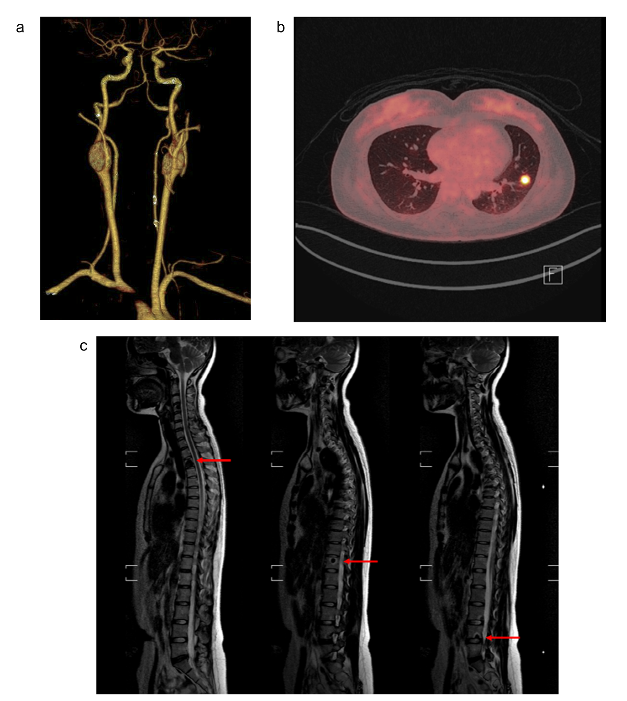
Among our cohort, one patient with bilateral CBP showed multiple distant metastases. In patients with bilateral CBPs, there has been concern that bilateral carotid body resection might damage carotid body functions critical for survival; however, published results show that mortality rates do not increase due to bilateral carotid body resection despite the preexisting severe comorbidity [23]. The progressive behavior of CBPs has a diverse morphological presentation in the form of increased mitotic activity, vascular invasion, neural peripheral invasion, and cell polymorphism; however, none of these morphological features has been found to accurately predict these tumors’ behavior. Therefore, surgical extirpation remains the standard treatment for CBPs to decrease morbidity and mortality in these patients.
This study has some limitations. First, it was a retrospective study with a small sample size due to the rarity of the disease. Second, the follow-up period was short; however, 100% of the patients underwent the outpatient follow-up to detect tumor recurrence or metastasis. Though there was no recurrence during the follow-up period, long-term follow-up is required to see distant metastasis of tumor recurrence. In the furure, tumor metastasis and GAPP score will be reported.


留言 (0)