
記住我


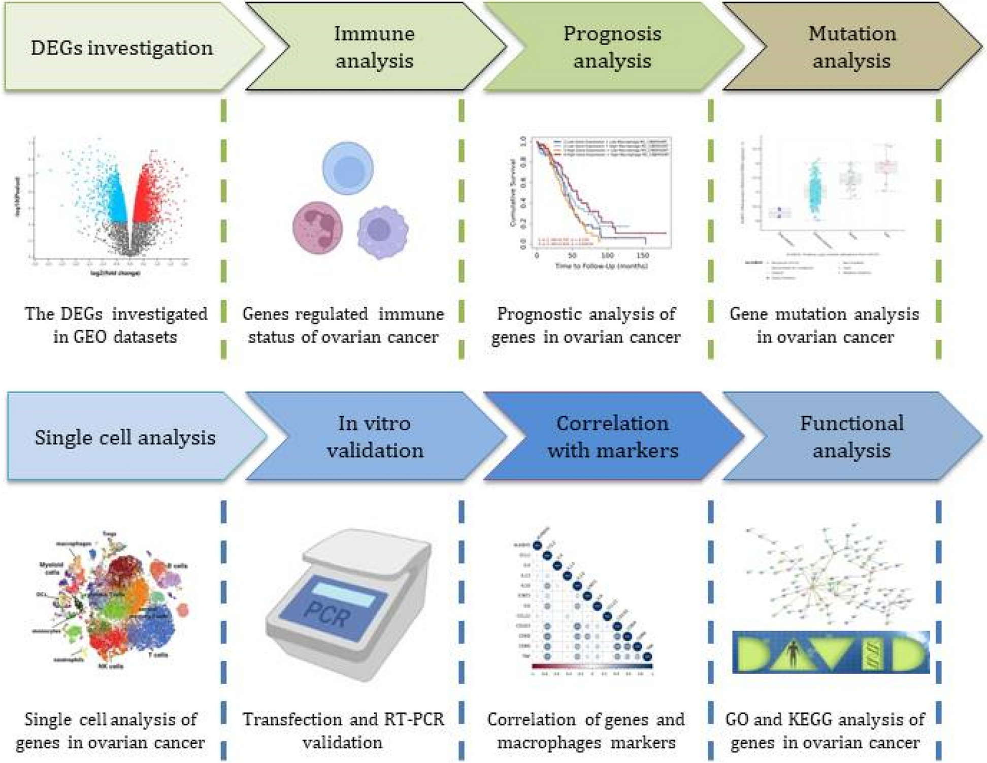
In Fig. 1, we constructed a flow diagram to represent our study design. In this study, we collected two independent GEO datasets, GSE35495 and GSE36537 to demonstrate the DEGs between M0 and M2 macrophages (Supplementary Fig. 1A-B). All the m6A methylation enzymes were analyzed to demonstrate differential expression in M0 and M2 macrophages, which showed most of the enzymes exhibited distinct expression between these two types of macrophages (Fig. 2A). However, the m6A methylation enzymes which were up-regulated in M2 macrophages were not that much. In the venn diagram, we found that only ALKBH5 and IGF2BP2 worked as the genes that up-regulated in M2 macrophages both in GSE36537 and GSE39495 (Fig. 2B). To further validate the relative expression of ALKBH5 and IGF2BP2 in M0 and M2 macrophages, GEO datasets GSE108312 and GSE35449 were used. The results showed that ALKBH5 and IGF2BP2 were both expressed highly in M2 macrophages (Fig. 2C-D). In Fig. 2E, we investigated the correlation between the m6A methylation enzymes in the two GEO datasets, which showed most of the enzymes had closely correlation. Heatmap plot was used to represent the m6A methylation enzymes in these two datasets (Supplementary Fig. 1C). We found that the expression of ALKBH5 was relatively high in macrophages, while IGF2BP2 was expressed at low levels in macrophages. Next, we would like to investigate the correlation of ALKBH5 and IGF2BP2 with immune cells expression in ovarian cancer microenvironment. Thus, we further investigated the role of ALKBH5 and IGF2BP2 played in immune cells using TIMER2.0 database. Results showed that the expression of ALKBH5 significantly correlated with macrophage, neutrophil, Tregs and endothelial cells expression, especially in macrophages had the closest correlation with R = 0.362 (Fig. 2F). In IGF2BP2 showed close correlation with monocytes, B cells, myeloid dendritic cells and macrophages, while the IGF2BP2 showed the closest correlation with monocytes with R = -0.353 (Fig. 2G). These results suggested that ALKBH5 and IGF2BP2 showed closely correlation with macrophage expression in ovarian cancer, which might participate in regulating the polarization of macrophages in ovarian cancer microenvironment.
Fig. 1
Flow diagram of the study design
Fig. 2
ALKBH5 and IGF2BP2 were up-regulated in M2 macrophages. A The differential expression of m6A methylation enzymes between M0 and M2 macrophages in GSE35495 and GSE36537, the left plot represented macrophage samples from GSE35495 with three M0 macrophage samples and three M2 macrophage samples, the right plot represented macrophage samples from GSE36537 with three M0 macrophage samples and three M2 macrophage samples. B Venn diagram to analyze co-expression genes in GSE35495 and GSE36537. C The relative expression of ALKBH5 and IGF2BP2 in M0 and M2 macrophages in GSE108312 with three M0 macrophage samples and three M2 macrophage samples. D The relative expression of ALKBH5 and IGF2BP2 in M0 and M2 macrophages in GSE35449 with seven M0 macrophage samples and seven M2 macrophage samples. E The correlation between m6A methylation enzymes in macrophages in GSE35495 and GSE36537. F The correlation between expression of ALKBH5 and different immune cells in ovarian cancer using TIMER2.0. G The correlation between expression of IGF2BP2 and different immune cells in ovarian cancer using TIMER2.0
ALKBH5 and IGF2BP2 correlated with the expression of immune cells in ovarian cancerFurther validated the roles of ALKBH5 and IGF2BP2 played in ovarian cancer, GSCA software were used. Results showed that ALKBH5 and IGF2BP2 mRNA levels correlated with CD8 naïve cells, macrophages and neutrophils expressions (Fig. 3A & C). However, there was no significant relationship between the CNV mutation of ALKBH5 and IGF2BP2 with the expression of those immune cells in ovarian cancer (Fig. 3B & D). Based on these analysis, we found that ALKBH5 and IGF2BP2 could regulate the immune status of ovarian cancer microenvironment mainly through its mRNA expression levels, the mutation status of ALKBH5 and IGF2BP2 might not influence the immune status of ovarian cancer. These results represented that ALKBH5 and IGF2BP2 may be involved in regulating the immune status based on their expression value in immune cells, especially in macrophages.
Fig. 3
ALKBH5 and IGF2BP2 correlated with immune cells in ovarian cancer. A The correlation between mRNA expression of ALKBH5 and different immune cells in ovarian cancer using GSCA. B The correlation between ALKBH5 CNV mutation and different immune cells in ovarian cancer using GSCA. C The correlation between mRNA expression of IGF2BP2 and different immune cells in ovarian cancer using GSCA. D The correlation between IGF2BP2 CNV mutation and different immune cells in ovarian cancer using GSCA
Overexpression of ALKBH5 and IGF2BP2 correlated with worse prognosis in ovarian cancerTo investigate the prognostic role of ALKBH5 and IGF2BP2 played in ovarian cancer, we used the online K-M plotter database. The results showed that high expression of ALKBH5 and IGF2BP2 was associated with a poorer prognosis both in OS and PFS analysis in ovarian cancer, which might be due to its high expression in M2 macrophages in ovarian cancer microenvironment (Fig. 4A-B). In cell localization, ALKBH5 was widely expressed in the nucleus and cytoplasm of cells, with a predominant expression in the nucleus, while IGF2BP2 was mainly expressed in cytoplasm of the cells (Fig. 4C).
Fig. 4
ALKBH5 and IGF2BP2 correlated with the prognosis of ovarian cancer. A OS and PFS analysis of ALKBH5 in ovarian cancer from K-M plotter. B OS and PFS analysis of IGF2BP2 in ovarian cancer from K-M plotter. C The localization of ALKBH5 and IGF2BP2 in A-431 cell line. Green color represented target protein, blue color represented cell nucleus, and red color represented microtubulues. Therefore, the upper left figure represented ALKBH5 expression, the upper middle figure represented colocalization of ALKBH5 and cell nucleus, the upper right figure represented colocalization of ALKBH5, cell nucleus and microtubulues. The lower left figure represented IGF2BP2 expression, the lower middle figure represented colocalization of IGF2BP2 and cell nucleus, the lower right figure represented colocalization of IGF2BP2, cell nucleus and microtubulues
ALKBH5 correlated with M2 macrophages markers in ovarian cancerFurthermore, we tried to investigate the role of ALKBH5 and IGF2BP2 played in macrophages in ovarian cancer. In order to investigate whether ALKBH5 and IGF2BP2 regulated the immune status of ovarian cancer by influencing the polarization of macrophages, we investigated that the correlation between ALKBH5 and IGF2BP2 with M2 macrophage polarization-related genes. In Fig. 5A, we found that ALKBH5 positively correlated with the expression of M2 macrophages markers IL-10 (p-value = 0.0012) and MRC1 (p-value = 0.026), which indicated ALKBH5 might participate in promoting M2 macrophages polarization. However, there was no significant correlation between IGF2BP2 with the polarization markers IL-10 and MRC1. To further prove the relationship between ALKBH5 and M2 macrophage markers in TME of ovarian cancer, we found that ALKBH5 positively correlated with M2 macrophage markers IL-10 and CD163 through Pearson analysis in ovarian cancer related dataset GSE158739 (Fig. 5B). In addition, to investigate the relationship between M2 macrophages with ALKBH5 and IGF2BP2 in ovarian cancer, two independent datasets from GEO database using CIBERSORT method were searched for further study. In both GSE44104 and GSE65986, the results showed that M2 macrophages positively correlated with ALKBH5 in serous ovarian cancer, while no significant correlation between M2 macrophages and IGF2BP2 was found (Fig. 5C-D). Thus, we assumed that ALKBH5, but not IGF2BP2 might participate in regulation the macrophages in ovarian cancer, especially by promoting the M2 polarization of macrophages. Furthermore, we analyzed the prognostic value of ovarian cancer based on the expression level of ALKBH5 and IGF2BP2 with distinct phenotypes of macrophage expressions (Fig. 5E-F). The results showed that in M1 macrophage group, patients with high expression of ALKBH5 and low expression of M1 macrophages had the worst prognosis. While in M2 macrophage group, patients with low expression of ALKBH5 and low level of M2 macrophages exhibited the best prognosis. However, no obvious survival differences were observed in IGF2BP2 combined analysis with macrophages. The small molecules or drugs targeting ALKBH5 or IGF2BP2 were investigated using GDSC and CTRP database, which might help demonstrate the potential drugs that target ALKBH5 or IGF2BP2 in ovarian cancer (Fig. 5G-H).
Fig. 5
ALKBH5 correlated with M2 macrophage markers in ovarian cancer. A The correlation between ALKBH5 and IGF2BP2 with M2 polarization markers MRC1 and IL-10 in ovarian cancer. B The correlation between ALKBH5 and M2 macrophage markers IL-10 and CD163 in macrophages derived from TME of ovarian cancer in GSE158739. C The correlation between ALKBH5 and IGF2BP2 with M2 macrophages in serous ovarian cancer through GSE44104. D The correlation between ALKBH5 and IGF2BP2 with M2 macrophages in serous ovarian cancer through GSE65986. E Combined OS analysis of ALKHB5 and expression of M1 and M2 macrophages in ovarian cancer from TIMER2.0. F Combined OS analysis of IGF2BP2 and expression of M1 and M2 macrophages in ovarian cancer from TIMER2.0. G The small molecules or drugs targeting ALKBH5 in ovarian cancer. H The small molecules or drugs targeting IGF2BP2 in ovarian cancer
The mutation analysis of ALKBH5 and IGF2BP2 in ovarian cancerIn cBioPortal database, we analyzed the copy number alterations of ALKBH5 and IGF2BP2 in ovarian cancer. The copy-number alterations of ALKBH5 and IGF2BP2 in ovarian cancer were shown in Fig. 6A. The results showed that distinct copy-number alteration of ALKBH5 represented different levels of ALKBH5 mRNA. However, no obvious mRNA level of IGF2BP2 was observed in different copy-number alteration of IGF2BP2. Similarly, in GSCA database, we analyzed the correlation between CNV status of ALKBH5 and IGF2BP2 with mRNA levels. We found that ALKBH5 mRNA level showed a significant correlation with CNV status, however, IGF2BP2 didn’t show the obvious correlation (Fig. 6B). It is worth noting that we described the correlation between ALKBH5 or IGF2BP2 CNV status and immune cells infiltration in Fig. 3. Different from Fig. 3, we described the correlation between ALKBH5 CNV status with its mRNA expression levels in Fig. 6. In total, we found that mutation of ALKBH5 is only 1.5% in ovarian cancer, however, the mutation of IGF2BP2 accounts for 27% of ovarian cancer patients (Fig. 6C). Finally, the top 15 genes correlated with ALKBH5 and IGF2BP2 in ovarian cancer were shown in Fig. 6D.
Fig. 6
The mutation status of ALKBH5 and IGF2BP2 in ovarian cancer. A The copy number alteration of ALKBH5 and IGF2BP2 in ovarian cancer. B The correlation between copy number alteration of ALKBH5 and IGF2BP2 with mRNA expression. C The mutation status of ALKBH5 and IGF2BP2 in ovarian cancer. D The 15 top genes correlated with ALKBH5 and IGF2BP2 in ovarian cancer. The left figure represented ALKBH5, and the right figure represented IGF2BP2
The single-cell analysis of ALKBH5 and IGF2BP2 in immune cells in ovarian cancerTo explore the role of ALKBH5 and IGF2BP2 in immune cells at single-cell level in ovarian cancer, we found that ALKBH5 was widely expressed in immune cells, but its expression was highly expressed in macrophages (Fig. 7A). However, the expression of IGF2BP2 was very low in immune cells in ovarian cancer microenvironment. In the single-cell analysis of ovary tissues, we found that ALKBH5 was expressed highest in the blood and immune cells (Fig. 7B). Further investigated the expression of ALKBH5 in immune cells in detail, we found ALKBH5 was expressed at the highest levels in macrophages and T cells, especially in macrophages, which showed positive correlation with the macrophages markers CD163, CD68, MARCO, MRC1 and MSR1 (Fig. 7C). These results suggested that ALKBH5 also showed significantly correlation with immune cell, especially macrophages at the single-cell level in ovarian cancer. However, we found that the expression of IGF2BP2 was mainly expressed in granulosa cells and smooth muscle cells, its expression in the macrophages of ovarian tissues and ovarian cancer tissues in single-cell level analysis were relative very low (Fig. 7D-E). What’s more, in Supplementary Fig. 2A, we also found the expression of ALKBH5 was relatively high in most tissues, especially in ovarian tissues. However, the expression of IGF2BP2 was very low in most of the tissues, including ovarian tissues. The expression of ALKBH5 in distinct subtypes of ovarian cancer cells was also much higher than IGF2BP2, including differentiated, immunoreactive, mesenchymal and proliferative ovarian cancer (Supplementary Fig. 2B). Thus, we paid more attention to investigating the role that ALKBH5 played in ovarian cancer.
Fig. 7
Single-cell analysis of ALKBH5 and IGF2BP2 in ovary and ovarian cancer tissues. A The expression of ALKBH5 and IGF2BP2 in distinct immune cells in ovarian cancer microenvironment using sc-TIME. The orange one represented ascites DCs, the green one represented ascites macrophages, the red one and the blue one represented the monocytes, while the pink one represented tonsil DCs. The middle one represented ALKBH5, while the right one represented IGF2BP2. B Single-cell analysis of ALKBH5 in distinct immune cells in ovary tissues. C Correlation between ALKBH5 and distinct immune cells markers in ovary tissues. D Single-cell analysis of IGF2BP2 in distinct immune cells in ovary tissues. E Correlation between IGF2BP2 and distinct immune cells markers in ovary tissues
ALKBH5 promoted the M2 polarization of macrophages in ovarian cancerTo investigate the distinct roles of ALKBH5 played in ovarian cancer and normal ovary immune microenvironment. In Fig. 8A, we found that the immune cells expressions in these two immune microenvironments showed significant differences. Most importantly, the expression of M1 and M2 macrophages were significantly higher expressed in ovarian cancer. However, the expressions of monocytes were significantly higher expressed in normal ovary tissues. We used PMA to differentiate THP-1 cells into macrophages and then we transfected lentivirus into macrophages, which could overexpress or inhibit the expression of ALKBH5 in macrophages. In Fig. 8B, when we overexpressed ALKBH5 in macrophages, the results showed that down-regulation of M1 macrophage marker TNF-α, up-regulation of M2 macrophage marker IL-10 and ARG-1. In Fig. 8C, when we inhibited the expression of ALKBH5 in macrophages, the results showed that up-regulation of M1 macrophage marker CD86, down-regulation of M2 macrophage marker CD163 and CCL22. These results indicated that ALKBH5 could promote the M2 polarization of macrophages. Thus, we explored the correlation between ALKBH5 and immune inhibitors, which also showed ALKBH5 might influence the immune response in the ovarian cancer immune microenvironment (Fig. 8D). Finally, the correlation between ALKBH5 and macrophage polarization markers in ovarian cancer was investigated using the Pearson method, as shown in Fig. 8E. Results showed that ALKBH5 correlated with most of the macrophage markers, which meant that ALKBH5 closely correlation with macrophages polarization. Due to the expression value of ALKBH5, ovarian cancer patients were divided into two groups. The DEGs were collected to investigate the biological role of ALKBH5 played in ovarian cancer (Supplementary Table 2). A PPI network was constructed through STRING software and Cytoscape based on DEGs (Fig. 8F). Top 20 hub genes were identified using cytoHubba (Suppelementary Fig. 3A). All the DEGs were collected to investigate GO and KEGG analysis (Table 1). Results represented that ALKBH5 was associated with immune response and inflammatory response in ovarian cancer (Fig. 8G-H). The pathways that ALKBH5 correlated in ovarian cancer were also immune-related pathways, such as Th17 cell differentiation, NOD-like receptor signaling pathway and NF-κB signaling pathway (Table 2). In Supplementary Fig. 2B-C, we found that ALKBH5, but not IGF2BP2 correlated with the apoptosis pathways and MAPK pathways in ovarian cancer.
Fig. 8
ALKHB5 promoted M2 macrophage polarization in ovarian cancer. A Differential expression of ALKBH5 in normal ovary and ovarian cancer immune cells. B Overexpression of ALKBH5 in macrophages inhibited TNF-α expression and promoted IL-10, ARG-1 expression. C Inhibition of ALKBH5 in macrophages promoted CD86 expression and inhibited CD163, CCL22 expression. D Correlation between ALBKH5 and immune inhibitors in ovarian cancer. E Correlation between ALBKH5 and macrophages polarization markers in ovarian cancer. F PPI network constructed according to the DEGs of ALKBH5 in ovarian cancer using cytoscape. G Bubble plots showed GO and KEGG analysis of DEGs based on TCGA. H Circos plots showed the GO and KEGG analysis of DEGs based on TCGA
Table 1 GO analysis of DEGs according to the expression of ALKBH5 in ovarian cancerTable 2 KEGG analysis of DEGs according to the expression of ALKBH5 in ovarian cancer
留言 (0)