
記住我
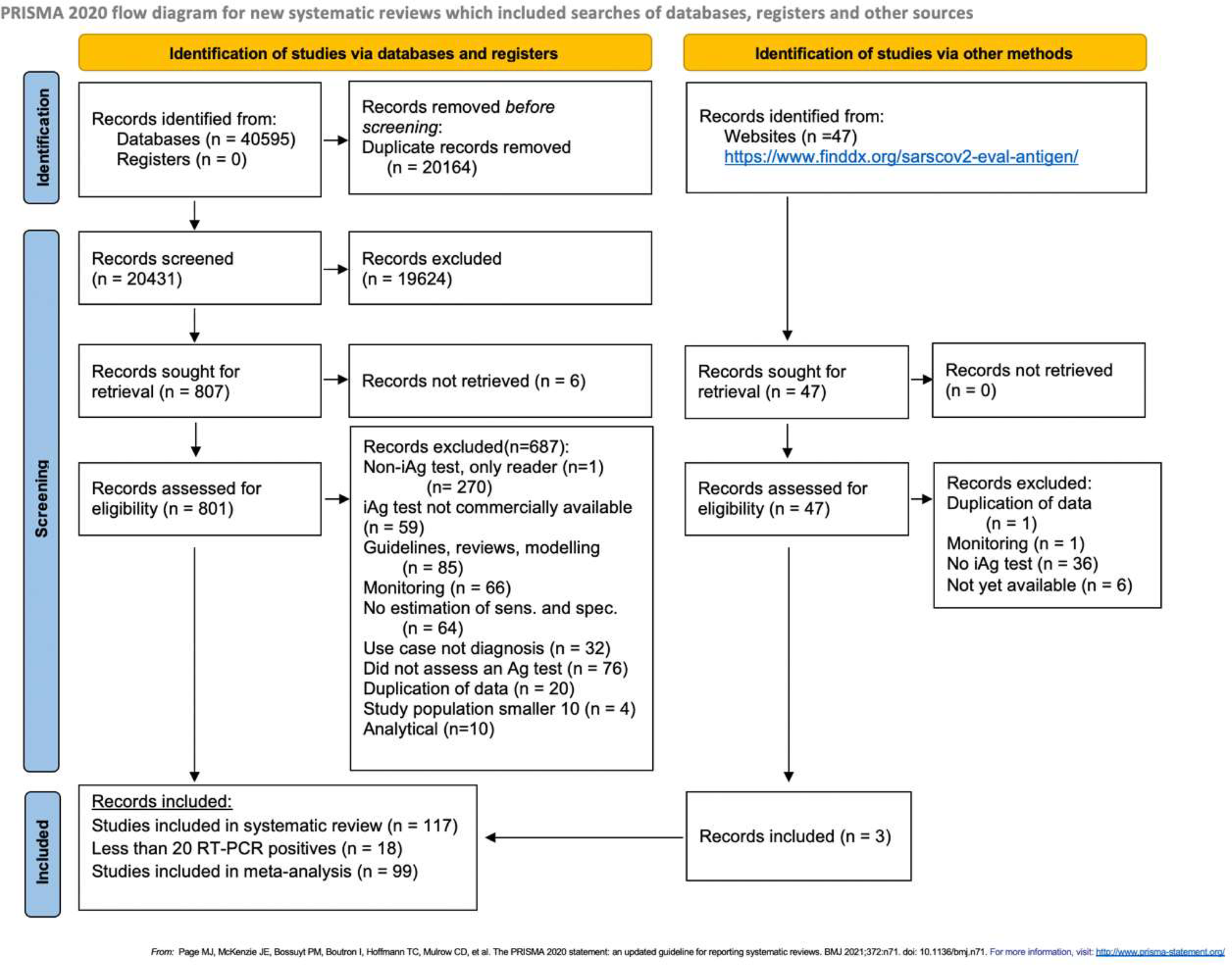
The aim of this study was to assess the role of VP1 in VLPs stability and assembly and to investigate whether a single structural protein (VP2) or a combination of VP2 and VP1 could be employed to successfully construct BPV VLPs. Furthermore, we experimentally verified the bioinformatically predicted physicochemical parameters (pH, ionic strength, and temperature) that most affect assembly yield and VLP stability. Details of the experiments are illustrated in the following architectural research process flow chart Fig. 1.
Fig. 1
This diagram is a simplified representation of the research process as a whole
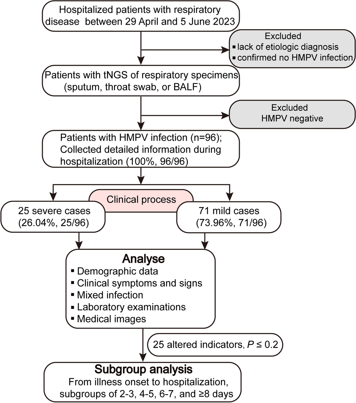
Physico-chemical analysis of the capsid proteins of BPV (dry lab)Using the ProtParam website, we determined the expected physicochemical properties of the VLP protein sequences in Table 1. It was calculated that the resulting protein would have a molecular weight (MW) of 75.1 kDa for VP1 and 60.2 kDa for VP2 alone, with theoretical isoelectric point (pI) values of 9.27 and 8.18, respectively. Based on the pI, the protein is expected to have an alkaline pH. Both capsid proteins were estimated to have a half-life of 30 hours in human reticulocytes in vitro, 20 hours in yeast, and 10 hours in E. coli in vivo [16]. The anticipated instability index (II) for VP2 alone was 33.7, while for VP1VP2, it was 32.61, making the protein stable (II >40 implies instability). Expression of the protein was anticipated to result in an insoluble form (solubility score of 0.238) [38] and proteins with a higher scaled solubility value (value greater than 0.45) are predicted to be more soluble [20]. VP1VP2 and VP2 had aliphatic indices of 70.15 and 72.3%, respectively, indicating thermostability [20]. Aliphatic indices of 70.15 and 72.3% for VP1VP2 and VP2, respectively, were calculated, indicating that the capsid proteins are stable at high temperatures [20]. The predicted grand average of hydropathicity (GRAVY) was − 0.407. A negative value indicates that the protein is hydrophilic in nature and can interact with water molecules [27]. A comparison of the predicted secondary structures of BPV VP1VP2 and BPV VP2 proteins (Fig. 2) reveals that they are very similar, with the exception of the number of alpha-helixes.
Table 1 Bioinformatic prediction of the physicochemical properties of the structural proteins of BPV that form VLPSFig. 2
The graphic depicts PSIPRED 4.0's secondary structure prediction for the amino acid sequences VP1 and VP2 of BPV; A illustrates the various structural forms of the amino acid sequence of BPV-VP2, as indicated by colours, including helical, strand, random coil, and others. B represents the BPV-VP1VP2 amino acid sequence in various structural forms, as shown by different colours, including helices, strands, random coils, and others
Chemicals, biological and synthetic biochemicalsForeign and domestic commercial sources were used to acquire biological components and chemical reagents of analytical quality. DNA polymerase and T4 DNA ligase were acquired from TaKaRa Bio Inc. (Dalian, China), as well as the DNA marker, DNA restriction enzymes, E. coli DH10Bac cells, and Escherichia coli DH5 cells. Fetal bovine serum (FBS), GibcoTM Sf-900TM III SFM, and the pure LinkTM Quick Plasmid Miniprep Kit were all purchased from Thermo Fisher, USA). We used ExpiSf9TM cells that we originally purchased from Thermo Fisher, USA). Anti-His-Tag mouse monoclonal antibody and goat anti-mouse secondary antibody labeled with horseradish peroxidase (HRP) (Abcam, Cambridge, UK) Monoclonal rabbit antiDE-Loop BPV-VP2 (SanQ Biotechnology Co.Ltd., Beijing) and HRP-labeled anti-rabbit IgG (from Sigma-Aldrich;St.Louis, Missouri, USA) were used.
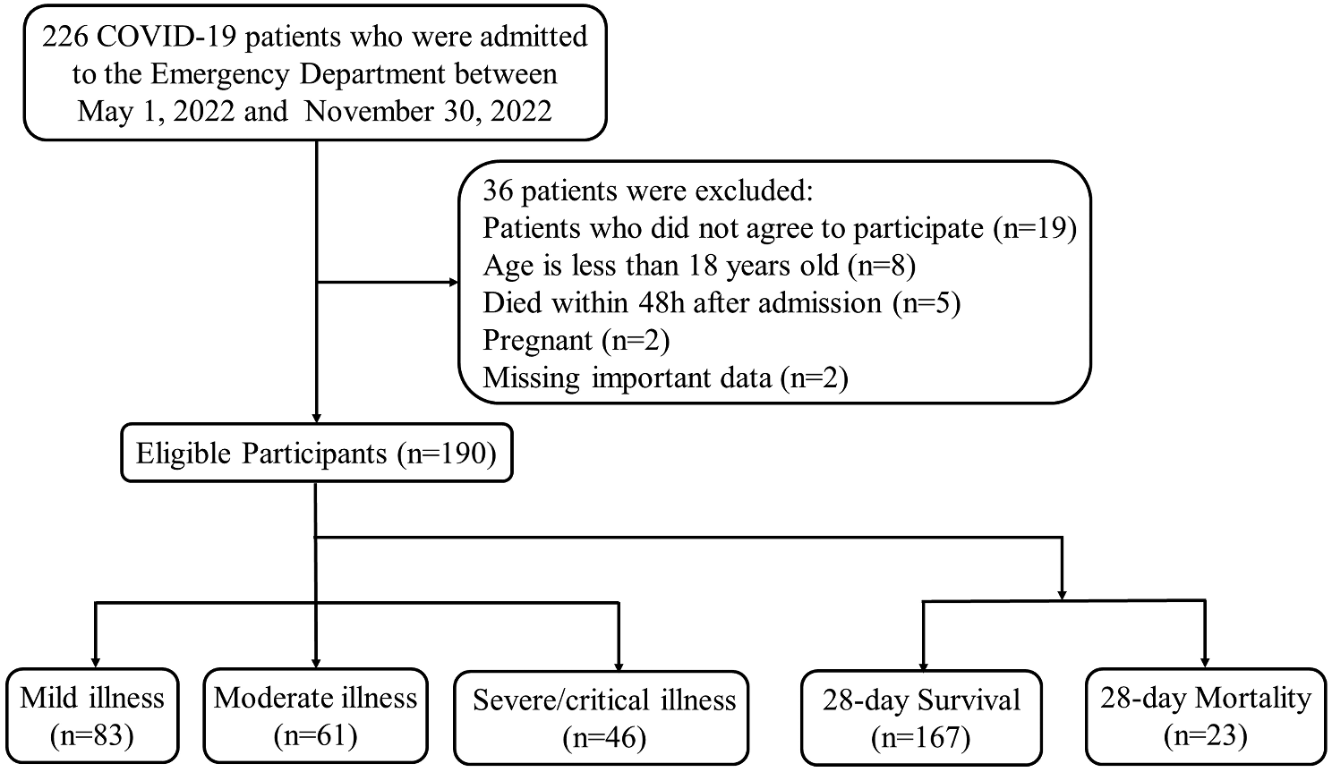
Monoclonal antibodyA DE loop joins two strands, βD and βE, and found on the surface capsid. Protein similarity analysis (job-ncbiblast-I20231106-061600-0533-17915353-p1m) shows that the DE-loop has 100% identity among all bovine parvovirus type strains but 73.5% sequence similarity with human bocavirus 1 type 1. The DE loop amino acids have been shown to be involved in VP1/VP2 externalization and antibody recognition [3]; Fig. 3). The IEDPA results showed that the antigenicity score calculated using the methods of Kolaskar and Tongaonkar was higher than the typical cutoff value of 1.013. Based on the results of the Bepipred Linear Epitope Prediction technique, the DE loop region is classified as a linear epitope with a prediction score greater than the cutoff value of 0.500. Therefore, we sent the aa sequence coding to the DE-loop region to a company (SanQ Biotechnology Co. Ltd., Beijing), and rabbit anti-DE-loop monoclonal antibody (mAb) was synthesized.
Fig. 3
Tertiary structure and functional prediction of the DE-loop region of BPV-VP2; a schematic representation of the tertiary structure of BPV-VP2 is depicted above, specifically the location of the DE-loop between the D and E strands in the variable region (VRII) and surface topology. Blended and improved diagram adopted from [11, 32], b shows the result of the functional prediction of the 49 aa sequence of the bovine parvovirus type1
Construction of the recombinant pFastBac dual vectorIn this study, we used recombinant bacmids BPV VP2 and VP1VP2 expressed in Sf9 cells to generate nonenveloped VLPs using the baculovirus vector expression system adhering to the protocols outlined in the Bac-to-Bac® system manual. Full-length capsid genes (BPV-VP1VP2 and VP2) were retrieved from GenBank under the accession number JN191349.1. In silico design by the Snap Gene program (https://www.snapgene.com/) was first applied to generate the recombinant BPV capsid proteins into vectors. The VP1VP2 (2022 bp) and VP2 (1611 bp) coding sequences were optimized in Vector Builder (https://en.vectorbuilder.com/tool/com) and amplified using primers with a His-tag and without a His-tag at the N-terminal fragment table. Four separate inserts were created using BamHI and PstI and cloned and inserted into pFastbacdual at matching restriction sites (Fig. 4). The anticipated completed architectural structures of the recombinant genes were used as a backdrop in the liquid experiment.
Fig. 4
Insilco's building design was simulated using molecular architecture; a shows BPV-VP2 with His-tag b represents BPV-VP2 without His-tag c depicts BPV-VP1VP2 with His-tag d illustrates BPV-VP1VP2 without His-tag
Codon use optimization and gene synthesis of designed recombinants were performed at Gene Script (Piscataway, New Jersey, USA). Accordingly, the BPV VP2 and VP1VP2 segments were amplified (Fig. 5) using pairs of primers listed in Table 2 and structural genes built into the pUC vector as a template. The nucleotide primers were designed with restriction sites upstream (BamHI) and downstream (PstI). To generate the recombinant pFastBac BPV-VP2 and pFastBac BPV-VP1VP2 plasmids, the PCR products were digested with BamHI and PstI and then cloned and inserted into the appropriate restriction sites of the pFastBac dual vector. DNA sequencing and double enzyme digestion validated the insert of the recombinant plasmid. Transposition of the recombinant pFast donor plasmid into the bacmid needed the transformation of DH10BacTM. Bac-to-Bac® (Invitrogen Life Sciences) protocols were followed for both the transposition experiment and the subsequent transfection operations. To isolate recombinant bacmid DNA, only white colonies harboring the correct recombinant bacmid were chosen using the white‒blue screening method. To verify that the white colonies were indeed white, they were subcultured again on a new petri plate with the same antibiotics. A single pure white bacterial colony was inoculated into 2 mL of this LB medium, and the culture was left to grow overnight at 37 °C in a shaking water bath at 250 rpm. To purify and preserve (in 50% glycerol) the right recombinant DH10Bac for future use, a 2% inoculum (100 μL) was inoculated into 5 ml of LB medium containing 3R+ (penicillin, gentamicin and chloramphenicol) for purification purposes. The PureLinkTM HiPure Plasmid DNA Miniprep Kit (InvitrogenTM, Life Sciences, USA) was used to isolate high-quality Bacmid DNA (r-pFastBPV VP2 and VP1VP2) from the white colonies for transfection purposes. A polymerase chain reaction (PCR) employing M13 primers and gene-specific primers was subsequently performed on the recombinant Bacmid (pFastBPV VP2 and VP1VP2).
Fig. 5
The results from PCR, colony PCR, and double enzyme digestion (a, b and c, respectively) were electrophoresed on agarose gels
Table 2 The primers were designed using BamHI (upstream) and PstI (downstream) to amplify BPV-VP1VP2 and BPV-VP2 with and without His-tagsTransfection and expression of the recombinant proteinWith the use of cellfectin® reagent, SF9 cells were transfected with purified and validated DNA inoculums of His-tagged r-BacVP2 and r-BacBPV1VP2 and His-tagged free r-BacVP2 and r-BacVP1VP2. Cells in a 6-well plate were monitored 24 hours after infection (pi) under an inverted microscope (EVOS) for any signs of CPE (Fig. 5). Hence, common symptoms of infection (CPEs, if any) were documented at 24, 48, 72, and 96 hours postinfection (pi). After confirming regular CPE and viability, we proceeded to harvest viral stocks. We optimized infection settings in Sf9 cells to obtain the highest possible production of VLPs. The MOI (5 pfu/cells) [7] of the recombinant baculovirus P2 stock of P1 and P3 stocks of P2 was found to be optimal for the infection of Sf9 insect cells and for the entire expression. For further expression, a pool of the viral stocks was clarified (at 500 rpm for 5 minutes), filtered (0.22 μm), and then stored at 4 degrees Celsius. Filtered P1 and P2 stocks were aliquoted and frozen at − 80 degrees Celsius in a black 1.5 milliliter centrifuge tube. After 4–6 days (cell viability <60–80% and cell size 18.5–21 µm), infected cells were frozen and thawed three times at intervals of 3–4 hours and centrifuged at low speed (10,000 r/min) for 30 minutes at 4 °C to remove the cell debris. The recombinant proteins that had been expressed were then harvested from the supernatant.
VLPs purification methodsOvernight, the supernatant was stirred on multipoint magnetic stirrers (Thermo Fisher, USA) with 8% (8 g/1 L) polyethylene glycol 8000 and 0.5 M NaCl (0.5x 1Lx58.44 = 29.22 g) in a 4 °C cold environment. After 45 minutes of centrifugation at 12,000 rpm and 4 degrees Celsius, the pellet (precipitate) was collected and then resuspended in cold, sterile 1xPBS. Debris and insoluble proteins were then removed by centrifugation at a low speed. Ultracentrifugation with a 10–40% sucrose gradient at 36,000 rpm for 3 hours at 4 °C in a Beckman Optima L100XP centrifuge using a SW32 Ti rotor (Beckman Coulter, Thermo Fisher Science, USA) was used to further purify the clear supernatant, as described by two other authors [7, 18]. Ultracentrifuged VLP protein content (mg/ml) and purity (260/280 wavelength) were determined using a UV‒Vis spectrophotometer (Thermo Scientific NanoDropTM One, Rockford, USA) throughout 11 ml, with 1 ml in each 1.5 ml centrifuge tube. IFAT, SDS‒PAGE, Western blotting, and TEM negative staining were used to determine the protein's intracellular location, expression level, purity, immunogenicity, and VLP (rec-VP2 and rec-VP1VP2) integrity.
SDS‒PAGE and western blottingUndiluted pure recombinant proteins (0.35 mg–5.57 mg/ml) were separated by 12% SDS‒PAGE, and their protein bands were visualized using Coomassie Brilliant Blue R250 (Sigma‒Aldrich, St. Louis, USA). For western blotting, purified proteins were diluted to 20–30 µg/ml, separated by 12% SDS‒PAGE, transferred to PVDF membranes, and blocked with 5% (w/v) nonfat milk for 1 hour at room temperature (RT). The immunodominant conserved DE-loop at the N-terminus of the VP2 BPV protein bioinformatically predicts immunogenic function [18]. To our understanding, we synthesized this region for the first time as a monoclonal antibody for target protein detection. To detect the four recombinants, we used a primary antibody of rabbit anti-DE-loop monoclonal IgG (1:5000) (SanQ Biotechnology Co.Ltd., Beijing) and a secondary antibody of mouse anti-rabbit IgG (1:5000) (SIGMA), with three washes in phosphate-buffered saline-Tween solution (PBST) at five-minute intervals between each step. In addition, mouse anti-His-tag monoclonal antibody (diluted at 1:2000; Abcam, Cambridge, UK) and rabbit anti-mouse IgG antibody (diluted at 1:5000) were employed to detect His-tagged recombinants using the same methods described above. Recombinant protein bands were characterized using an ECL machine (GE IA600, AmershamTM, He Nan De Quan Xingye, Co. Ltd., China) and a chemiluminescence kit (Yeasen Biotechnology Co. Ltd, Shanghai).
IFAT (indirect immunofluorescence assay)Sf9 cells (2x10⁶) were transfected with four BPV-VP2 recombinants at m.o.i of 5, bacmid alone as a positive control, and mock-infected cells as a negative control in a 6-well plate with a cell density of 1x10⁶. At 24, 48, 72, and 96 hours postinfection, cells were fixed with 4% paraformaldehyde in PBS pH 7.4 for 10 minutes at room temperature to assess the level of expression of all recombinants. After blocking for an hour with 1% BSA/milk at 37 °C, all wells were rinsed three times for five minutes in PBS. After incubation with DE-loop mAb (1:5000) specific for BPV-VP2 protein, the cells were washed. Red fluorescent protein (RFP) Alexa Fluor® 594-conjugated anti-rabbit IgG antibodies were diluted to 1:800 and incubated in the dark at room temperature for 60 minutes. Fluorescence microscopy (EVOS FL Cell Imaging System, Thermo Scientific, USA) was used to observe the reaction.
Transmission electron microscopy (TEM)Lysates from rBac-VP1VP2- and VP2-infected Sf9 cells were separated by sucrose density gradient centrifugation, as described previously [23]. The samples were diluted to a concentration of 0.2–0.3 mg/L (100 ng). Approximately 10 µL of the purified fractions were incubated for 5 minutes at room temperature on a formvar-carbon film-coated copper grid (400 mesh, Pelco, CA, USA). The grid was dyed with 10 µL of 3% ammonium phosphotungstic acid (Ph = 6.5) after being rinsed three times in PBS. The grids were prepared for analysis by carefully removing any excess sample, PBS, and dye with filter paper or tissue paper. A Hitachi Limited (Japan) HT-7700 120 kV TEM was used to observe the assembly of recombinant BPV virus-like particles (VLPS).
Stability and secondary structure analysis of VLPs of BPV (VP1+VP2) and (VP2)VP1 is a small, structural, and nonessential protein of BPV. This may help keep the BPV particles stable. To determine what role VP1 plays in keeping BPV particles stable, two types of VLPs were studied at various temperatures and pH levels. These were the VP1 and VP1+VP2 VLPs Two kinds of VLPs were treated with different phosphate buffers (pH 4.5 to pH 8); the secondary structure was then analyzed by far UV circular dichroism (CD) spectroscopy. The percentages of the secondary structures α-helix, β-sheet, and β-turn, as well as the percentage of coil in VLPs (VP1+VP2) and (VP2) after being treated with different PBS (phosphate buffers, pH 4 to pH 8), were analyzed using USB circular dichroism (CD) spectroscopy. The thermal stability of VLPs produced in PBS at 4 °C was assessed to determine the integrity of the particle or if morphological disruption or changes existed while exposed to different temperatures (20 °C, 40 °C, 60 °C, and 80 °C) for 30 minutes. Changes in the morphology or integrity of the VLPS while incubating VLPs at different temperatures were also analyzed by TEM.
留言 (0)