
記住我


Diffuse pleural mesothelioma (DPM) is an aggressive malignant neoplasm involving the pleural linings and is associated with industrial pollutants with a strong link to asbestos exposure.1 DPM affects >30,000 individuals globally, with an annual incidence of ~3300 cases per year in the United States.2,3 DPM has 3 subtypes: epithelioid, sarcomatoid (including desmoplastic), and biphasic, with the epithelioid subtype comprising the majority of cases. The prognosis of DPM is poor, with an average overall survival of ~18 months from the time of diagnosis.4 However, it has been well recognized that epithelioid mesotheliomas follow a more favorable clinical course than biphasic and sarcomatoid mesotheliomas.1,5
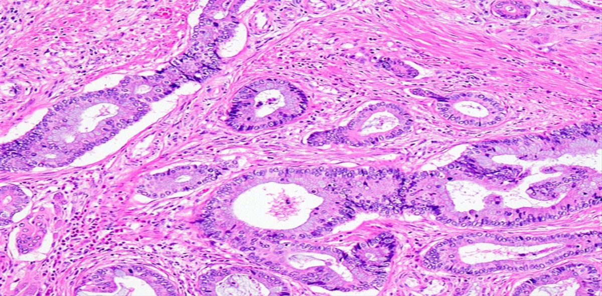
Although pleural epithelioid mesotheliomas (PEMs) typically have bland appearing cytologic features, some PEMs may show marked cytologic atypia, variable mitotic activity, and an array of growth patterns with or without necrosis.6,7 To separate the distinct subgroups within PEM cases, the 2021 WHO classification implemented a grading system for PEMs as low and high grade, based on the nuclear grade (a combination of mitotic score and nuclear atypia score) and the presence or absence of necrosis. There is limited information in the literature on the reproducibility of this grading system, although it is well recognized that the assessment of histologic parameters (especially nuclear atypia) tends to be subjective.8 Moreover, counting mitoses is highly labor-intensive and potentially also subjective, though to a lesser degree than scoring nuclear atypia. Thus, one can consider the Ki-67 proliferation index via digital image analysis (DIA) as an efficient and likely more objective surrogate for the tumor grade determined by the current grading system. Ki-67 proliferation index has been reported as an independent prognostic factor in epithelioid type (but not in biphasic or sarcomatoid types) of pleural mesotheliomas.9 Ki-67 index has also been used as an important parameter in clinical decision-making for various surgical procedures (eg, extrapleural pneumonectomy or extended pleurectomy and decortication).10,11 Ki-67 index is currently used as the parameter for tumor grading in parallel with mitotic count in other types of tumors, such as neuroendocrine tumors of gastrointestinal and pancreaticobiliary tracts.
The main purpose of this study is to evaluate the possibility of Ki-67 index via automated DIA as an effective surrogate for tumor grade and/or mitotic score in PEM cases. We also examined the reproducibility of the 2021 WHO tumor grading and individual parameters for PEMs to further determine the role of Ki-67 as an objective parameter for tumor grading.
MATERIALS AND METHODS Case CohortAn electronic search was performed using the keyword “mesothelioma” for all surgical pathology cases that underwent biopsy or surgical resection at Mayo Clinic, Rochester, MN, from 2000 to 2021. All cases were further reviewed for the primary location of mesothelioma and specimen type. Cases with only cytology specimens or nondiffuse pleural mesotheliomas were excluded. The remaining cases underwent a histopathologic review of all available glass slides. This study was approved by the Institutional Review Board at Mayo Clinic (IRB 21-009026).
Slide ReviewTwo thoracic pathologists independently reviewed H&E-stained slides from each case and rendered a diagnosis. Cases diagnosed as PEM by both pathologists were included in the study. Cases in disagreement between the 2 pathologists were evaluated by a third thoracic pathologist. If the third pathologist diagnosed the case as PEM, the case was included in the study. All cases included in the study were then independently evaluated by 2 pathologists for nuclear atypia, mitotic count, and necrosis and the overall tumor grade according to the current WHO criteria. Mitoses were counted in “hotspots” in contiguous fields when possible.
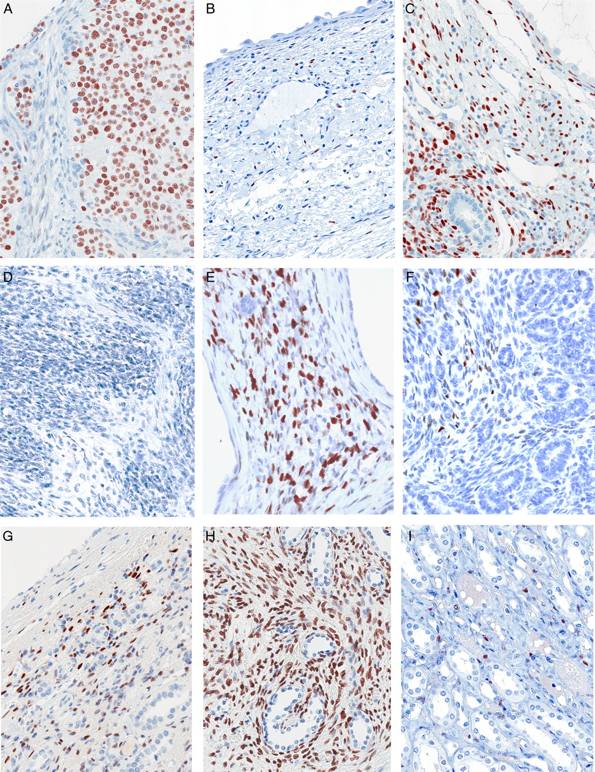
Ki-67 IndexA most representative block was selected for Ki-67 immunohistochemical testing (MIB-1 clone, DAKO). DIA was performed using Aperio ImageScope Software (Leica Biosystems). The Ki-67 index for each slide was evaluated by a cytotechnologist by identifying the region of interest with the highest percentage of Ki-67–positive mesothelioma cells (“hot spot”) and placing 10 fix-sized tiles on that region, avoiding nontumor entities and staining artifacts. The tiles were analyzed using a nuclear algorithm. A minimum of 500 cells were analyzed. Additional tiles were placed until 500 cells were achieved if 500 cells were not analyzed in the first 10 tiles. All cases were reviewed by a pathologist (E.S.Y.) to approve tile placement and analysis results.
Clinical DataMedical oncologists reviewed the electronic medical records to abstract pertinent clinical information, date of diagnosis, type and intent (diagnostic or therapeutic) of surgery, treatment history including chemotherapy or radiation, and survival data.
Statistical AnalysisData were summarized with frequencies and percentages, means and SD, or medians and ranges, as appropriate. Kappa (κ) statistics were calculated to assess reviewer agreement for necrosis (no/yes) and tumor grade (low/high), using weighted kappa for mitotic count score (0 to 1/2 to 4/5+), nuclear atypia score (1/2/3), and nuclear grade (1/2/3). Reviewer agreement was calculated based on the data from all specimens; some patients underwent more than 1 biopsy and all biopsies were included for mitotic count, tumor grading, and Ki-67 DIA. Kappa values of 0 to 0.20 were regarded as slight agreement, 0.21 to 0.40 fair, 0.41 to 0.60 moderate, 0.61 to 0.80 substantial, and 0.81 to 1 as almost perfect agreement.12 Correlation between mitotic count and Ki-67 index was calculated using the Spearman-rank method. The area under the curve (AUC) for predicting tumor grade based on mitotic count and Ki-67 index was calculated with logistic regression models. Overall survival (OS) was estimated with the Kaplan-Meier method and was compared between mitotic count and Ki-67 index using Cox proportional hazards regression. Hazard ratio (HR), median OS, 1- and 3-year OS, and 95% CI were reported. The concordance (C-index) was also reported. The continuous effects of mitotic count and Ki-67 index on the HR of death were explored with penalized splines, and categorizations for each variable were informed by those findings. P-values <0.05 were considered statistically significant. For patients with more than 1 procedure, the biopsy used at initial diagnosis, or the earliest date of accession, was used for the overall survival analysis. Analyses were performed using SAS version 9.4 (SAS Institute Inc., Cary, NC) and R version 4.2.2 (https://www.R-project.org).
RESULTS Demographic CharacteristicsA total of 96 specimens from 85 unique patients met the inclusion criteria with complete sets of data (histologic review, Ki-67 index by DIA, clinical information). During the initial selection process, 8 cases underwent review by a third pulmonary pathologist (reviewer 3) due to the discrepancy in the classification of mesothelioma (epithelioid vs. biphasic type) between the 2 reviewers. Reviewer 3 determined 3 of 8 discrepant cases as epithelioid type, and these 3 cases were included in the final cohort for this study.
Demographics are summarized in Table 1. The mean age at diagnosis was 67.2 years (SD 11.2 years) and ranged from 27 to 89 years. Males comprised 77.6% (n=67) of patients. Seventy-four of 85 patients (87.1%) underwent 1 procedure, and 11 patients (12.9%) underwent 2 procedures. Considering the first specimen in each patient, the most common type of procedure was a diagnostic biopsy (64.7%, 55/85), and the remainder were obtained at the time of surgery during treatment (35.3%, 30/85). Specimen types included biopsy (n=53), decortication (n=31), and other type (n=1, lymph node excision). Of the 85 patients, the median survival was 1.5 years (95% CI: 1.2-1.6 years) from the date of diagnosis, with 1-year survival of 71.1% (95% CI: 61.1%-81.1%) and 3-year survival of 18.4% (95% CI: 9.7%-27.1%).
TABLE 1 - Patient Demographics Patients (n=85) Age at diagnosis (years) Mean (SD) 67.2 (11.2) Range (26.6–88.6) Sex, N (%) F 19 (22.4) M 66 (77.6) Total specimens per patient, N (%) 1 74 (87.1) 2 11 (12.0)Nuclear atypia grading had only fair agreement between the 2 reviewers (κ value of 0.37), while there was moderate agreement for mitotic count, nuclear grade, necrosis, and overall tumor grade according to the current WHO criteria (κ values of 0.53, 0.49, 0.56, and 0.47, respectively). Details of these results are summarized in Table 2. Reviewer agreement for noncategorized mitotic counts is shown in Figure 1. Considering OS, the HR for tumor grade (low vs high) was similar in reviewer 1 and reviewer 2 (HR=1.27 [95% CI: 0.80-2.03] and HR=1.55 [95% CI: 0.95-2.49], respectively).
TABLE 2 - Interobserver Agreement Using WHO Grading Guidelines (N=96 Specimens) WHO parameter Reviewer 1, N (%) Reviewer 2, N (%) Kappa (95% CI)* Mitotic count 0–1 24 (25.0) 34 (35.4) 0.53 (0.39, 0.66) 2–4 26 (27.1) 18 (18.8) — 5+ 46 (47.9) 44 (45.8) — 0–4 50 (52.1) 52 (54.2) 0.52 (0.38, 0.66) 5–9 15 (15.6) 21 (21.9) — 10+ 31 (32.3) 23 (24.0) — Nuclear atypia score 1 5 (5.2) 5 (5.2) 0.37 (0.22, 0.52) 2 30 (31.3) 54 (56.3) — 3 61 (63.5) 37 (38.5) — Nuclear grade 1 16 (16.7) 23 (24.0) 0.49 (0.35, 0.63) 2 44 (45.8) 53 (55.2) — 3 36 (37.5) 20 (20.8) — Necrosis No 73 (76.0) 71 (74.0) 0.56 (0.36, 0.75) Yes 23 (24.0) 25 (26.0) — Tumor grade Low 53 (55.2) 60 (62.5) 0.47 (0.29, 0.64) High 43 (44.8) 36 (37.5) —*Weighted kappa presented for mitotic score, nuclear atypia score, and nuclear grade. Kappa values assess the amount of agreement expected above and beyond chance alone.
 FIGURE 1:
 Interobserver Agreement for Mitotic Counts. Scatter plot demonstrating interobserver correlation (reviewer 2 x-axis, reviewer 1 y-axis) based on mitotic counts for 96 specimens. The red diagonal line (identity line) demonstrates perfect agreement between reviewers.
A data-driven penalized spline analysis illustrated no apparent change below a mitotic count of 5 and then a steady increase in the risk of death after the mitotic counts reached 5 or higher (Supplemental Fig. 1, Supplemental Digital Content 1, https://links.lww.com/PAS/B770). Since there was no significant change between 0 to 1 and 2 to 4 (representing the current mitotic scores 1 and 2, respectively), we explored an alternative scoring system: 0 to 4, 5 to 9, and ≥10, as mitotic scores 1, 2, and 3. These new categories for mitotic count (0 to 4, 5 to 9, 10+) elicited comparable agreement between the 2 reviewers (κ value of 0.52).
Association Between Ki-67 Index and Tumor GradeThe Ki-67 index was compared with the tumor grade that was assigned by each reviewer. The Ki-67 index was significantly higher in high-grade tumors as compared with low-grade tumors (P ≤ 0.0001), and this finding was consistent within each reviewer (median Ki-67: 29.5 vs. 13.5 [reviewer 1], 31.4 vs. 13.7 [reviewer 2]) (Table 3).
TABLE 3 - Association Between Ki-67 Index and Tumor Grade in 2 reviewers Tumor grade Low High P Ki-67: reviewer 1 N 53 43 0.0001 Median 13.5 29.5 — Q1, Q3 8.2, 25.5 19.2, 42.1 — Range (1.8–76.6) (1.1–70.5) — Ki-67: reviewer 2 N 60 36 <0.0001 Median 13.7 31.4 — Q1, Q3 8.4, 24.5 25.6, 44.3 — Range (1.1–76.6) (6.0–70.5) —The correlation between Ki-67 index and mitotic count as well as the predictability of Ki-67 index versus mitotic count for tumor grade (assigned by each reviewer) and OS were evaluated. The correlation between mitotic count (reviewer averaged) and Ki-67 index was 0.65 (Fig. 2A). The AUCs for predicting tumor grade by mitotic count and Ki-67 index were 0.84 and 0.74 (reviewer 1) and 0.85 and 0.81 (reviewer 2), respectively (Fig. 2B).
 FIGURE 2:
 A, Correlation between mitotic count (reviewer averaged) with Ki-67%. Scatter plot demonstrating moderate correlation (0.645) between reviewer-averaged mitotic count (x-axis) and Ki-67% positivity (y-axis) for 96 specimens. B, ROC curves predicting tumor grade based on mitotic count or Ki-67%. ROC curve for tumor grade versus mitotic count for each reviewer (left). ROC curve for tumor grade versus Ki-67% for each reviewer (right). AUC indicates area under the curve; ROC, reviewer operating characteristic.
Both increased mitotic count (reviewer averaged) and Ki-67 index were associated with worse OS. Supplemental Figure 1, Supplemental Digital Content 1, https://links.lww.com/PAS/B770 shows the change in HR for death as mitotic count and Ki-67 proliferation index increase, based on penalized splines. For mitotic count, the HR stayed relatively low for counts <5, at which point the hazard ratio began to steadily increase. For Ki-67 index, the HR began to steadily increase after 10%.
On the basis of these findings, together with the distributions, different categorizations for each variable were explored. Mitotic counts of >10 per 2 mm2 had significantly worse OS compared with mitotic counts of <10 (HR=3.21, 1-year OS 43.8% vs. 78.0%, P=0.0005). Of note, the OS of ≥10 per 2 mm2 differed significantly from those with 5 to 9 (HR=2.72, 1-year OS 43.8% vs. 81.0%, P=0.006), despite the fact that both groups (5 to 9 and ≥10 per 2 mm2) belong to the same mitotic score of 3 under the current WHO classification system. Also, there was no significant change between 0 to 1 and 2 to 4, which are in the range of the current mitotic score 1 and 2, respectively (HR for 2 to 4 vs. 0 to 1: 0.83 [95% CI: 0.44-1.59], based on reviewer-averaged mitotic counts). Higher Ki-67 index (≥30% vs. <30%) showed an association with worse OS (HR 1.79, 1-year OS 60.0% vs. 76.2%, respectively, P=0.03). These results are summarized in Table 4 and illustrated in Figure 3. Also, OS by WHO tumor grade (by each observer) was shown in Figure 3. Eleven patients underwent 2 procedures. Mitotic count, Ki-67 index, and WHO tumor grade in each patient are listed in Table 5.
TABLE 4 - Overall Survival (OS) by Mitotic Count and Ki-67 Index Variable Patients, N Deaths, N 1-y OS (95% CI) 3-y OS (95% CI) Hazard ratio (95% CI) P C Mitotic count (reviewer averaged) 0–4 43 38 76.5 (63.8, 89.3) 26.3 (13.0, 39.6) Reference 0.001* 0.587 5–9 22 19 81.0 (64.2, 97.7) 15.2 (0.0, 32.5) 1.31 (0.72, 2.30) — — ≥10 20 16 43.8 (19.4, 68.1) 0.0 (non-Est.†) 3.55 (1.83, 6.66) — — <10 65 57 78.0 (67.8, 88.2) 23.2 (12.6, 33.8) Reference 0.0005 0.585 ≥10 20 16 43.8 (19.4, 68.1) 0.0 (non-Est.†) 3.21 (1.72, 5.72) — — Ki-67 Index 0–<10 18 15 77.4 (57.8, 96.9) 23.8 (3.5, 44.1) Reference 0.14 0.554 10–<20 25 21 74.1 (56.2, 91.9) 30.5 (11.7, 49.3) 1.05 (0.54, 2.07) — — 20–<30 15 13 78.6 (57.1, 100.0) 10.7 (0.0, 29.0) 1.37 (0.63, 2.97) — — ≥30 27 24 60.0 (40.8, 79.2) 5.3 (0.0, 15.1) 2.00 (1.03, 4.04) — — <30 58 49 76.2 (64.9, 87.5) 23.9 (12.4, 35.4) Reference 0.03 0.556 ≥30 27 24 60.0 (40.8, 79.2) 5.3 (0.0, 15.1) 1.79 (1.06, 2.96) — —*Pairwise P-values: 5 to 9 versus 0 to 4 (P=0.37), ≥10 versus 0 to 4 (P=0.0003), ≥10 versus 5-9 (HR 2.72, 95% CI: 1.35-5.39, P=0.006). Of note, OS did not differ significantly when comparing 2 to 4 versus 0 to 1 mitotic count (HR=0.83, 95% CI: 0.44-1.59).
†CI nonestimable due to low or no variability.
 FIGURE 3:
 Overall survival by WHO tumor grade, mitotic count, and Ki-67 proliferation index. Kaplan-Meier plots illustrating overall survival for 85 patients with diffuse pleural epithelioid mesothelioma. Proposed mitotic count categorizations of 0 to 4, 5 to 9, and >10 mitoses show poorer survival probability with higher mitotic counts (lower left). Ki-67 proliferation index with a delineation of 30% positivity (lower right), showing similar performance to the tumor grade by reviewer 2.
TABLE 5 - Mitotic count, Ki-67 Index, and Overall Tumor Grade in the Cases With 2 Procedures Patients with 2 biopsies (n=11) Specimens Days after the first biopsy Procedure type Mitotic count (average by 2 reviewers) Ki-67 index (%) Overall tumor grade (reviewer 1) Overall tumor grade (reviewer 2) 1 1 0 Biopsy 0.5 8.6 Low Low 2 118 Resection 0.0 1.8 Low Low 2 1 0 Biopsy 6.5 38.1 High High 2 145 Pneumonectomy 2.0 22.3 Low Low 3 1 0 Biopsy 6.5 36.4 Low High 2 157 Excision 17.5 76.6 Low Low 4 1 0 Biopsy 5.5 31.3 Low High 2 89 Excision 1.5 7.0 Low Low 5 1 0 Excision 20.0 30.3 High High 2 212 Biopsy 0.5 8.7 Low Low 6 1 0 Biopsy 1.5 8.2 Low Low 2 100 Resection 0.5 25.6 Low Low 7 1 0 Biopsy 6.5 30.8 High High 2 329 Biopsy 33.0 21.1 High High 8 1 0 Biopsy 5.5 7.4 Low High 2 496 Resection 15.0 44.4 High High 9 1 0 Biopsy 12.5 47.7 High High 2 112 Pneumonectomy 0.5 22.1 High High 10 1 0 Resection 8.0 31.7 High Low 2 79 Resection 8.5 47.8 High High 11 1 0 Biopsy 4.0 13.2 High Low 2 155 Biopsy 5.5 15.4 Low LowMultiple methods have been proposed to stratify patients with PEM according to risk and to guide therapy, mainly using histopathologic features.6,8,13,14 A 2-tiered grading system for PEM has been implemented in the fifth edition of the WHO Classification of Tumors.15 The 2 major challenges with this new grading system are interobserver variability and limited efficiency, as has been recognized in the past.7,16 Ki-67 proliferation index could be supplementary or even an alternative to the histopathologic grading system; in fact, it has been adopted as an integral part of grading other types of tumors in different organs such as neuroendocrine tumors in gastrointestinal and pancreaticobiliary tracts.17–19
The main purpose of this study was to evaluate the role of Ki-67 proliferation index by well-established DIA method in comparison with tumor grade and mitotic counts according to the current WHO classification system. As it has not been widely reported to date in the literature, the interobserver agreement for grading PEMs was also studied via blinded review by 2 pulmonary pathologists. Although the number of cases in our cohort is relatively low, it is from one institution during the era when the treatment modality has not changed significantly. Thus, there is probably no significant bias caused by differing treatment modalities that could have affected OS in PEM patients. There were some differences in mitotic count, Ki-67 index and WHO tumor grade between the 2 specimens from 11 patients who underwent 2 procedures (Table 5). Due to the small number of cases with highly variable intervals between the 2 procedures, it is difficult to determine the cause(s) of discrepancies, however.
Ki-67 proliferation index had a strong association with tumor grade. Furthermore, Ki-67 index was associated with OS, especially when the cutoff was optimized to 30%, according to the results shown in Figure 3 and Table 4. Ki-67 index also associated with numeric mitotic counts as well as categorized mitotic scores. Since Ki-67 index by DIA method has been well established in most surgical pathology services for the evaluation of various types of tumors (breast cancers, neuroendocrine tumors of gastrointestinal and pancreaticobiliary tracts, etc.), it should be easily applicable to the setting of PEMs. Our results demonstrated that the Ki-67 index has a very good association with tumor grade and showed its comparable predictive value for OS (C-index=0.56) to that of mitotic counts (C-index=0.59).
Manual evaluation of Ki-67 index can be done if DIA method is not available. The most accurate way would be selecting and photographing the tumor area of the highest Ki-67 positivity, followed by printing the image on a color printer and manually ticking off each immunopositive and immunonegative tumor cell with a pen until at least 500 neoplastic cells are counted. A comparable visual evaluation without photographing or printing could be acceptable after appropriate validation processes in individual laboratory. A rigorous process has been described in a previous study to improve interobserver agreement in grading well-differentiated neuroendocrine tumors of gastrointestinal neuroendocrine tumors by using manual counting method for Ki-67 index.20
We explored alternative mitotic scoring systems with different ranges of mitosis counts for each mitotic score to compare with the performance of the current scoring system. A data-driven penalized spline analysis illustrated no apparent change before the mitotic counts of 5 and then a steady increase in the risk of death after the mitotic counts
留言 (0)