Ligament samples collection
The LF tissue of OLF samples was collected from ten patients (aged 58–82) diagnosed with OLF in The First Hospital of Tianshui and The Third Xiangya Hospital of Central South University; four females and six males were involved in this study, while normal LF samples were obtained from seven volunteers (aged 42–68) diagnosed with spinal trauma or disc herniation, including three females and four males. Prior to participation, informed consent was acquired from all patients. The Ethics Committee of Third Xiangya Hospital, Central South University, approved this research protocol. After the surgical excursions, we immediately preserved the collected ligament tissues in liquid nitrogen, typically within a thirty-minute post-surgical window, to maintain their integrity.
Primary LF fibroblasts culture and osteogenic differentiation
Primary LF fibroblasts were isolated via the tissue explant technique following a protocol outlined in previous literature [2]. Initially, ligament samples were cleared of non-ligamentous tissue under a dissecting microscope and then subjected to a PBS rinse. The cleaned ligament tissues were then finely diced into 0.5 mm3 pieces and underwent a dual PBS wash to remove residual debris. These prepared tissues were enzymatically dissociated first with 0.25% trypsin (Gibco, USA) for one hour at 37℃ and then with 200 U/mL of type I collagenase (Sigma-Aldrich, USA) for an additional four hours at the same temperature. Following digestion, the tissue fragments were cultured in DMEM (Hyclone, USA) that was fortified with 10% FBS, 100 U/mL penicillin, and 100 µg/mL streptomycin (Gibco, USA), maintained at 37 °C in a 5% CO2 humidified environment.
To induce osteogenic differentiation, we cultured fibroblasts in an osteogenic medium formulated with DMEM enriched with 10% FBS, 0.1 mM dexamethasone, 0.2 mM ascorbic acid, and 10 mM β-glycerophosphate (Sigma-Aldrich). This specialized medium supports the cells’ progression toward an osteogenic phenotype.
RNA interference
KLF5 siRNA is purchased from Ribobio (Guangzhou, China). Fibroblasts were placed into 24-well plates and cultured in a complete medium for roughly 24 h until they reached around 60–70% confluence. Following this, these fibroblasts are transfected with the siRNA at the described concentrations, using lipofection with the agent Lipofectamine 3000.
Lentiviral infection of LF fibroblasts
Lentiviral vectors for the overexpression of KLF5 and CX43 were sourced from Ribobio (China). Corresponding empty vectors served as negative controls (Lenti-NC). In preparation for infection, fibroblasts were plated at 5 × 10⁵ cells per well in 100 mm culture dishes to establish a suitable environment for viral infection. On the second day post-seeding, we added a 100 µl infection mixture containing 5 × 10⁵ PFU of the lentivirus directly to the culture medium of the fibroblasts. Post-infection, cells were maintained in an incubator at 37 °C with a humidified atmosphere of 5% CO2 for optimal growth conditions.
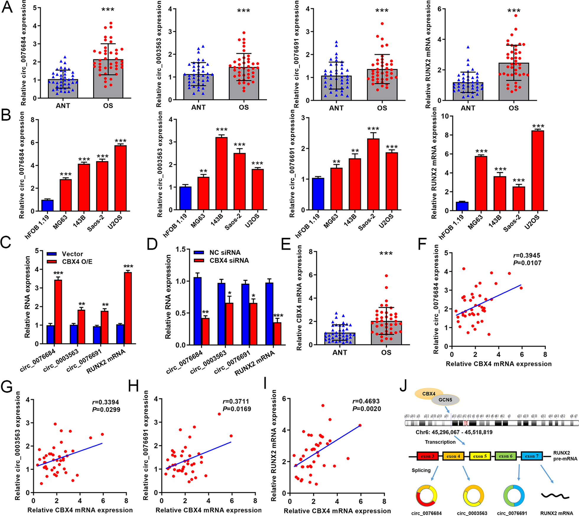
Quantitative real-time PCR (qRT-PCR)
Total RNA was extracted from cultured fibroblasts utilizing TRIzol reagent (Invitrogen Life Technologies, Carlsbad, CA, USA), adhering to a standard protocol [22]. The purity and integrity of the extracted RNA were verified through Nanodrop 2000 (Thermo Scientific, USA). Subsequently, 1 µg of total RNA was reverse transcribed to cDNA using the HiScrip III 1st Strand cDNA Synthesis Kit (Vazyme, China). Primer Design and PCR Amplification Primers specific to the genes of interest, KLF5, and CX43, were formulated using Primer 3.0 software (ABI, USA) and synthesized by Tsingke Biotech (Beijing, China). The primer sequences were as follows: KLF5-F, 5′-ATGGAGAAGTATCTGACACCTCA-3′; KLF5-R, 5′-TCAGTTCTGGTGCCTCTTCATATG-3′; CX43-F, 5′-CTTCACTACTTTTAAGCAAAAGAG-3′; CX43-R, 5′-TCCCTCCAGCAGTTGAG-3′; GAPDH-F, 5′-ATGGTTTACATGTTCCAATATGA-3′; GAPDH-R, 5′-TTACTCCTTGGAGGCCATGTGG-3′. The qRT-PCR assays were carried out in triplicate, employing SYBR-Green Master mix (Vazyme, China) on a LightCycler 480 (Roche, USA). The amplification protocol was initiated with a 95 °C denaturation step for 30 s, followed by 40 cycles of 95 °C for 10 s, and a combined annealing/extension phase at 60 °C for 30 s. Data Analysis The quantification of mRNA levels was based on the 2^-ΔΔCt method. Expression levels of the target genes were normalized using the endogenous control GAPDH.
Immunofluorescent staining
Fibroblasts were fixed using 4% paraformaldehyde for 15 min at 4℃, followed by permeabilization with 0.1% Triton X-100 for 5 min at room temperature, then washed with PBS and blocked with 5% BSA (Solarbio, China) for 30 min at room temperature to prevent non-specific binding. After blocking, fibroblasts were incubated with a primary antibody targeting KLF5 (1:200 dilution, Abcam, USA) overnight at 4 ℃ and then incubated with Dylight Fluor-conjugated secondary antibodies for 1 h at 37 ℃. DAPI was applied to stain the cells for 5 min at room temperature to label the nucleus. Imaging Immunofluorescence signals were detected and imaged using a confocal microscope (Leica TCS SP8, GER).
Western blot
Fibroblasts were lysed using pre-chilled lysis buffer at 4℃. The BCA assay kit (Abcam, USA) was employed to quantify the total protein content in each sample. Measured aliquots of 30 µg total protein per sample were subjected to electrophoresis on a 4-20% gradient SDS-PAGE gel and transferred onto PVDF membranes. Membranes were blocked using a solution of 5% skim milk in TBS-T and incubated with primary antibodies, all from Abcam, against KLF5 (1:200), Runt-related transcription factor 2 (RUNX2, 1:1000), osteocalcin (OCN, 1:1000), osteopontin (OPN, 1:1000), CX43 (1:1000), and β-actin (1:200) at 4℃ overnight. Following primary antibody incubation, membranes were treated with peroxidase-conjugated anti-rabbit IgG secondary antibody (1:5000, Millipore Corporation). Enhanced chemiluminescence reagent (Merck Millipore, Billerica, MA, USA) and a chemiluminescence imaging system (Bio-Rad Laboratories, Inc) were used for visualization. Protein bands were quantified using ImageJ software version 1.52 (National Institutes of Health, USA), with β-actin as the loading control.
Alkaline phosphatase (ALP) activity assay
Approximately 105 cells per well were plated into six-well plates to assess osteogenic differentiation. Upon reaching 65% confluence, the cells were subjected to an osteogenic induction medium to initiate differentiation. After seven days of osteogenic induction, cells were rinsed with PBS and fixed with 4% formaldehyde for 15 min. The detection of ALP activity was performed using an ALP staining kit (Stemgent 00–0055; Mito Biological technology Co,.LTD, Shanghai, China) following the manufacturer’s instructions, then washed with PBS and examined under a microscope to visualize ALP activity.
Alizarin red staining (ARS)
Following the osteogenic induction of primary LF fibroblasts, the cells were fixed using 95% ethanol for 30 min to preserve cellular structures for staining. After fixation, the cells underwent two PBS washes to remove any traces of fixative. Staining was carried out with a 0.2% ARS solution (Biosharp, China) at 37 °C for 30 min to label calcium deposits indicative of mineralization. Post-staining, the fibroblasts were rinsed with distilled water to remove excess dye. The formation of red mineralized nodules, a hallmark of osteogenic differentiation, was observed under a TS100 microscope (Nikon).
Luciferase activity assay
The wild-type (WT) full-length promoter region of the CX43 gene and its site-directed mutagenesis variant (MUT) were cloned into the psiCheck2 reporter vector (Genechem, Shanghai, China) to construct luciferase reporter plasmids for transcriptional activity analysis. HEK 293T cells were co-transfected with the constructed luciferase reporter plasmids and KLF5 overexpression plasmids using Lipofectamine 3000 reagent (Invitrogen, CA, USA) following the manufacturer’s protocol. After 48 h, luciferase activity was assessed using the Dual-Luciferase Reporter Assay System (Promega, WI, USA).
Chromatin immunoprecipitation (ChIP) assay
SimpleChIP Enzymatic Chromatin IP Kit (Catalog #9003s, Cell Signaling Technology) was used following the provided instructions. Fibroblasts underwent cross-linking with 1% formaldehyde for 10 min, quenching with glycine for 5 min at room temperature, then collected for subsequent steps. Sonication was performed to further fragments the DNA, resulting in extracts ready for immunoprecipitation. The sonicated extracts were incubated with a specific KLF5 antibody (Abcam, USA) or control IgG (Abcam, USA) at 4℃ overnight. Then, it was followed by a reversal of cross-links at 65℃ for 8–10 h to free the associated DNA. The DNA recovered post-immunoprecipitation was then analyzed by PCR to determine the presence and enrichment of target sequences bound by KLF5.
Statistical analysis
All The analyses were performed using SPSS version 21.0 statistical software (IBM Corp, Armonk, NY), and experimental data are presented as the mean ± SD. The independent samples t-test was used to compare the differences between the two groups. One-way analysis of variance was used to compare more than two groups, followed by Tukey’s post hoc test to account for multiple testing adjustments. A p < 0.05 was considered as the threshold for statistical significance.


留言 (0)