
記住我


As the population ages, the incidence of Lumbar Degenerative Diseases (LDD) is steadily increase [12]. LDD lead to lower back pain, limb numbness, and reduced lower limb muscle strength, significantly impacting patients' quality of life. Moreover, LDD poses a substantial economic burden on both individuals and society [13].
Minimally invasive spinal techniques, especially spinal endoscopy, have become increasingly popular for treating LDD, owing to their significant advantages in surgical outcomes and patient recovery [14,15,16,17]. Among these, percutaneous endoscopic lumbar interbody fusion (PE-LIF) is favored for minimal surgical trauma, safety, reduced postoperative pain, less hidden blood loss, and quicker rehabilitation, alongside robust internal fixation [18,19,20,21,22,23,24]. Complementing these techniques, the unilateral laminotomy for bilateral decompression (ULBD) facilitates sufficient decompression through a single incision, minimizing spinal muscle damage and contributing to the overall minimally invasive approach [8, 9, 25, 26]. In previous studies, surgeons have utilized the traditional small endoscope for ULBD by directly traversing the epidural space [8, 9, 25,26,27]. For example, the protective sleeve of the uniportal bilateral endoscopy (UBE) system’s high-speed drill has been employed to compress the LF, facilitating direct decompression of the contralateral LR [28, 29]. Similarly, a traditional 7.2 mm or smaller uniportal endoscope (UE) can achieve contralateral LR decompression by traversing the epidural space [8]. However, in cases of lumbar vertebral instability or spondylolisthesis, interbody fusion becomes a necessary adjunct to ensure stability.
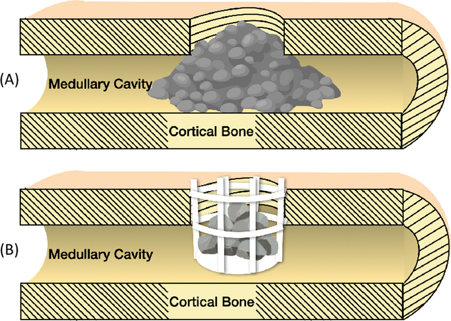
Traditional UE and UBE systems have been less effective in performing endoscopic interbody fusion procedures due to their narrow field of view and limited Working Channel Diameter (WCD). This raises a critical question: how can we efficiently execute both endoscopic interbody fusion and ULBD in patients with bilateral symptomatic LSS and lumbar spinal instability? The large-channel endoscope, with its wider WCD (Table 1), is adept at performing unilateral endoscopic decompression and fusion. However, its use in ULBD is challenging due to the potential risk of nerve root and dural sac damage when traversing the epidural space, given its large Outer Diameter (Table 1). To date, there have been few studies on performing ULBD with a large endoscope. To address this gap, we have developed the ‘Non-touch Over-Top’ technique. This innovative method allows minimally instruments traversing the space between dural sac and contralateral lamina. It ensures that neither the LF nor the dural sac is compressed by the endoscope or the working sleeve, which does not traverse this space at any point (Fig. 1). The typical six steps of the ‘Non-touch Over-Top’ technique have been illustrated in Fig. 3.
Fig. 1
Endoscopic view of the ‘Non-touch Over-Top’ technique. a, b The interlaminar window was expanded using an endoscopic trepine, and the upper and lower endpoints of the LF were detached. c Initially performing ULBD using the endoscopic high-speed drill. d, e Decompression of contralateral LR. f DS and bilateral exiting nerve roots after ULBD. g Intervertebral space after discectomy. h Intervertebral bone grafting and cage implantation. The blue dashed line represents the median of the spinal canal, the blue triangle represents the cranial side, the blue square represents the cauda side. ULBD unilateral laminotomy for bilateral decompression, LR lateral recess, DS dural sac, LF ligamentum flavum, BSP base of spinous process, IL inferior lamina, SL superior lamina, NR nerve root, NP nucleus pulposus
The key steps of the ‘Non-touch Over-Top’ technique are as follows. Firstly, the base of spinous process (BSP) is regarded as the anatomical marker of the starting point of performing ULBD, and the bony structure of BSP and the contralateral cranial lamina should be removed as much as possible. Decompression is performed by drilling between the laminae, with an effort to preserve the cortical bone near the LF as much as possible (Fig. 1). A sensation of sudden give is felt upon reaching the superior articular surface, after which this layer of cortical bone is removed. Next, the contralateral LF should be progressively removed using the endoscopic Kerrison rongeur (Fig. 2). Afterwards, the water pressure was reset at 100 mmHg. Through the combined effect of the irrigation water pressure and the dural sac’s tension, a spacious area is created between the dural sac and the contralateral lamina, and this space allows to observe and depress the contralateral LR easily. Meanwhile, through the collaborative effects of sturdy fixation of the pedicle screws and solid intervertebral fusion, spinal instability will not occur. Kim et al. [30] introduced specific twelve steps of ULBD under the traditional endoscope. Hua et al. [8, 9], whose study mentioned that preserving the LF would reduce damage and irritation to the dural sac and nerve roots during ULBD, claimed satisfactory clinical efficacy using the traditional small working channel endoscope when performing ULBD as well. However, we hold the perspective that the contralateral LF should be initially removed to expand the operative space and endoscopic sight during compressed with efficiency by large-channel endoscope.
Fig. 2
Six steps of the ‘Over-Top’ technique. A Ipsilateral laminectomy. B Decompression of the contralateral spinal canal while preserving the partial cortical bone near the LF. C Removal of the contralateral LF and the residual bone of lamina. D Decompression of contralateral LR. E Removal of the ipsilateral LF and discectomy. F Vertebral space bone grafting, cage and pedicle screws implantation. LF ligamentum flavum. LF ligamentum flavum, LR lateral recess
In this study, surgical efficiency and clinical efficacy has been significantly improved by the Non-touch Over-Top technique, which both ipsilateral and contralateral LR was completely decompressed simultaneously, 41 patients were followed for at least 6 months after surgery and none experienced spinal instability or internal fixation failure. The back VAS scores, leg pain VAS scores, ODI and JOA scores were recorded at four preoperative and postoperative time intervals to assess clinical efficacy. Back pain VAS decreased from 5.56 ± 0.20 to 0.20 ± 0.06, leg pain VAS scores decreased from 6.95 ± 0.24 to 0.12 ± 0.05, ODI decreased from 58.68 ± 0.80% to 8.10 ± 0.49%, and JOA scores increased from 9.37 ± 0.37 to 25.07 ± 0.38, which means these clinical indicators continued to improve significantly after twelve months of follow-up observation (P < 0.05) and the symptoms of the patients were significantly improved after surgery (Fig. 3). The fusion rate was 100% at the last follow-up, and the high fusion rate should be attributed to cleanly bone implanting beds (Fig. 1g).
Fig. 3
Visualization of clinical data of functional outcomes. Visual analogue scale (VAS) scores for back pain a, VAS scores for leg pain b, Oswestry disability index (ODI) scores c, and Japanese Orthopaedic Association (JOA) scores d showed a significantly improvement trend postoperatively compared with preoperative values
The Cage subsidence rate was 2.44% (1/41) at both 6 months and 1 year postoperatively. It may be related to severe osteoporosis (T = − 4.7) in this patient [31, 32]. In addition, one patient suffered dural tear and postoperative cerebrospinal fluid leakage, and one patient suffered incision infection. Dural tear and CSF leakage occurred in the same patient during the early stages of applying this technique, likely due to improper use of the visualized trephine with excessive force.
Radiological examinations were performed before and after surgery to evaluate changes in various parameters. As shown in Table 4, LLA increased from 34.04 ± 0.25° to 45.56 ± 0.17°, SLA increased from 9.50 ± 0.11° to 20.71 ± 0.11°, and DH increased from 7.19 ± 0.13 mm to 9.15 ± 0.02 mm. This technique can effectively improve lumbar lordosis and intervertebral disc height. In addition, the results of axial CT examination showed that the CSAC increased from 98.20 ± 2.57 mm to 200.49 ± 4.73 mm, which was significantly improved compared with pre-operative results (P < 0.05, Fig. 4). The changes in CSAC indicate that PE-PLIF with ULBD can effectively improve the vertebral canal volume.
Fig. 4
A 68-year-old male patient with severe intermittent claudication in both legs underwent PE-PLIF combined with the ‘Non-touch Over-Top’ technique. a preoperative AP X-ray fluoroscopy, and the green arrow showed I° spondylolisthesis at L4-5 segment. b preoperative MRI, the green arrow showed central canal stenosis at the L4-5 segment. c preoperative axial CT, and the green arrows showed bilateral LR stenosis at the L4-5 segment. d 1d postoperative AP X-ray fluoroscopy showed solid fixation of the implant and the successful reduction of the L4-5 segment. e 3d postoperative CT clearly demonstrated adequate spinal canal decompression. PE-PLIF percutaneous endoscopic posterior lumbar interbody fusion, AP anterior–posterior, ULBD unilateral laminotomy for bilateral decompression
In conclusion, our findings suggest that the integration of PE-PLIF with the novel ‘Non-touch Over-Top’ ULBD technique via a large-channel endoscope is highly effective in the short-term management of LDD. It can not only significantly expand the spinal canal volume and fully decompress the nerve roots and dural sac, but also improve the lumbar sagittal parameters and stabilize the vertebral column. This novel technique markedly ameliorates the symptoms, boosting LSS patients’ quality of life and spinal stability. With minimal complications observed, this integrated technique emerges as a promising option for LDD treatment, warranting further exploration and optimization in future research.
留言 (0)