Since Masson’s tumours were first described in 1923 [1], cases affecting almost every part of the human body have been reported. This tumour is often subcutaneous or dermal, with a predilection for the head, neck, fingers, and torso [2]. It is currently considered a reactive intravascular proliferation that develops in the lumen of a dilated vessel, hematoma, or preexisting vascular lesion [2, 3].
Additionally, approximately one-third of all cases originate in the head and neck, particularly in the neck, orbit, lip, pharynx, and mandible [3]. Tumours occurring in the paranasal sinuses or nasal cavity are rare, with only 14 reported cases to the best of our knowledge [4,5,6,7,8,9,10,11,12,13,14,15].
Symptoms vary and often include a combination of nasal congestion, epistaxis, postnasal drip, cheek pain, and frontal headaches.
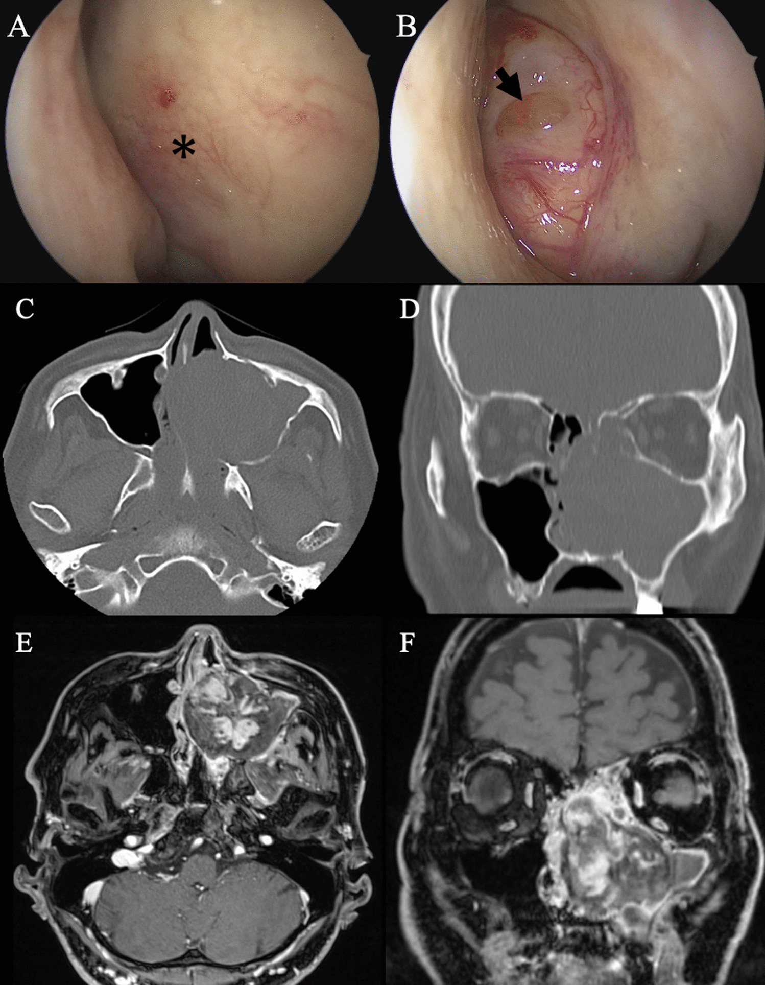
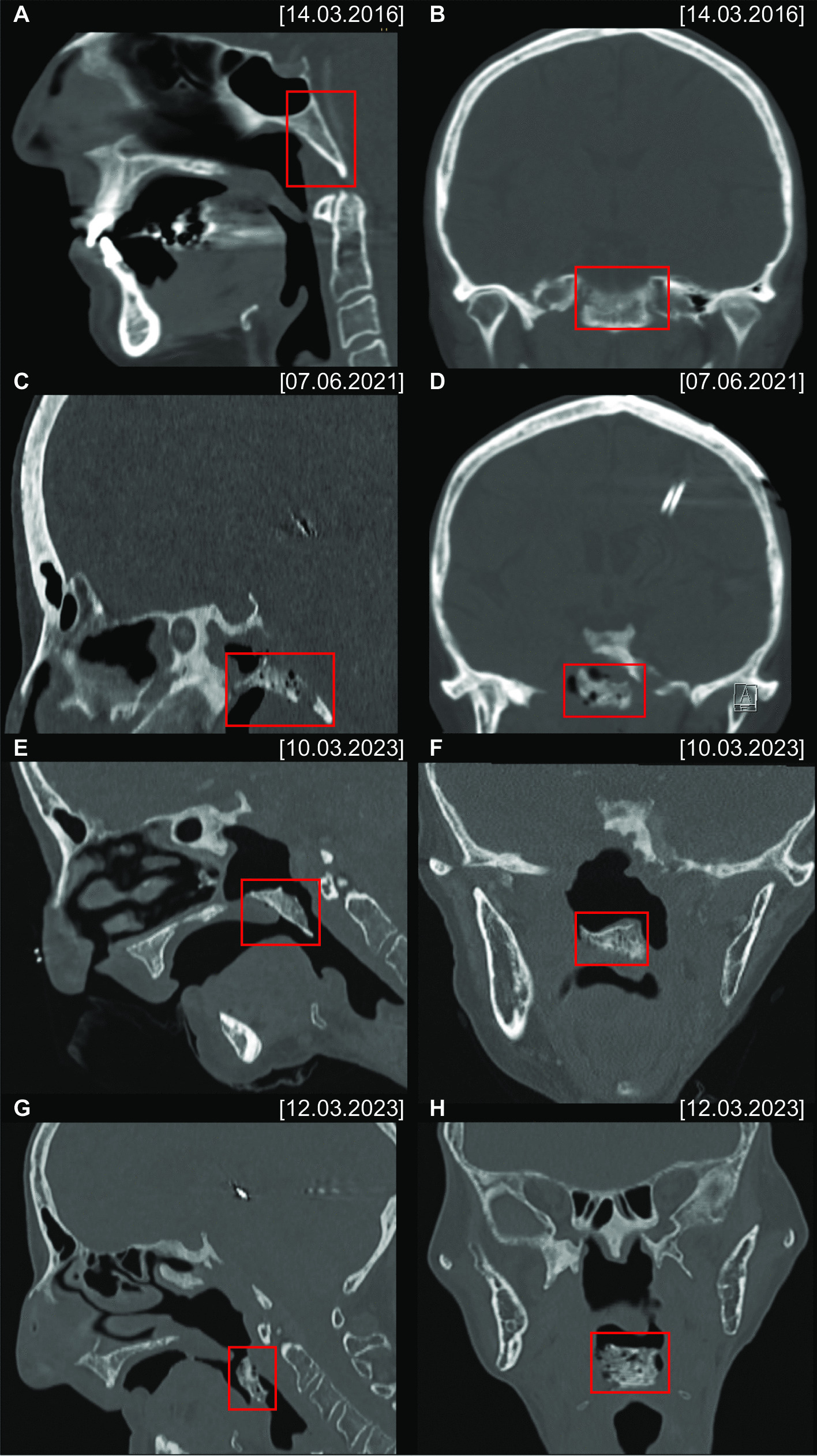
These symptoms are attributed to lesions occurring in the nasal and sinus cavities that spread with bone demineralization, causing anatomical changes in the surrounding area. Of the 15 cases of sinonasal IPEH, including our report, nine were of the maxillary sinus [5, 7, 11, 13,14,15]. Among them, four cases complained of nasal obstruction [7, 11, 14, 15], and in addition to our case, only one case developed bilateral nasal obstruction [14]. Our patient complained of bilateral nasal obstruction because the tumour compressed the nasal septum and extended into the contralateral nasal cavity. Moreover, epiphora occurred due to obstruction of the left nasolacrimal duct. These symptoms disappeared entirely after tumour excision.
Radiologically, IPEH closely resembles angiosarcoma but has a much more favourable prognosis [16]. The differential diagnosis of IPEH is challenging primarily due to its resemblance to angiosarcoma; however, several key histologic features differentiate the two: (1) IPEH resides intravascularly, while angiosarcomas invade the surrounding tissue; (2) IPEH is often closely associated with a thrombus; (3) IPEH lacks necrosis; and (4) IPEH has limited mitotic activity [5, 14, 16]. All these characteristics correspond to our case. Accurate histopathological diagnosis is crucial to prevent overtreatment due to suspicion of angiosarcoma. Among the eight reported cases of IPEH of the maxillary sinus, three were suspected to be malignant [9, 13, 15]. In two reports, malignancy was ruled out by perioperative biopsy [11, 13]. Through intraoperative frozen section analysis, we could also rule out malignancy and avoid overly aggressive treatment.
Several surgical approaches have been proposed for IPEH. Endoscopic excision has been reported in a few studies [7, 11, 14, 15], Table 1. The open approach using the Caldwell-Luc procedure has been described to access the maxillary sinus to eradicate the disease [5]. In the case of large lesions, a combined approach, open and endoscopic [13] or additional skull base repair by the neurosurgery team, may be performed to eradicate the disease safely [14]. We confirmed with preoperative CT and MRI that there was no bone destruction or infiltration in the lateral maxillary sinus or skull base. Furthermore, intraoperative frozen section analysis ruled out malignancy. We were able to entirely remove the tumor using endoscopic surgery alone, even though it was a large lesion.
Table 1 Review of all reported cases of IPEH of the maxillary sinus in the English literatureThe aetiology of IPEH has not yet been fully elucidated; however, many investigators have suggested that changes in the thrombotic process can lead to lesion development [3]. Any potential underlying cofactors promoting IPEH formation should be explored, given the diverse presentations in different cases. Recurrence of IPEH following incomplete excision of the lesion was documented as a result of poor exposure and visualization of the lesion has been reported [4, 6]. For postoperative follow-up of sinonasal IPEH, transnasal endoscopy is useful. Additionally, there is a possibility of submucosal recurrence, so if symptoms of bleeding or pain appear, an MRI should be performed. This report adds to the limited literature on IPEH by demonstrating its potential to mimic malignant processes and the critical role of accurate diagnosis through multidisciplinary collaboration. Complete resection remains a definitive treatment option. Recognizing this rare condition may prevent unnecessary overtreatment.


留言 (0)