
記住我


In this investigation, nine adult male New Zealand rabbits (Liaoning Changsheng Biotechnology Co., LTD. ) were two months old and weighed between 2 and 3 kg [12,13,14]. All experiments were conducted in accordance with the Association for Research in Vision and Ophthalmology Statement for the Use of Animals in Ophthalmic and Vision Research and the guidelines of the institution. The rabbits were housed on a light/dark cycle of 12/12 hours, free of food and water, for 8 h before the start of the experiment. All rabbits received anesthesia via subcutaneous injection with a blend of xylazine hydrochloride and ketamine hydrochloride. The pupil was dilated using topical eye drops comprising 0.5% tropicamide and 0.5% phenylephrine hydrochloride. After induction of general and topical eye (0.4% hydrochloride) anesthesia, the rabbits underwent slit-lamp, intraocular pressure (IOP), color fundus photography (Fig. 1A), optical coherence tomography (OCT) and an ultrasound B scan before Nd:YAG laser vitreolysis and 1 day, 4 weeks, and 12 weeks after treatment to determine a potential change in IOP and transient effects on the vitreous and retina. Three rabbits were euthanized by injecting sodium pentobarbital intravenously (100 mg/kg) 1 day, 4 weeks, and 12 weeks after laser treatment, respectively. A 21-gauge needle was attached to a 2.0-ml tuberculin syringe and injected 2 mm posterior to the limbus into the mid-vitreous cavity. A sample of undiluted vitreous humor (0.5 ml) was manually aspirated and immediately transferred to a sterile tube, which was stored at − 80 °C until the assay was performed. Then the eyes were enucleated cautiously to avoid probable globe violations and stored in Davidson’s fixative solution for 24 h [15].
Fig. 1
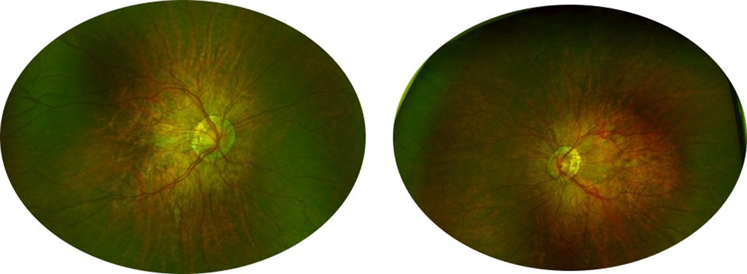
Retinal images following Nd:YAG laser vitreolysis. A. Color photo of the retina before Nd:YAG laser vitreolysis; B. Color photo of the retina after YAG laser vitreolysis, gas bubbles (black arrow) were noted above the optic disc; C. Retinal OCT of the treated eye three months after Nd:YAG laser vitreolysis, clear retinal layers were noted without any damage; D. Ultrasound B scan of the treated eye three months after Nd:YAG laser vitreolysis, no unmistakable echo of the vitreous cavity was found
Nd:YAG laser vitreolysisRabbits’ left eyes were treated with Nd:YAG laser vitreolysis, whereas the right eyes were used as controls. A Q-switched Nd:YAG laser (Ultra Q reflex, Ellex Medical Lasers, Adelaide, AU) was used in this experiment. The laser has a wavelength of 1064 nm and a pulse width of 4 ns. A mid-vitreous laser lens (Volk Singh Mid-Vitreous Lens, Volk Optical, US) was applied to the eye with gel (Ofloxacin Eye Ointment, Shenyang Sinqi Pharmaceutical, Shenyang, CN). The middle vitreous humor anterior to the visual streak area was focused with 15 degrees of oblique axis irradiation in reference to the thickness of the lens and the blurring retinal background. Each eye received a total of 500 spots with 10 mJ of pulse energy. Visible gas bubbles formed in the vitreous (Fig. 1B).
Vitreous humor analysesThe concentrations of vascular endothelial growth factor (VEGF), interferon inducible protein 10 (IP-10), monocyte chemoattractant protein 1 (MCP-1) and interlenkin 6 (IL-6) were determined using a multiplex cytokine assay (Shanghai Enzyme-Linked Biotechnology, Shanghai, CN) per the manufacturer’s user guide. Each sample was measured three times, and an average was then determined.
AsA concentrations were determined using a colorimetric technique based on its decrease from Fe3+ to Fe2+ upon reaction with 2,2’-dipyridyl. Modifications to the assay allowed for the examination of 10 µl samples, and all measurements utilized a standard curve. As a control for AsA specificity, two units of ascorbate oxidase (Shanghai Enzyme-linked Biotechnology, Shanghai, CN) were added to extra 10 µl samples, thoroughly mixed at room temperature, and then AsA measurements were performed in triplicate.
Using the trolox equivalent antioxidant capacity (TEAC) method on a sample made in the same way as for thiobarbituric acid reactive substances (TBARS) measurements, the total reactive antioxidant potential (TRAP) of vitreous humor fed a diet high in verbascosides was found. Specifically, using the radical cation decolorization assay established by Re et al. [16], the total antioxidant activity of the samples was determined. A chemical reaction with potassium persulfate (K2S2O8) generated the 2,2’-azino-bis (3-ethylbenzothiazoline-6-sulfonic acid) (ABTS+) radical. The sample of 25 ml of ABTS (7 mM) was spiked with 440 ml of K2S2O8 (140 mM) and placed in the dark at room temperature for 12–16 h for the radicals to form. The concoction of the working solution was prepared by diluting a volume of the previously mentioned solution with ethanol until its absorbance at λ = 734 nm reached 0.70 ± 0.02.
The measurement was carried out with a Varian Cary 100UV-VIS spectrophotometer, and this instrument was then connected to a 25 °C thermostat basin. The reaction took place directly in the measuring cuvette. Here, 2 ml of the ABTS+ radical was added, the absorbance (A0) was measured, and promptly 100 µl of the sample or standard was added. The radical was inhibited by the antioxidants in the sample, resulting in a decrease in absorbance proportional to the antioxidant concentration in the sample. At each standard and sample dilution, triplicate measurements were performed, and the calculation for percentage inhibition was relative to the absorbance of the blank at 734 nm. The definition of total antioxidant activity of samples was the concentration of Trolox equating to mol/g sample.
Hematoxylin–eosin (H&E) stainingAfter fixation, the eyes were cut in half along the anterior-posterior axis. Following the completion of histological processing, H&E was used to stain the 4 μm-thick specimens, which were viewed with the help of a light microscope (Fig. 2A). A minimum of three retinal tissue sections were evaluated, with one of them exhibiting a visual streak [15]. The morphologies of the retinal pigment epithelium (RPE), photoreceptors, and ganglion cells were carefully examined. The same pathologist (Y.L.A.) examined each specimen.
Fig. 2
Retinal histological examination following Nd:YAG laser vitreolysis. A. H&E-stained retinal histological specimen of the control eye; B. H&E-stained retinal histological specimen of the treated eye; C. TUNEL-stained retinal histological specimen of the treated eye. NFL: Nerve Fiber Layer; GCL: Ganglion Cells Layer: IPL, Inner Plexiform Layer: INL, Inner Nuclear Layer: OPL, Outer Plexiform Layer: ONL: Outer Nuclear Layer; IS: Inner Segment and OS: Outer Segment
Terminal deoxynucleotidyl transferase dUTP nick end labeling (TUNEL) stainingIn summary, after the retinal tissues were fixed in paraffin, slices of 4 μm thickness were stretched at 42 °C water to be set and baked. Next, the subsequent sections were added dropwise with the TdT reaction solution, and the reaction took place in the darkroom for one hour. Thenceforward, the reaction was stopped by incubation in deionized water for 15 min. Following the use of hydrogen peroxide to block the activity of endogenous peroxidase, the sections were added dropwise to the working solution. After the reaction was left for 1 h, the sections were washed, 3,3′-Diaminobenzidine (DAB) solution was added in drops for the color to develop, counterstained with hematoxylin for 20–30 s, and then washed again in running water. Finally, they were dehydrated at increasing alcohol concentrations and watched while sealed. The apoptotic index would be calculated if positive cells were noted.
Statistical analysisAll statistical analyses were performed on SPSS version 27.0 (SPSS Inc., USA). The mean and standard error of the mean were used to present the data in this study. The Dunnett-t test was used to analyze the differences between the two groups, and one-way analysis of variance (ANOVA) was utilized to calculate multiple comparisons. P < 0.05 indicated statistical significance.
留言 (0)