Culture and identification of hUC-MSCs
hUC-MSCs were obtained from Guangzhou DuDe Biotechnology Co., Ltd. They were cultured in α-MEM medium (Gibco, USA) supplemented with 10% fetal bovine serum from South America (Gibco, USA), 1% penicillin/streptomycin (Gibco, USA), and 0.05% bFGF. The fourth passage hUC-MSCs were selected and identified by flow cytometry. Cell surface markers including CD34 (Elabscience), CD45 (Elabscience), CD73 (Elabscience), CD90 (Biolegend), and CD105 (Endoglin) were used for MSC characterization.
Identification of hUC-MSCs by adipogenic differentiation
The 4th-generation hUC-MSCs with a cell density of 80-90% were seeded into a six-well plate and cultured at 37℃, 5%CO2 and saturated humidity. When the cells reach 100% confluence, add adipogenesis induction medium A; replace it with adipogenesis induction medium B after 3 days of culture, and replace it with A after 24 h of culture. Repeat 4 more cycles. Add solution B and culture for 7 days, changing the medium every 3 days. Control cells grew normally. After 23 days of induction, cells were stained with Oil Red O. The cells were washed with PBS, observed and photographed under an inverted microscope.
Identification of hUC-MSCs by osteogenic differentiation
Pluripotency assay was used to assess the pluripotent ability of MSCs to generate osteoblasts using commercially available differentiation media (StemPro Differentiation Kit, Thermo Fisher Scientific). To this end, different groups of mesenchymal stem cells were cultured in 6-well slides and evaluated histologically. Briefly, cells were cultured in osteogenic differentiation medium for osteogenic differentiation. Change differentiation medium twice weekly. After 21 days, differentiation evaluation and calcium deposition quantification was assessed by alizarin red staining.
Isolation and identification of hUMSC-Exos
The 5th passage of hUC-MSCs, grown to 80–90% confluency, were cultured in α-MEM medium supplemented with 1% exosome-depleted serum and 0.05% bFGF for 48 h. The cell culture supernatant was collected and filtered through a 0.22 μm filter to remove cells, apoptotic bodies, and cellular debris. Sequential centrifugation at 300 g, 2000 g for 10 min, and 10,000 g for 30 min at 4 °C was performed to further eliminate contaminants. Subsequently, ultracentrifugation at 110,000 g for 70 min was carried out to isolate the exosomes. Finally, PBS was used for resuspending the exosomes. Nanoparticle tracking analysis was employed to determine the size of the exosomes, while transmission electron microscopy was utilized for exosome identification.
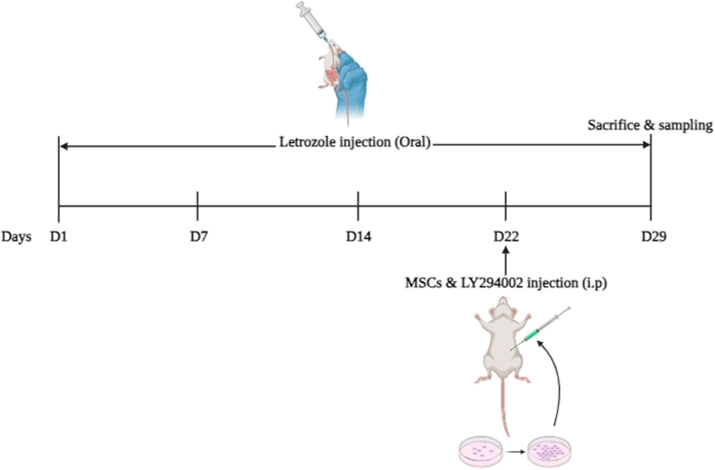
Establishment of POI mouse model and treatment with hUMSC-Exos
6-week-old C57BL/6 female mice were purchased from Guangdong Yaoke Biotechnology Co., Ltd. (Guangdong, China). The mice were maintained at a temperature of 22 ± 1 °C, a relative humidity of 50 ± 1%, and a 12/12-hour light/dark cycle. Sterilized food and water were provided ad libitum. All animal experiments were conducted in accordance with the guidelines and regulations of Shunde Hospital, Southern Medical University, and the AAALAC and IACUC guidelines.
The mice were divided into three groups: control group, POI group (n = 10), and POI + Exos group (n = 10). To establish the POI model, mice received intraperitoneal injections of CTX (50 mg/kg) daily for 14 consecutive days, while the control group received an equivalent volume of PBS. In the POI + Exos group, mice were transplanted with 30 µl of hUMSC-exos (0.015 µg/µl) into each ovary one week after the POI model was established. Meanwhile, mice in the POI group were injected with an equivalent volume of PBS. At the end of the 2nd and 4th week after treatment, five mice from each group were euthanized under anesthesia, and serum and ovarian tissues were collected.
Hematoxylin and eosin staining
The staining procedure for hematoxylin and eosin (H&E) was performed following the established protocol described in previous publications [16]. Briefly, ovarian tissues fixed with 4% paraformaldehyde (Servicebio, China) were dehydrated, embedded, and sectioned (The maximum transverse section of the ovary wasserially sliced into 4 μm sections). The sections were stained with hematoxylin and eosin. The morphology of each ovarian section was observed and photographed using an inverted microscope (Leica DMI1, Germany). The number of follicles at different stages was counted.
Serum hormone levels determination by ELISA
Serum for hormone measurements was obtained by centrifuging mouse blood at 5000 rpm for 10 min. The levels of anti-Müllerian hormone (AMH), estradiol (E2), and follicle-stimulating hormone (FSH) in serum were determined using ELISA kits (Lengton, China). The optical density (OD) values were read at 450 nm using an ELISA reader.
Culture and treatment of human granular tumor cells
KGN cells (Procell CL-0603) were provided by Wuhan Procell Life Science & Technology Co., Ltd. and cultured in DMEM/F12 medium (Gibco, USA) supplemented with 10% fetal bovine serum (Gibco, USA) and 1% penicillin/streptomycin (Gibco, USA). The cells were divided into four groups: the control group (Nc), the POI group (CTX, 1 mg/mL, POI), the hUMSC-derived exosomes (0.015 µg/µl) treatment group (POI + Exos), and the hUMSC-derived exosomes + NRF2 inhibitor treatment group (ML385, 1 µmol/L, MCE) (POI + Exos + ML385).
EdU staining
Cell proliferation was assessed by EdU staining after both 24 and 48 h of culture in each group. Cells were treated with EdU working solution for 4 h, fixed with 4% paraformaldehyde, and then stained with EdU mixture and DAPI. Cell apoptosis was evaluated using Hoechst staining. After 24 and 48 h of cell culture, cells were incubated with Hoechst/PI staining working solution in the dark for 20 min. Finally, assessment was performed using a fluorescence microscope (Leica DMI8, Germany).
Reactive oxygen species detection
The levels of reactive oxygen species in cells were measured using a ROS detection kit (Beyotime, Shanghai, China) following the manufacturer’s instructions. The probe for intracellular ROS (DCFH-DA) was diluted to a concentration of 10 µmol/L in serum-free medium. KGN cells were then incubated with the diluted DCFH-DA at 37 °C for 30 min. Fluorescence was detected using an inverted fluorescence microscope.
Ferrous ion detection
A fluorescent probe for ferrous ions (FeRhoNox-1) detection kit (MKBio, China) was used to measure Fe2 + levels in cells. FeRhoNox-1 was diluted to a concentration of 5 µM in PBS (prepared fresh) and incubated with KGN cells at 37 °C for 60 min. Fluorescence was detected using an inverted fluorescence microscope.
Cellular lipid peroxidation detection
A lipid peroxidation sensor (BODIPYTM 581/591 C11) (Shanghai BioHub Biotechnology Co., Ltd.) was employed to indicate intracellular lipid peroxidation and antioxidant status. Treated cells were stained with 5 µM BODIPYTM 581/591 C11 at 37 °C for 30 min. Fluorescence images of cells were captured using a fluorescence microscope.
Western blotting
Western blotting was performed using the Multistrip Western blotting protocol as previously described [16]. The following antibodies were utilized: The exosomes were identified by CD63 (1:1000; Abcam), CD9 (1:1000; Abcam), TSG101 (1:1000; Abcam). The expression of Nrf2/GPX4 pathway was detected by Nrf2 (1:1000;Cell Signaling Technology), xCT (1:1000; Abcam), GPX4 (1:1000; Abcam), and Alpha Tubulin (1:200; Proteintech). After incubation with secondary antibodies at room temperature for 30 min and subsequent washes, the bands were visualized using enhanced chemiluminescence (Abcam, USA).
Statistical analysis
All quantitative data are presented as mean ± standard deviation (n ≥ 3). Statistical analysis was conducted using GraphPad Prism 9.0 software, and significant differences were evaluated using one-way analysis of variance (ANOVA) and t-tests; p < 0.05 was considered statistically significant.


留言 (0)