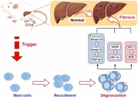
Worldwide, more than one billion people suffer from chronic liver disease caused by different triggers (e.g. alcohol, hepatitis B or C virus infection) and is accompanied by liver fibrosis (Zhang et al., 2019). Liver fibrosis is a reversible wound-healing response to acute or chronic liver injury (Zong et al., 2021). Liver fibrosis is typically characterized by the accumulation of excess extracellular matrix (ECM) in the liver (Zhao et al., 2019). Continued progression of fibrosis can lead to structural and functional damage to the liver, leading to cirrhosis and even liver failure (Caligiuri et al., 2021). Disadvantages of anti-hepatic fibrosis drugs include poor specificity and high toxicity (Peng et al., 2021). There are no effective antifibrotic therapies or medications (Zhang et al., 2022), and there is an urgent need to develop antifibrotic drugs or treatment pathways.
Mast cells (MCs) have been recognized as immune cells that play a role in the process of tissue fibrosis (He et al., 2017), including liver fibrosis, (Weiskirchen et al., 2019) pulmonary fibrosis (Veerappan et al., 2013), intestinal fibrosis (Liu et al., 2021), adipose tissue fibrosis (Silver, 2013), renal fibrosis (Veerappan et al., 2012), and cardiac fibrosis (Legere et al., 2019). MCs are observed in connective and mucosal tissues throughout the body. They originate from hematopoietic stem cells in the bone marrow and circulate through the bloodstream, where they differentiate and mature after migrating to different tissues (Weiskirchen et al., 2019). MCs are present throughout the body in small numbers under normal physiological conditions. However, when stimulated, MCs are activated and release cytokines, vasoactive amines, and lipid mediators (Gao et al., 2023, Li et al., 2021). Notably, cytokines released by MCs, such as histamine, chymases, tryptases, leukotrienes, heparin, TNF-α, renin, and TGF-β1 (Li et al., 2023) are inflammatory and pro-fibrotic mediators that contribute to the histological alterations by triggering a variety of immune responses (Choi et al., 2015). Kyritsi et al. demonstrated that the introduction of MCs into mice with normal livers or those lacking MCs induced ductular reaction, inflammation, biliary senescence and hepatic fibrosis, and TGF-β1 was demonstrated to be a strong driver of these pathological changes (Kyritsi et al., 2021).
Stem cell factor (SCF), produced by fibroblasts or endothelial cells, plays an important role in the survival, proliferation, and differentiation of MCs (Ding et al., 2013). SCF binds to the tyrosine kinase membrane receptor kit (c-kit) on the surface of MCs, leading to the recruitment and activation of MCs in tissues (Weiskirchen et al., 2019). The SCF/c-kit axis is closely associated with the processes of tissue remodeling and fibrosis (Ding et al., 2013). MCs are key cells involved in allergic and inflammatory diseases and regulate cellular biological processes through the SCF/c-kit axis (Franke et al., 2023). MCs maintain high levels of c-kit throughout their development (Tsai et al., 2022). Inappropriate c-kit activation leads to the recruitment of MCs in tissues (El-Agamy, 2012). It has been reported that SCF/c-kit may be a potential therapeutic target for the regulation of numbers and activation of MCs and eosinophils in inflammatory diseases (Reber et al., 2006). Moreover, MCs infiltration promoted renal interstitial fibrosis through the SCF/c-kit pathway, and inhibition of SCF/c-kit signaling prevented the development of fibrosis (Yin et al., 2018).
The level of oxidative stress is a reflection of the balance of the pro-oxidant/antioxidant capacity of the body (Sadasivam et al., 2022). Numerous studies have shown that oxidative stress plays an undeniable role in promoting liver fibrosis and has been considered as an effective target for the treatment of liver fibrosis (Gu et al., 2020). Oxidative stress occurs with the dysregulation, overproduction, and decreased elimination of reactive oxygen species (ROS) in the liver and dysfunction of enzymes and non-enzymatic antioxidants in the liver, leading to hepatocellular dysfunction and ultimately hepatic fibrosis (Seen, 2021). In response to oxidative stress, the slow rate of Nrf2 degradation leads to the accumulation of Nrf2 in the cytoplasm, which results in the translocation of Nrf2 to the nucleus (Hassanein et al., 2023). Nrf2 binds to the antioxidant response element (ARE) present in the promoter region of target genes that encode various antioxidant and detoxifying enzymes, including glutathione S-transferase A1 (GST), NAD(P)H quinone oxidoreductase 1 (NQO1), heme oxygenase 1 (HO-1), glutathione peroxidase (GSH-Px), and superoxide dismutase (SOD), all of which enhance cellular defense against oxidative stress (Mapuskar et al., 2023).
In this study, we first established a CCl4-induced liver fibrosis model in rats to investigate the association between MCs and liver fibrosis. The expression of hepatic SCF/c-kit, associated fibrotic factors (e.g., TGF-β1 and α-SMA), and the Nrf2/HO-1 pathway were investigated, along with the level of hepatic oxidative stress. The purpose of our study was to explore the relationship between MCs and liver fibrosis with the goal of identifying new ideas and therapeutic approaches for the treatment of liver fibrosis.


留言 (0)