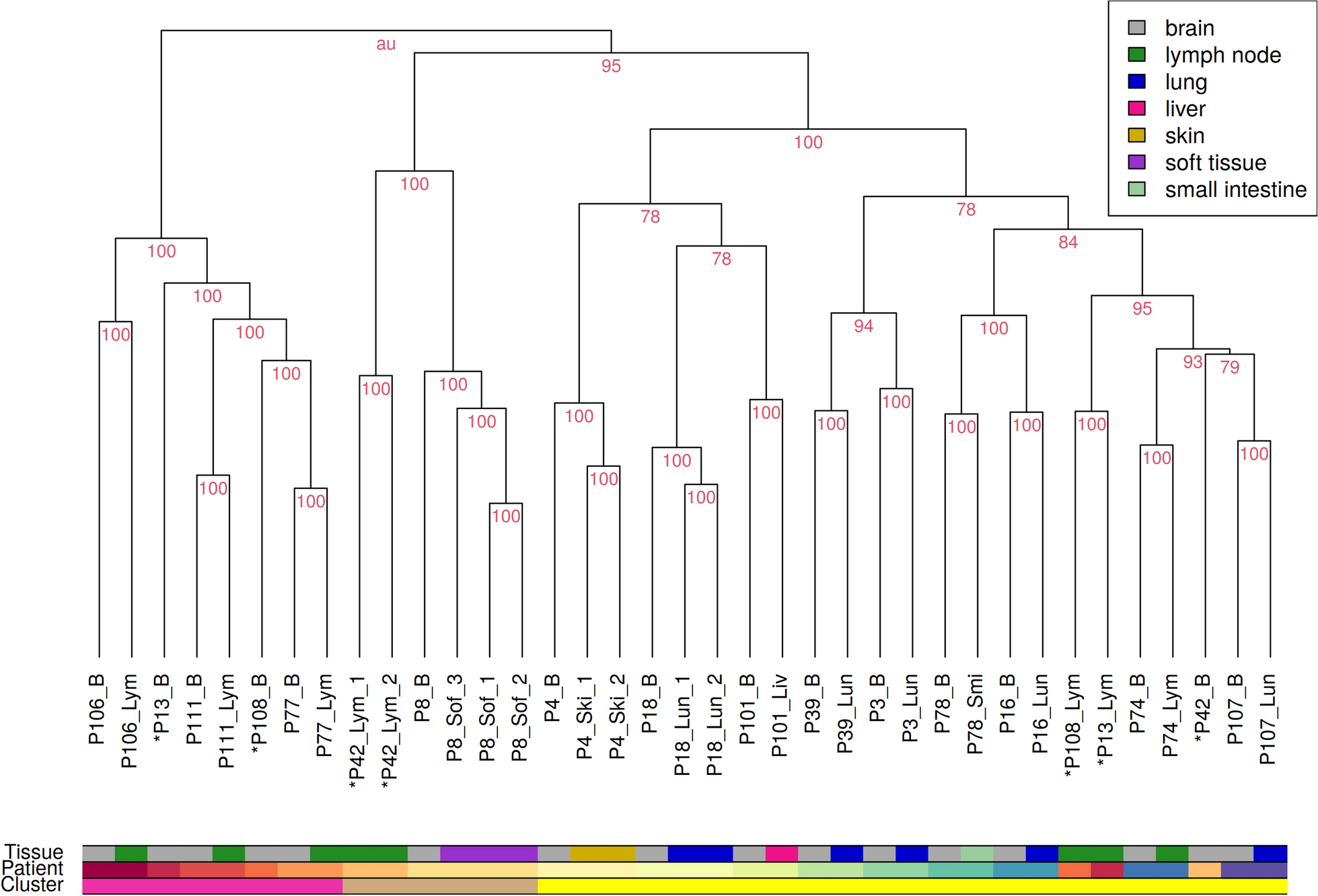
In this study, we investigated the clinical course and molecular features of high-grade gliomas that developed in three adult patients with LFS. All patients showed an aggressive clinical course despite multidisciplinary treatment. The location and radiographic features varied among the cases. In Cases 1 and 3, MRI revealed infratentorial lesions without contrast enhancement, which is not typical of adult-type glioblastoma, IDH-wildtype. In contrast, all tumors possessed typical histopathological features of high-grade gliomas, and two tumors did those of glioblastoma such as mitosis, palisading necrosis, and microvascular proliferation. WES of DNA derived from tumor tissues revealed no mutations in the IDH or histone H3 genes. Intriguingly, WES data also revealed no typical molecular features of glioblastoma, IDH-wildtype (TERTp mutation, EGFR amplification, and 7 +/10−) in all tumor tissues, whereas all tumor tissues harbored TP53 mutation and PDGFRA amplification. Additionally, all tumor tissues clustered within pHGG H3-/IDH-wt RTK1 subtype in t-SNE analysis based on DNA methylation status, although Cases 1 and 2 did not exhibit significantly high calibrated scores for pHGG H3-/IDH-wt, RTK1 subtype using DKFZ classification system. Lower tumor cellularity is a major cause of lower calibrated score in the DNA methylation-based classification [18]. However, tumor cellularity of our cases was higher than 75% in all tissues, implying the presence of other reasons for the unmatched scores of Cases 1 and 2. Capper et al. reported that 12% of analyzed tumors could not be classified by DNA methylation profiling and this subset was highly enriched for unusual syndrome-associated tumors [14]. Other studies have also reported that a significant proportion (6–17%) of tumors, including pediatric or adolescent CNS tumors, could not be assigned to a classifier diagnosis [19,20,21]. These publications suggest that some cases of syndrome-associated tumors or pediatric-type tumors may not be accurately classifiable by DNA methylation profiling, although the entity of pHGG H3-/IDH-wt was not included in version 11.4 of the classifier. Since the introduction of the entity pHGG H3-/IDH-wt, Drexler et al. reported that 33.3% of cases that could not be classified by version 11.4 of the classifier, were not classified even by version 12.8, although only a small number of cases were classified as pHGG H3-/IDH-wt using version 12.8, including the entity of pHGG H3-/IDH-wt. They also reported that the t-SNE plot is also quite effective for the diagnosis of these unclassifiable tumors, beyond the matching score alone [22]. In addition, Guerrini-Rousseau et al. reported that tumor tissues from pediatric patients with LFS did not match the methylation classification but clustered in pHGG MYCN subtype using t-SNE analysis, as we did [23]. These findings suggest that for diagnosis based on the DNA methylation status of tumors arising in the context of a germline disorder, such as LFS, not only the DKFZ classification system but also t-SNE plot analysis is useful.
pHGG H3-/IDH-wt is a newly defined tumor type in the WHO CNS 5. pHGG H3-/IDH-wt comprises three subtypes (MYCN, RTK1, and RTK2). The RTK1 subtype has an intermediate prognosis (median OS, 21 months). The RTK1 subgroup is characterized by ahigh frequency of PDGFRA amplification. In The Cancer Genome Atlas (TCGA) database [24], among 362 adult GBM cases that were defined using the 4th edition of WHO classification whose gene mutations and copy numbers were available, only 14 cases (3.9%) harbored TP53 mutation and PDGFRA amplification. These data might also support our findings that gliomas developing in adult patients with LFS have molecular profiles not like those of glioblastoma, IDH-wildtype but rather like those of pHGG H3-/IDH-wt.
There are a few other studies reporting that gliomas developed in adult patients with LFS. Tian et al. reported six IDH-mutant glioma cases and 13 IDH-wildtype glioma cases that developed in adult patients with LFS in a Chinese cohort, including three cases with EGFR mutation or amplification, and six cases with PTEN mutation, implicating glioblastoma, IDH-wildtype [25]. Among the other adult IDH-wildtype cases, no tumors harbored PDGFRA amplification. Two other studies have reported IDH-mutant astrocytoma cases in adult patients with LFS [7, 8]. Wu et al. reported a case of glioma in an adult patient with LFS, who possessed wild-type IDH, H3F3A, TERT and EGFR, and mutant NF1 and PDGFRB, suggesting the potential of pHGG H3-/IDH-wt [26].
We also compared the TP53 mutation status of tumor tissues with that of germline cells in each case. Mutation spots, copy number, and BAF of chromosome 17 suggested that UPD is associated with the development of gliomas in our cases. Recently, a high rate of copy number gains in TP53 mutations was detected in tumors of patients with LFS [27]. This study revealed that the copy number gain of TP53 variants occurs many years before tumor diagnosis, followed by accumulation of additional driver mutations that result in tumor development. Some studies have reported MYCN amplification in gliomas harboring wild-type IDH arising in pediatric LFS cases [8, 9, 23]. Our cases suggest that the copy number gain of mutant TP53 obtained by UPD and PDGFRA amplification may play pivotal roles in the development of gliomas in adult patients with LFS.
In general, many mutant TP53 proteins exhibit a dominant-negative effect on wild-type TP53, mostly by forming mixed tetramers with diminished DNA binding and transactivation activities [2, 28]. The mutation spots of TP53 in our cases were not as frequent in LFS or brain tumors. The variants in Cases 1 (Y220C) and 2 (Y234H) have been reported to be associated with reduced transcriptional activity in the IARC TP53 database (http://p53.iarc.fr). Although the variant in Case 3 (H214R) has also been associated with reduced transcriptional activity, an increased ability to transactivate GADD45 has been reported in a subset of breast cancer with this mutation [29]. However, no study has revealed an association between this mutation and gliomas. Our cases suggest that homozygous alteration of TP53 by UPD might play a pivotal role in the oncogenic effects of some types of TP53 variants, especially in the setting of LFS.
There is very little literature on pHGG H3-/IDH-wt because this is a rare tumor type. Additionally, it is quite difficult to distinguish this tumor subtype from glioblastoma, IDH-wildtype by histopathological and DNA mutation analyses. DNA methylation analysis is a unique and powerful tool for distinguishing this subtype from glioblastoma, IDH-wildtype. Although tumor predisposition syndromes are associated with pediatric-type cancers, the pathophysiology of adult-onset cancers in these patients is poorly understood. Our cases suggest that adult LFS patients also develop pediatric-type high-grade gliomas, and that PDGFRA amplification may have a high affinity for this type of glioma. Overall, this study revealed that atypical high-grade gliomas in adult patients with tumor predisposition syndromes, such as LFS, might be pediatric-type gliomas (Additional files 1 and 2).


留言 (0)