PJK is the most common complication after DLS surgery, which can lead to progressive sagittal decompensation, nerve damage, revision surgery, and worse clinical outcomes [12, 13]. The incidence of PJK varies from one literature report to another but can typically reach between 10 and 30% within 12 months of surgery [14,15,16]. In our study, PJK was observed in 27 patients with a prevalence of 22.13%, which is similar to what Wang et al. [17, 18] reported in patients with DLS.
The development and progression of PJK are associated with multiple factors, as no study has shown that a single etiology is closely and consistently associated with their development. Relevant studies have identified risk factors associated with patients, procedures, and imaging. In this study, preoperative TK did not differ significantly between PJK and non-PJK groups. It was mentioned that there is no consensus on standard values for TK and that thoracic compensatory capacity may manifest as kyphosis and degenerative spine. Meanwhile, the incidence of TK for PJK before surgery is controversial in the literature. Oe et al. [19] pointed out that both kyphosis and kyphosis excess of TK preoperatively contributed to the high incidence of PJK. Our results found that larger TLK (OR 1.191, 95% CI 1.041–1.362, p = 0.011) was an independent risk factor for postoperative PJK in patients with DLS. According to previous literature [20, 21], PJK may be a compensatory mechanism resulting from changes in spinal balance, and patients with higher TLK are at greater risk for PJK. Greater TLK may lead to sagittal imbalance, and the occurrence of PJK after corrective surgery is a compensatory mechanism of sagittal imbalance, consistent with our findings. Age, BMI, and osteoporosis were considered risk factors associated with patients. Kim et al. [22] also found older patients with PJK requiring revision. However, in our study, age and BMI were similar. A decrease in bone mineral density (BMD) is another crucial risk factor for PJK and can lead to loosening of pedicle screws and compression fractures in UIV.
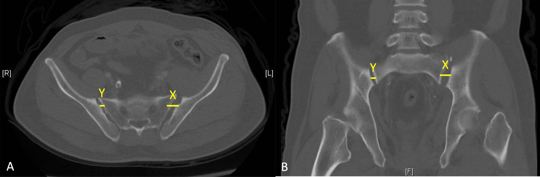
DEXA is currently considered the gold standard for osteoporosis screening. However, due to osteophyte proliferation, the accuracy of traditional DEXA measurements for certain degenerative diseases has also been questioned. Factors such as degenerative changes in the lumbar spine or superimposed calcified tissue may cause bone mineral density in patients with osteoporosis to be incorrectly overestimated, influencing surgeons to make the best decisions. Computed tomography-based Hounsfeld unit (HU) measurements have been reported as an alternative method for accurately assessing bone mass. Pickhardt et al. [23] confirmed the diagnostic value of L1 HUs, while Zou et al. [24] further evaluated the use of S1 HUs, providing a simple method for directly assessing S1 BMD.
Recently, MRI-based bone mass measurements have increasingly been applied to lumbar spine BMD assessment to reduce patient radiation exposure [25,26,27]. MRI bone analysis shows that when vertebral BMD decreases, trabecular bone increases due to fat infiltration and higher signal on T1-weighted imaging. This method has been shown to accurately assess vertebral bone mass, an essential predictor of osteopenia and osteoporosis. Kuo et al. [28] reported that the higher VBQ was independently associated with PJK in patients undergoing DLS correction. VBQ score measured by preoperative MRI may be a valuable adjunct to DLS surgical planning. However, accurate measurement of VBQ score using conventional measurement methods is variable due to the lumbar spine profile of DLS. Therefore, Huang et al. [26] proposed a new simplified S1 VBQ score concerning the measurement method of S1 HU. The results showed that the diagnostic accuracy of S1 VBQ method was comparable to the results of L1-4 VBQ or S1 HU, and the measurement method was more straightforward to ensure reliability within/among raters. The S1 VBQ score is a promising alternative to assessing BMD in patients with DLS. As in previous studies, our results show that S1 VBQ score is an independent risk factor for PJK after DLS, and S1 VBQ score is a good predictor of PJK (AUC = 0.721). The ideal limit of VBQ score was 3.205 (sensitivity: 77.8%, specificity: 81.4%), indicating that VBQ score is still a very sensitive parameter for assessing PJK risk after excluding other factors.
At present, the choice of distal fixation fusion segment, whether fusion to S1 or not, is still the focus of debate. LIV located at L5 can preserve more motor function of the active segment and improve the quality of life of patients. However, fusion to L5 alone accelerates degeneration of the L5-S1 disc, which may lead to further scoliosis and sagittal imbalance. Fusion to S1 results in the loss of L5-S1 motion segments and changes in spinal-pelvic biomechanics, which may exacerbate sacroiliac joint pain and accelerate sacroiliac joint degeneration. Furthermore, fusion to the sacrum increases surgical exposure, prolongs surgical time, and may increase the incidence of complications. Liu et al. [29] conducted a meta-analysis of 14 studies and showed that fusion to the sacrum was a risk factor for PJK. Yagi et al. [13] also found that the incidence of PJK was twice as high in patients with LIV fixation to the sacrum as in patients with LIV fixation at the superior sacrum. Surgical risk factors should also consider factors such as the number of fusion levels and interbody fusion. Bridwell et al. [30] found that too short fusion segment is also a risk factor for PJK. In this study, by comparing the surgical data of PJK group and non-PJK group, it was found that the fixed position, fixed level and interbody fusion of UIV and LIV were not risk factors for PJK. Due to the small sample size of this study, further accumulation of cases needs to be further analyzed.
Limitations
The study also has some limitations. First, this is a single-center retrospective study with a relatively limited sample size, and further, more extensive multicenter studies are needed to help validate and reinforce these findings. Our analysis did not include preoperative laboratory values such as calcium, phosphorus, glomerular filtration rate, and bone metabolism, leading to a more comprehensive assessment of osteoporosis risk. This work will help improve the potential complementary role of S1 VBQ in predicting osteoporosis.


留言 (0)