The femoral head is located in the hip joint, and the hip capsule must be opened intraoperatively to remove the femoral head lesion [13]. For most pediatric surgeons, they usually choose the Simith-Peterson approach, which allows clear exposure of the surgical area [18], but the approach involves a larger incision and often requires severing the rectus femoris muscle, which takes a longer time to suture. In addition, due to postoperative damage to the rectus femoris muscle, the patient needs to wait for the muscle to fully heal before appropriate joint activity can be initiated. If a femoral neck fracture or lesion is encountered, the Watson-Jones approach is usually used. This approach, which is entered from the lateral side of the hip, can easily treat the basal lesions of the femoral neck with relatively good results [8]. However, if there is a lesion on the inner side of the femoral neck, a major trochanteric osteotomy is required. After lesion removal, the major trochanter needs to be fixed with screws, which involves a larger trauma and may require secondary surgery for removal of the internal fixation.
The DAA isfirst described by German surgeon Carl Hueter in the nineteenth century and published in Der Grundriss der Chirurgie (The Compendium of Surgery). This surgical approach is also known as the "Hueter approach" [4]. It was not until 1917, after a report by Smith-Peterson, that the surgical approach became widely known. In 1950, French doctor Judet also reported hip replacement by anterior approach [19], but with the emergence of new artificial joints, this approach gradually decreases and was only occasionally used in the treatment of hip infection in children [4]. In 1980, Light and Keggi reported the experience of 104 patients of modern total hip arthroplasty using the anterior approach. This surgical approach has the advantages of the short operation time, less bleeding, no intraoperative complications, short hospitalization time, fast functional recovery, etc., which has aroused the attention of the medical community again. Become one of the surgical approaches for Total Hip Arthroplasty (THA) [20]. But what really brought it to the forefront of clinicians' discussions was the popularity of minimally invasive surgery in recent decades.
Compared with Simth-Peterson and Watson-Jones, the DAA is not familiar to pediatric surgeons and has been rarely reported in the field of pediatric orthopedics. In fact, this approach has the advantages of less trauma, easy exposure and shorter operation time, which is more suitable for children. In addition, the DAA also relatively "protects" the vessels where the base of the femoral neck is located, which can effectively avoid vascular damage [21, 22]. If the lesion removal in this area causes excessive trauma and disrupts the blood supply, it may increase the risk of complications such as avascular necrosis of the femoral head. Therefore, minimally invasive surgery is the key to avoiding this serious complication. In contrast, the Watson-Jones or Simith-Peterson approaches often result in large surgical trauma and a relatively higher risk of complications. Surgical dislocation approaches involve even greater trauma and may require secondary surgeries.
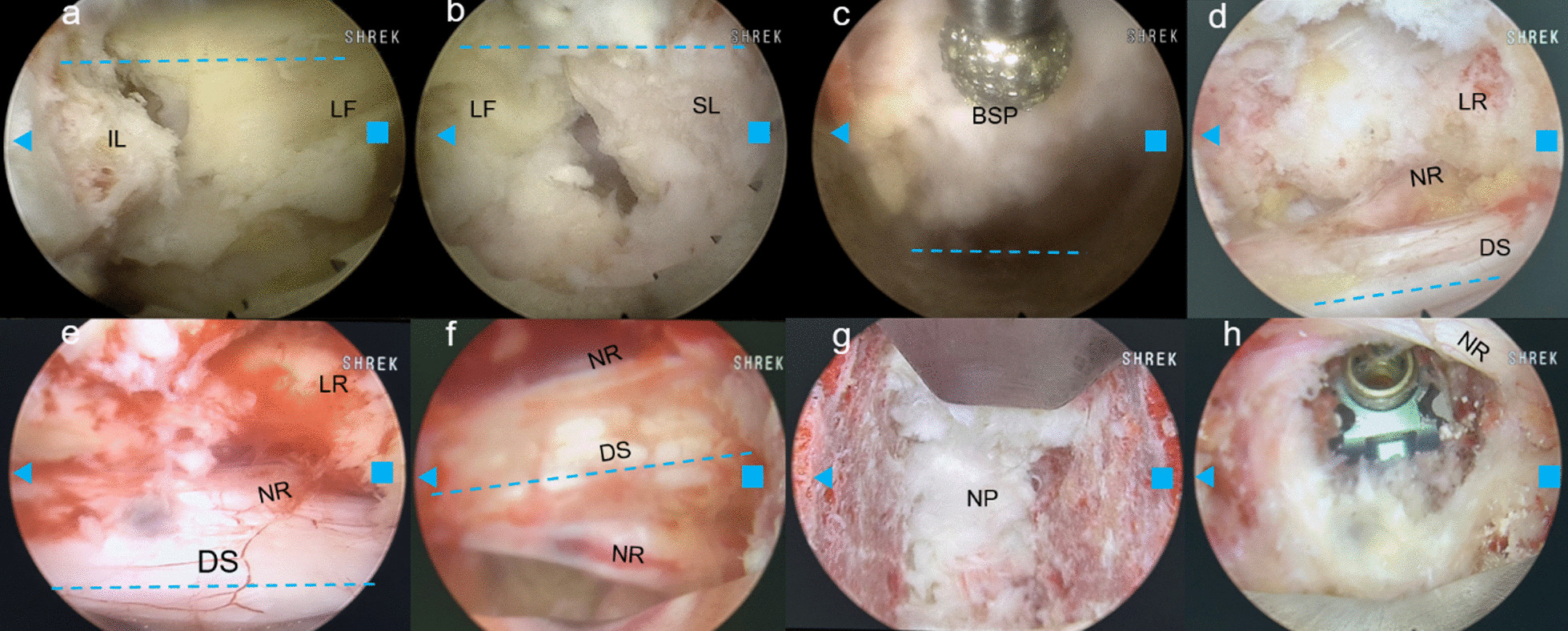
The DAA involves dissecting through the superficial interval between the sartorius muscle and the tensor fasciae latae, as well as the deep interval between the rectus femoris and gluteus medius muscles. This approach allows for good exposure of the hip joint while avoiding the “extensive dissection” required by other approaches (15). Thus, there is minimal bleeding during hip joint exposure, with an average blood loss of only 23.9 mL in our case series. In addition, the DAA can expose the femoral neck and femoral head without cutting any muscles and tendons and is a scheme to expose the surgical field solely through the space between muscles, basically without damaging or affecting the muscles of the lower extremities. In rare patients, during the final articulation of the hip joint, it is necessary to cut off part of the inverted head of the rectus femoris in order to facilitate the articulation of the hip joint (in most patients, it is not necessary to cut off). The anatomical advantages of this surgical approach allow for rapid recovery of muscle and joint function after surgery. In our case series,unless there were a concern about the fracture, it would be rare for patients to need to remain in bed for rest. Because this approach can directly expose the hip joint, the exposed incision is small and there is no muscle disconnection, so the incision suture is fast. The average suture time in this group is 40.3 min, which relatively shortens the entire operation time. However, the amount of bleeding can vary significantly among different diseases, and therefore the bleeding volume between patients may not be directly comparable. For example, aneurysmal bone cyst bleeding is more, and Kapo's sarcoma bleeding is also a lot, but simple bone cyst bleeding will be less. Therefore, the characteristics of different diseases need to be taken into account when assessing and comparing the amount of blood loss.
This retrospective study has limitations due to its limited sample size. Specifically, the low incidence of femoral head ischaemic necrosis does not completely demonstrate the benefits of the access route, which requires verification in a larger study. Additionally, alternative approaches to removing femoral neck lesions exist, but we have focused solely on the method used in this study.


留言 (0)