
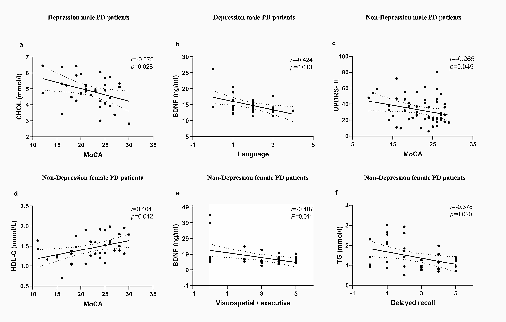
記住我


All experiments described were performed on animals, with both young (3–5 months) and aged (20–22 months) Sprague–Dawley rats. Based on our experience, 17 (weight: 500–650 g) aged animals will be required per group for the behavioural analysis of neurological recovery after stroke. This calculation is founded on an expected effect size of 25% of mean values, which will yield statistically significant effects (α error < 0.05) with a power of 0.80. This assumption considers that common standard deviations are 25% of mean values. Given the smaller parameter variability in young and sham animals, we opted for 10 animals per group for both young sham (weight: 310–400 g), aged sham (weight: 500–650 g) and young animals (weight: 310–400 g).
The animals were kept on a 12 h light/dark cycle in a temperature-controlled room with free access to water and food. After randomization, the animals were transferred to the testing location and given 48 h to acclimate. All experiments were approved by the Institutional Animal Care and Use Committee of the University of Medicine and Pharmacy Craiova (approval nr. 2.5 from 29.10.2020), and by the National Sanitary Veterinary and Food Safety Authority Dolj (approval nr. 12 from 29.03.2022). All animal experiments complied with the ARRIVE guidelines and were carried out in accordance with the EU Directive 2010/63/EU for animal experiments.
Beam narrowing walking testAll behavioural tests were performed by two investigators blinded to the identity of the animal groups.
Animal trainingFirst, the animals were given three to five days of training. This involved positioning the animals on a narrowing beam 135 cm in length and encouraging them to walk towards the end of the beam for three to five trials until continuous walking was achieved. During the training period the goal is to have animals capable of walking, without stopping, from one part to another of the beam. While some animals have a good performance from the start, other animals are more anxious and will need more patience and encouragement. To minimize the stress of the animals we recommend the first placement of the animal, within the testing period, to be in front of the resting chamber and not at the starting position, in order for the animal to easily be able to enter the “safe space”. The placement distance should gradually be increased, either from trial to trial or day by day, depending on the performance of each animal. Regardless of the individual variability, young animals perform the task within 3 days, while older animals will be able to perform it with around 5 days of training. Once the animal reached the resting cage, a period of at least 30 s was allowed between trials. This period of relaxation should not exceed one minute before the start of a new trial or before the animal was returned to its normal environment. To further encourage the animals to reach the resting area, small amounts of peanut butter were concealed within the cage. Following the examination of each animal, the apparatus was disinfected with 70% ethanol. Animals unable to perform this task within 3 days for young animals and within 5 days for aged animals should be excluded from the study group.
Recording the testWhen an animal was able to cross the beam without hesitation, it was considered as being trained. Due to the shape of the beam (Fig. 1), at the starting point the animals had a large area (5 cm width) to step on. However, once the animal begins to move toward the resting area, the beam’s width progressively decreases to 3 cm in the beam’s center and 1.5 cm just before the resting chamber. The beam and the resting cage are placed 50 cm above the ground. A mirror was also conveniently placed to enable simultaneous observation of all four paws. A video camera was used to record the trial. At the beginning of each trial, a bright light positioned above the starting point was activated to encourage the rats to cross the beam (Fig. 1).
Fig. 1
Narrow beam apparatus: side view of the beam apparatus and side view of the wooden beam. A fixed camera was used to record the narrowing beam crossing, which is 1.35 m in length, with a starting width of 5 cm and a finishing width of 1.5 cm
Evaluation of the testAnimal performance was recorded and analyzed. For each trial, the number of missteps for each paw (Fig. 2) and the crossing time were recorded. The average value of each parameter over the course of each day was used in the analysis. At the end of the day, a performance index was calculated using the average time required to complete the test, multiplied by the arithmetic mean of the slips made by the animal for each member (Table 1). A lower index reflects better performance. For each individual, the data obtained were used to calculate the daily performance index using the following formula:
$$\left( *1/4} \right)*\left( } \right)$$
Fig. 2
Examples of experimental animals misstepping during the recording. Due to the setup, the beam causes the left front paw to slip (A), the left back paw to slip (B), the right front paw to slip (C), and the right back paw to slip (D)
Table 1 Example of results obtained for one animalExcept for the training sessions, all other trials were recorded. Prior to stroke surgery, the baseline performance index was calculated for each animal. Following the stroke surgery, behavioural evaluations were conducted every 7 days until day 28, at which point the animals were euthanized, and their brains were analyzed (Fig. 3).
Fig. 3
Timeline of the Motor Performance Index experiment. After acclimatization and training, a baseline was established for all animals used in the current experiment. Under identical recording conditions, testing was repeated at 7, 14, and 28 days post-stroke
Induction of focal cerebral ischemiaAll animals were subjected to either middle cerebral artery occlusion (MCAo) or sham surgery. MCAo was induced as previously described (Gresita et al. 2022). In brief, after anesthesia (I.P. mix of ketamine (60 mg/kg) and xylazine (8 mg/kg)), a small craniotomy was made above the right somatosensory cortex. Using a laser Doppler device (Perimed, Stockholm, Sweden), the normal blood flow was measured for 20 s. Following this, the right MCA was thermocoagulated. The occlusion was first visually confirmed, and then the blood flow was measured and compared to the normal flow. An 80% drop in blood flow was considered successful. Bone wax was used to seal the brain, and the muscle/skin was sutured. Throughout the procedure, the body temperature of the animal was maintained at 37 °C using a Homeothermic Blanket System (Harvard Apparatus).
Infarct volume determinationAt the end of the experiment, the animals were anesthetized using a mix of Ketamine and Xylazine (60 mg/kg, 8 mg/kg), and perfused with neutral buffered saline followed by buffered 4% freshly depolymerized paraformaldehyde. The brain was removed, post-fixed in 4% buffered paraformaldehyde for 24 h, cryoprotected in 15% glycerol prepared in 10 mmol/l phosphate buffered saline, flash-frozen in isopentane, and stored at − 70 °C until sectioning.
To assess the size of the infarct induced by permanent focal cerebral ischemia, brain sections at 500 µm intervals (i.e., every 20th section) were stained with methyl green/pyronine Y. Images of the stained sections were captured, and the infarct areas were measured using ImageJ. Infarct areas at various rostrocaudal levels were manually delineated and used to calculate partial infarct volumes by multiplying by the section thickness and accounting for the number of discarded sections in between. By integrating partial infarct volumes across the brain, the total infarct volume was determined.
Power and sample size determinationThe power of the performance index and the sample size were calculated using GraphPad StatMate 2.00 for Windows (GraphPad Software, www.graphpad.com). We were able to determine the power of the experiment through this software. For this purpose, we employed an unpaired t-test approach that utilized the numbers of animals and their standard deviation for each group. By identifying the difference between the means of the Sham and MCAo groups, we determined the power for a significance level of 0.05 in a two-tailed analysis. The normal Gaussian distribution of animal scores was verified using the Kolmogorov–Smirnov test. We plotted the data for a power percentage interval ranging from 10 to 99%. Using the same software, we determined the future sample size, assuming the use of an unpaired t-test by comparing two means based on the differences between the standard deviations of each group, with a significance level of 0.05, two-tailed.
留言 (0)