Bioinformatic analysis
Transcriptome data and clinical data of TNBC patients were obtained from The Cancer Genome Atlas (TCGA) database and Gene Expression Omnibus (GEO) database (Accession number = GSE58812). The Estimate the Proportion of Immune and Cancer cells (EPIC), Microenvironment Cell Populations-counter (MCP-counter) algorithms, and Tumor Immune Dysfunction and Exclusion (TIDE) website (http://tide.dfci.harvard.edu) were applied to calculate the cancer-associated fibroblast scores in dataset. Then, the relationship of CAFs’ scores with TNBC patients’ survival was evaluated using Cox regression model. The expression of CXCR4 and FAP in normal tissues and tumor tissues was appraised using R software (R version 4.2.3).
Cell line
4T1 mouse breast cancer cells (a kind gift from Dr. Xiang Ma, College of Pharmacy, Tongji Medical College, Huazhong University of Science and Technology) were cultured in complete RPMI 1640 medium supplemented with 10% fetal bovine serum and 100 U/ml penicillin–streptomycin at 37 °C in a humidified atmosphere with 5% CO2. BALB/3T3 clone A31 fibroblasts were purchased from Procell Life Science&Technology Co. Ltd, China, and cultured in Dulbecco’s modified eagle medium (DMEM).
Radiolabeling
The radiosynthesis of [18F]AlF-NOTA-FAPI-04 was performed following a previous report [17]. Briefly, [18F]fluoride was trapped by a Sep-Pak light QMA cartridge and eluted by 0.2 mL saline. Then, 50 nmol NOTA-FAPI-04 (Nanchang Tanzhen Bio Co., Ltd) and 40 nmol AlCl3 were added into the vial; the reaction pH was adjusted to 4 using acetate buffer. After reaction at 110 °C for 15 min, the product was purified by passing through an activated C18 Sep-Pak Light solid-phase extraction cartridge (WAT023501, Waters, the USA). The purified product was characterized using high-performance liquid chromatography equipped with a radio-detector (radio-HPLC). The product was eluted using a gradient mobile phase from 8% solvent B at 0 min to 28% solvent B at 20 min: solvent A = 0.1% trifluoroacetic acid (TFA) in ultrapure H2O; solvent B = acetonitrile.
[68Ga]Ga-DOTA-Pentixafor radiolabeling was performed according to previous protocols [18]. Briefly, 2 ml of 0.05 M aqueous HCl containing 68Ga was transferred into a reactor vial containing 20 nmol of DOTA-Pentixafor (Nanchang Tanzhen Bio Co., Ltd) and 500 µL 0.25 M sodium acetate. The mixture was heated at 95 °C for 15 min with nitrogen slowly bubbled through the solution. The crude product was purified by passing through a C18 Sep-Pak cartridge. The radiolabeling efficiency was examined using radio-thin-layer chromatography using citrate solvent (0.1 M, pH 4.0) or 1 M ammonium acetate/methanol (50/50, v/v) as the mobile phase.
The process of 177Lu-labeling of DOTAGA.(SA.FAPi)2 (University Mainz, Germany) was the same as described in our earlier research [19]. After adding 150 MBq [177Lu]LuCl3 (Chengdu Xinke Pharmaceutical Co. Ltd, China) and 10 nmol DOTAGA.(SA.FAPi)2 to the vial, the reaction pH was adjusted to 5 and temperature increased to 95 °C and kept for 15 min. The crude product was purified by C18 Sep-Pak cartridge and sterilized for subsequent application. [68Ga]Ga-DOTAGA.(SA.FAPi)2 was prepared similarly to that of [177Lu]Lu-DOTAGA.(SA.FAPi)2.
Cell proliferation and migration detection
Cell proliferation was measured with 5-ethynyl-2′-deoxyuridine (EdU) staining. Briefly, 4T1 cells or co-culture preconditioned BALB/3T3 fibroblasts were seeded in 6-well plates overnight and allocated into 4 groups: vehicle, AMD3100 (10 µg/ml), [177Lu]Lu-DOTAGA.(SA.FAPi)2 (5 MBq), and the combination. After adding therapeutic reagent, 10 µM EdU was added, and immunofluorescent EdU (red) and Hoechst (blue) staining was performed on days 1, 3, and 5 after treatment.
Cell migration was measured using transwell chambers. Briefly, the BALB/3T3 fibroblasts (5 × 10⁵) were seeded in 24-well plates, and 4T1 cells (3 × 10⁴) were added into the apical chamber, and 1 ml of DMEM culture medium containing 10% FBS was added into the basolateral chamber of a 24‐well plate. Cells were treated as previously described, cultured for 16 h, fixed with methanol, and stained with crystal violet ammonium oxalate solution (G1062, Solarbio, China). Unmigrated 4T1 cells were wiped away using swab, and the numbers of cells passing through the transwell were counted and used as an index to evaluate the migrant ability of 4T1 cells.
Colony formation assay
For colony formation assays, co-culture preconditioned BALB/3T3 fibroblasts subjected to different treatment were plated into the 6-well plates at a density of 300 cells/well and then cultured in complete culture medium at 37 °C for 14 days. After gently washed in PBS twice, cells were fixed with 2% paraformaldehyde and stained with 0.1% (w/v) crystal violet. The numbers of colonies were counted and analyzed.
Animal models and treatment
All animal studies were conducted in accordance with the Guide for the Care and Use of Medical Laboratory Animals (Ministry of Health, China). 4T1 cells (1 × 10⁵) were subcutaneously inoculated at the right shoulder of 6-week-old BALB/c mice (SPF Biotechnology Co. Ltd, China). When tumors reached a volume of 50 mm3, all mice were randomized into the following: vehicle, AMD3100, [177Lu]Lu-DOTAGA.(SA.FAPi)2, and the combination therapy. AMD3100 was injected intraperitoneally at 5 mg/kg per injection, once per day for 2 weeks. [177Lu]Lu-DOTAGA.(SA.FAPi)2 was intravenously administered at 18.5 MBq on days 3 and 8. The tumor growth was monitored by vernier caliper and calculated as volume = (length × width2)/2.
Imaging
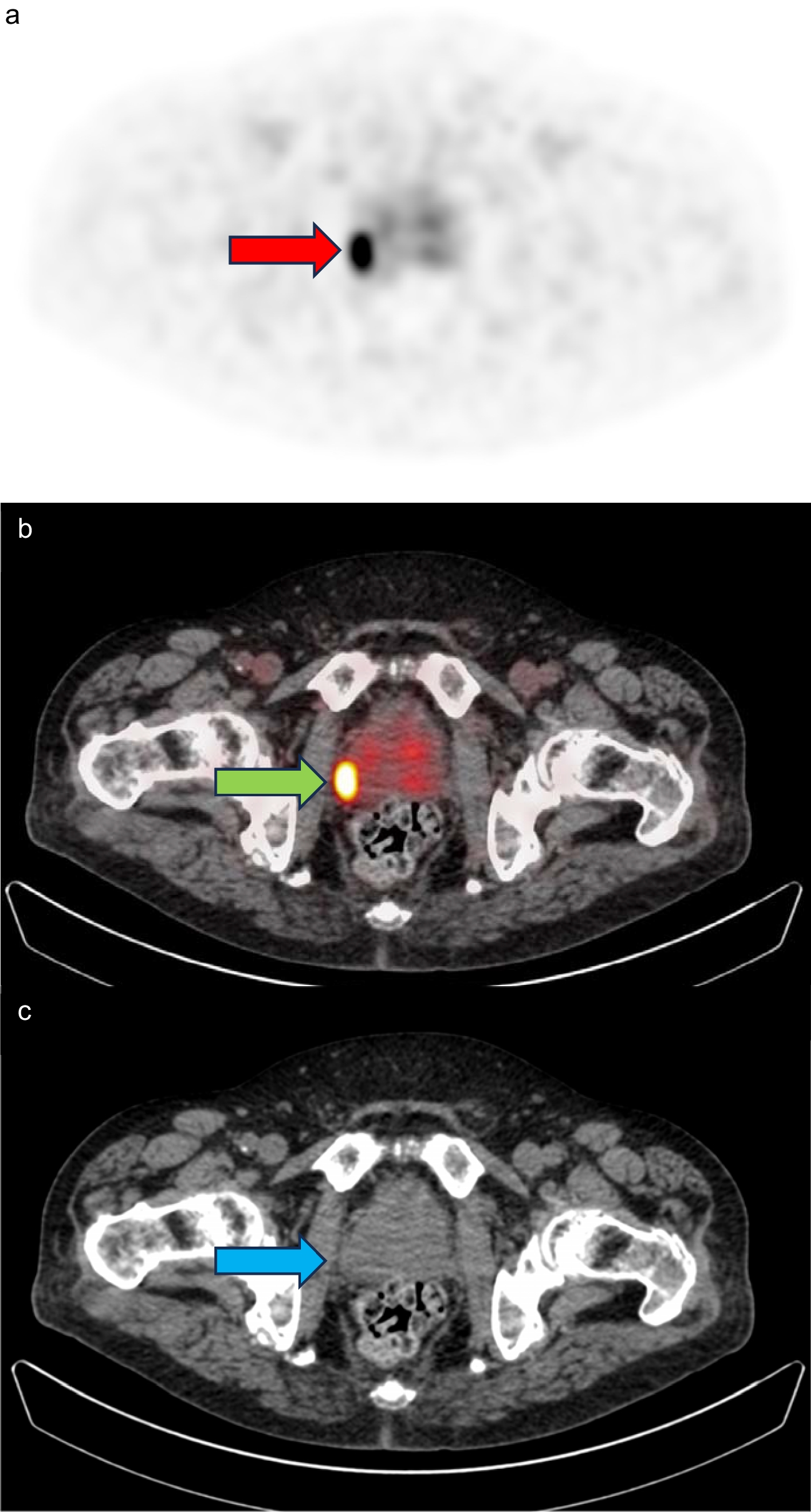
[68Ga]Ga-DOTAGA.(SA.FAPi)2 PET/CT imaging on a PET/CT scanner (uBio-EXPLORER, United Imaging, China) and [177Lu]Lu-DOTAGA.(SA.FAPi)2 single photon emission computed tomography (SPECT) imaging on a clinical SPECT/CT scanner (Discovery 670Pro, GE Healthcare, the USA) with intermediate energy collimator were performed to evaluate the biodistribution of [177Lu]Lu-DOTAGA.(SA.FAPi)2. 18F-FDG, [18F]AlF-NOTA-FAPI-04, and [68Ga]Ga-DOTA-Pentixafor PET/CT imaging were also performed on 6 tumor-bearing mice before treatment to evaluate their capacity to image 4T1 breast cancer models. Then, 4 mice in each group were randomly selected and received serial 18F-FDG, [18F]AlF-NOTA-FAPI-04, and [68Ga]Ga-DOTA-Pentixafor PET/CT scans on days 13-15, 18-20, 23-25, 28-30, and 33-35. All mice are treated as predetermined treatment plan. Given the large tumoral volumes of vehicle- and AMD3100-treated mice, the PET/CT scans for these two groups on days 28-30 and 33-35 were not performed. Owing to the death during imaging, there were only 3 mice in both groups left for PET/CT imaging on days 28-30 and 33-35. Mice were first intravenously injected with 1.85 MBq 18F-FDG, [18F]AlF-NOTA-FAPI-04, or 3.7 MBq [68Ga]Ga-DOTA-Pentixafor, respectively. If required (18F-FDG imaging), fast the mice for at least 6-8 h. One hour after tracer injection, the mice were anesthetized with 5% isoflurane and fixed at prone position. The anesthesia was maintained with 1% isoflurane during which a 10-min whole-body static PET/CT scan was performed. Data were reconstructed via using an ordered subsets expectation maximization (OSEM) algorithm (4 OSEM iterations, resolution: 1.4 mmHD) with scatter, attenuation, and decay corrections. The maximum standardized uptake value (SUVmax) was quantified and assessed among all groups.
Immunohistochemistry
Harvested tumors were fixed, embedded in paraffin, and cut into a 4-µm thick sections. The sections were then re-hydrated, subjected to antigen retrieval, blocked with 5% bovine serum albumin (BSA) in Tris-buffered saline with Tween-20 (TBST), and incubated with antibodies against α-SMA (1/200, bs-10196R, Bioss, China), Ki-67 (1/200, 12202, Cell Signaling Technology, the USA), CXCL12 (ER1902-35, Huabio, China), and anti-rabbit IgG horseradish peroxide. Positive staining was detected via a 3,3′-diaminobenzidine (DAB) reaction. The sections were then counterstained with hematoxylin and imaged on a bright-field microscope.
Flow cytometry
Tumors were cut into pieces and digested in a mixture comprising collagenase IV, hyaluronidase, and DNase I in DMEM medium at 37 °C under constant shaking for 30 min. After lysing red blood cells and removing debris, the cell mixture was washed and resuspended in PBS supplemented with 2% FBS for further analyses. Cells were counted and blocked with anti-mouse CD16/32, and then stained with the antibodies (anti-CD11b, anti-Gr-1). Samples were fixed and analyzed on a CytoFLEX-3 cytometer.
Toxicity analyses
Paraformaldehyde-fixed paraffin-embedded mouse organs on days 19 including heart, lung, liver, kidney, and spleen were stained with hematoxylin-eosin for evaluating toxicity.
Statistical analysis
Results were presented as mean ± standard error (SD). Significant difference among multiple data sets was determined using one-way analysis of variance (ANOVA). Log‐rank test was used for Kaplan‐Meier survival analysis. A p‐value less than 0.05 was considered statistically significant. Data analysis was conducted using Prism GraphPad 9 (GraphPad Software Inc., San Diego, CA, USA).


留言 (0)