Bacterial material
S. E. isolate 19-SA00115, E. coli isolate 20-AB00467 and C. jejuni isolate BFR-CA-19285 were obtained from the German Federal Institute for Risk Assessment (BfR, Berlin, Germany) and originated from chicken meat samples. They were cryopreserved at the Institute of Food Hygiene Leipzig at -80 °C (Cryobank, Mast Group Ltd., Germany). Fresh working cultures of S. E. and E. coli were obtained as described elsewhere (Schumann-Muck et al. 2023b) and used for a fortnight. Overnight cultures were grown to a cell density of about 108 cfu per mL as described previously (Schumann-Muck et al. 2023b) except for the addition of bovine serum albumin. C. jejuni working cultures were obtained by spreading cryopreserved beads on blood agar plates (Columbia Agar with Sheep Blood Plus; Oxoid GmbH, Germany) for each experimental approach and incubating them microaerobically (85% N2, 10% CO2, 5% O2) for 36 h at 42 °C. Therefore, the TRILAB system (TRILAB; Jenny Science AG, Switzerland) was used. The bacterial colonies from these plates were then stirred into Brain Heart Infusion Broth (BHI Broth; TN1216; sifin diagnostics GmbH, Germany) and incubated without agitation microaerobically for 16 h at 42 °C to a cell density of about 108 cfu per mL.
On-site at the pilot scale slaughtering unit, the suspensions of S. E., E. coli and C. jejuni were combined in a 1:1:1 ratio for every experimental approach to obtain a pool of target organisms.
Surfaces
The experiments were carried out in the technical centre of the German Federal Institute for Risk Assessment (BfR, Berlin, Germany). Commercial plucking fingers made of thermoplastic rubber with the following data were used: 20 mm bore diameter; hard version; total length, 97.5 mm; (Westfalia Werkzeug company GmbH & Co. KG, Germany). Before being used in the experiments, the plucking fingers were pretreated by cleaning and sterilisation. For this purpose, they were first placed in 5% Decon solution (Decon™ Decon90, Fisher Scientific GmbH, Germany), then rinsed with distilled water and finally degreased in a 95% 2-propanol solution (Carl Roth GmbH, Germany). After a further rinse with distilled water, they were first dried in a biosafety cabinet and then autoclaved. A more detailed description of this treatment can be found at Schumann-Muck et al. (2023a). In order to produce the coated plucking fingers used in this study, a nanoscale layer of silicon dioxide was applied by Nanopool GmbH (Germany) utilising their commercial product Liquid Glass Metall. Afterwards, the same number of coated and uncoated fingers were inserted into a drum plucker (plucking drum, 92 cm diameter, 42 cm height, equipped with 258 plucking fingers).
Two stainless-steel rods (type 304; X5CrNi1810) with a diameter of 30 mm and a length of 110 cm were custom-manufactured by SKS (Sondermaschinen- und Fördertechnikvertriebs-GmbH, Germany). They were divided into sections of 10 cm in length, and these sections were then alternately left uncoated or coated with a nanoscale layer of silicon dioxide applied by Nanopool GmbH, Germany, using its commercial product Liquid Glass Metall.
One rod was then fixed at a mobile carcass rack for attachment experiments in such a way that it was at a height of 140 cm parallel to the ground. For the detachment experiments, the second rod was attached parallel above the first one at a distance of 6 cm, but offset by one section, so that coated and uncoated sections were directly above each other.
Study design and recovery of the bacteria
The broilers used for the experiments were purchased already killed from a commercial poultry processor and used for experiments within a maximum of 2 h after killing.
Attachment and detachment tests with plucking fingers were repeated four times each using five uncoated and five coated fingers. Five broilers, taken from slaughter line after stunning and bleeding, were used for every finger experiment. These were scalded for 3 min in tap water at about 53 °C (Brühanlage Typ L1000, Westerhoff Geflügeltechnik GmbH, Germany). After that, each broiler was inoculated using a pipette with 10 mL mixed bacterial suspension, which was distributed over the entire carcass. Then, these five broilers were defeathered together for 30 to 40 s in the drum plucker equipped with coated and uncoated plucking fingers. Afterwards, the carcasses were removed and discarded, and the respective ten plucking fingers to be examined were taken from the machine. The detachment experiments followed subsequently. Therefore, the already used and, thus, contaminated plucking machine was first given a coarse cleaning with tap water at 15 °C for 15 s using a hand shower to soak and loose coarse organic soiling such as feathers. This was followed by the actual cleaning in form of high-pressure cleaning at 130 bar and a water temperature of 75 °C for 1 min (HDS 9/18–4 M, Alfred Kärcher Vertriebs-GmbH, Germany). Recovering bacteria from plucking fingers for attachment and detachment experiments was carried out following the method of Arnold. The tips of the fingers were cut off behind the third rib with sterile scissors, placed in a 50-mL centrifuge tube (centrifuge tube 50, TPP, Switzerland) filled with 10 mL sodium chloride peptone solution and manually shaken for 15 to 20 s. They were kept refrigerated at 5 °C for a maximum of 6 h until further processing. Then decimal dilutions of the liquids were spread on the appropriate selective nutrient media for detection of each of the three bacteria used and incubated for 24 h at the optimal growth temperatures respectively: E. coli on Tryptone Bile X Glucuronic Agar (TBX; TN1259; sifin diagnostics GmbH, Germany) at 42 °C, S. E. on Xylose Lysine Deoxycholate Agar (XLD; 85,864.5000; VWR International GmbH, Germany) at 37 °C and C. jejuni on modified Cefaperazone Charcoal Desoxycholate Agar plates (mCCDA, Oxoid GmbH, Germany) microaerobically at 42 °C.
Experiments for attachment to the stainless-steel rod were repeated four times and performed with three broiler carcasses each, which were taken from a slaughter line after plucking. These were inoculated in the breast area using a pipette with 3.0 mL of the bacterial suspension, which was distributed with sterile spatulas. Carcasses were then hooked into a slaughter belt, which moved at a speed of 0.1 m/s. This caused them to move along the stainless-steel rod, thereby touching it at the inoculated breast area. Then, six consecutive sections, and thus alternately coated and uncoated sections, were sampled on the rod. For detachment experiments, a broiler was inoculated in the breast area with 3.0 mL of the bacterial suspension and moved along the rods with the aid of the slaughter belt in such a way that both rods were touched simultaneously with the inoculated breast area. Afterwards, the broiler was inoculated a second time and passed along the rods again. The two rods attached one above the other were then rinsed with 1.5 l water (76–77 °C). For each test, the first touched section of each rod was sampled, and thus a coated and an uncoated section were sampled at the same time. This was repeated 10 times.
According to DIN EN ISO 18593, bacteria were recovered from the rod by sampling each section vertically and horizontally for 30 s by wiping with uniformly strong pressure using a sterile touch sponge (Polywipe with Peptone Saline; Medical Wire and Equipment, UK). The sponge was then placed in a plastic bag filled with 90 mL sodium chloride peptone solution, which was sealed to prevent leakage and kept refrigerated at 5° C for a maximum of 6 h until further processing. Afterwards, the bags were stomached for 1 min at 260 rpm (Stomacher 400 Circulator, Seward Ltd., UK). Decimal dilutions were then prepared from the liquids, which were plated and incubated on the appropriate selective culture media for the bacteria used, as described for the plucking fingers.
Statistics
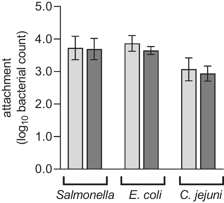
Bacterial counts were log10 transformed to perform the statistical analysis. Attachment and detachment of S. E., E. coli and C. jejuni from plucking fingers was calculated as the average of four replicates each from the recovered bacterial counts of five fingers per replicate. The attachment and detachment values represent the number of bacteria that were able to attach to the fingers or could not detach from them by the rinsing process. Differences between coated and uncoated plucking fingers per treatment were statistically analysed with an unpaired t-test at an alpha level of significance of 0.05.
For attachment of the target organisms to the stainless-steel rod, the section average bacterial counts of four replicates were used to calculate linear regression of fit. Linear regression lines of coated and uncoated steel were compared based on slope and intersect following the procedure described by Glantz (2012) to generate F values for reference at an alpha level of significance of 0.05.
Detachment of the bacteria from the stainless-steel rod was calculated as the average of 10 replicates from the recovered bacteria counts of one section per replicate. The detachment values represent the number of bacteria that were able to remain attached to the rod after rinsing. Differences between coated and uncoated rod sections per treatment were statistically analysed using an unpaired t-test at an alpha level of significance of 0.05.
Except comparison of linear regression lines, all statistical analyses were carried out using Prism 9 (GraphPad Software, LLC, USA).


留言 (0)