
記住我


Advancements in sequencing techniques and metabolomics combined with bioinformatic pipelines refined our knowledge of microbiota composition and function [1]. As a result, the gut microbiota has been increasingly recognized for their role in influencing various aspects of health, including metabolism and immune function. During infancy, the gut microbiota has yet to be established and this dynamic maturation is influenced by many different factors like maternal microbiota, delivery mode, feeding pattern, gestational age, and medication [2,3]. Disturbance of the initial microbial colonization has been associated with a variety of diseases in the neonatal period, like necrotizing enterocolitis and sepsis, and beyond, like atopy, allergies, obesity, and auto-immune disorders [4–6].
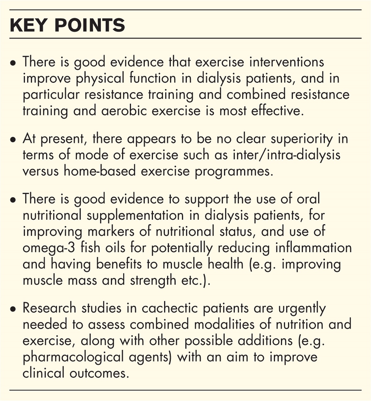
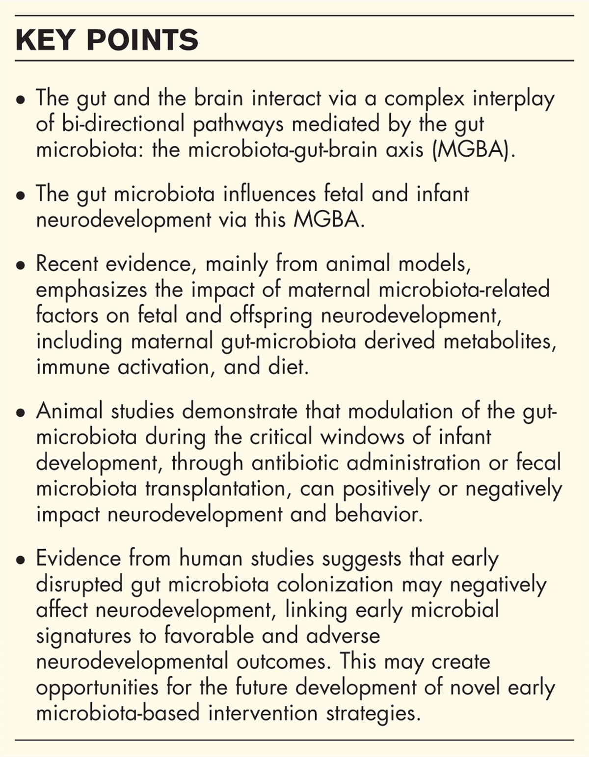
There has been accumulating evidence that the gut microbiota and its metabolites also regulate various aspects of neurodevelopment and cognitive functioning throughout life. It is thought that the gut influences the brain and vice versa via an interplay of bi-directional pathways, mediated by gut microbiota: the microbiota-gut-brain axis (MGBA) [1]. The MGBA is considered to play a key role in infancy when significant stages of neurodevelopment take place [3,7], especially in preterm infants, as these are at an increased risk of both dysbiosis and impaired neurodevelopment [2]. Defining the critical windows of early-life microbiota colonization and its effect on neurodevelopment is an important emerging translational research question since this can form the basis for future microbiota-based interventions to preserve neurocognitive development and function in infants.
In this review, we discuss the mechanisms of the MGBA and provide an update on the emerging evidence regarding the connection between the maternal, fetal, and neonatal microbiota and neurocognitive development and functioning. Furthermore, we will discuss the therapeutic options to modulate the gut microbiota for preservation of neurodevelopment.
 Box 1:
 no caption available
MICROBIOTA-GUT-BRAIN AXIS MECHANISMSThe vagal nerve is recognized as pivotal in facilitating bidirectional communication within the MGBA, constituting the primary conduit for microbiota-derived signals to interface with the brain. Although the vagal nerve itself is not in direct contact with the gut microbiota [8], it serves as a receptor for signals transmitted via microbial metabolites, inflammatory processes, or neuroendocrine cells which are mediated by gut microbiota [2,9] (Fig. 1). The microbiota exerts regulatory control over the neuroendocrine hypothalamic-pituitary axis (HPA). Experimentally induced gut dysbiosis modulates HPA-mediated cortisol release and consequent behavioral outcomes [1,9].
 FIGURE 1:
 Proposed mechanisms of the microbiota-gut-brain axis. The microbiota-gut-brain axis is established by different pathways that communicate bi-directionally via different pathways in a complex interplay. This is a simplified depiction showing the most important pathways. Created with Biorender. NTMs, neurotransmitters; SCFAs, short-chain fatty acids; Th, T-helper cells; TLR4, toll-like receptor 4; Treg, regulatory T cells.
The gut microbiota produces a plethora of metabolites such as short-chain fatty acids (SCFAs), tryptophan metabolites, and biliary acids. SCFAs, such as butyrate, propionate, and acetate, result from anaerobic microbial fermentation of indigestible polysaccharides in the colon [10]. SCFAs contribute to the maintenance and modulation of both gut and blood—brain barrier (BBB) integrity [11,12▪▪]. They can directly affect the brain compromising the BBB, binding to local receptors, inducing epigenetic modifications, contributing to brain homeostasis, and modulating neuro inflammation [13]. For instance, butyrate can possess direct anti-inflammatory effects on oligodendrocytes which are crucial for early brain development [11,14]. SCFAs can enhance regulatory T cell (Treg) function, vital for immune tolerance, and activate the sympathetic nervous system and vagal nerve [11,15]. Indole, a tryptophan derivative, exhibits beneficial effects on neuro inflammation, nerve signal transduction, and gut and BBB integrity [9,16]. Lastly, biliary acids play a role in the brain and HPA axis through specific local receptors [9].
Various neurotransmitters, and their compounds, such as serotonin (derived from tryptophan), dopamine, norepinephrine, histamine, and gamma-aminobutyric acid (GABA), can be directly produced by gut microbiota or indirectly through the stimulation of entero-endocrine cells in the gut epithelium. These processes can occur under the stimulation of SCFAs. For example, Lactobacilli, Bifidobacteriae, and Bacteroidetes are capable of producing GABA [8,15], while Escherichia coli (E. coli) and specific strains of Lactobacillus can contribute to serotonin production. These neurotransmitters can exert local effects by stimulating the vagal nerve or gut peristalsis [8], but can also traverse the SCFA weakened gut and BBB, exerting a more direct impact on the brain [11].
The gut microbiota can also influence the brain through immune signaling. Under eubiosis, immune cells in the gut-associated lymphoid tissue differentiate into anti-inflammatory Treg cells producing cytokines like interleukin (IL)-10 and TGF-β [17]. During dysbiosis, lymphocytes differentiate towards the Th1/Th17-subtype, resulting in the production of pro-inflammatory cytokines such as IL-2, IL-12, and TNF-α [17]. Furthermore, lipopolysaccharide (LPS) containing bacteria can induce an inflammatory cascade by TLR 4 signaling, with NF-kB induced release of pro-inflammatory cytokines such as TNF-α, IL-1β, and IL-6. These can directly affect the brain, by the weakening of the BBB, or via pathogenic signaling through the vagal nerve [11]. The resulting inflammatory processes damage the oligodendrocytes, pivotal for early brain development.
PRENATAL GUT MICROBIOTA INFLUENCES FETAL NEURODEVELOPMENTNeonates are not immunologically naive but primed to respond to exposure to environmental and microbial stimuli. Consequently, there has been debate about the presence of a fetal gut microbiome [18]. However, human and murine studies failed to provide evidence for the existence of a fetal gut microbiota [18,19]. More recently, it was postulated that the maternal gut microbiota influences fetal immune and neurodevelopment indirectly through microbiota-derived metabolites [20]. These metabolites cross the placenta during pregnancy, provoking a fetal immune response [12▪▪]. This is supported by studies demonstrating the human fetal intestine to exhibit a diverse metabolome at only 12 weeks gestation, with enrichment of metabolites and metabolic pathways associated with neurodevelopment [21]. Additionally, comparative studies between germ-free mice and specific-pathogen free (SPF) mice revealed higher levels of microbial-originating metabolites in SPF mice, showing the lack of a maternal microbiota can affect metabolite levels in fetal organs [22]. Furthermore, depletion of the maternal microbiome in germ-free mice impairs fetal thalamocortical axonogenesis, while maternal colonization with Clostridia-dominant spore-forming bacteria increases levels of specific bacterial metabolites, thereby preventing these axonal defects [23]. Despite these findings, the mechanisms underlying transportation across the placenta remain unclear, necessitating further exploration.
Maternal immune activation (MIA) during pregnancy can have detrimental impacts on neurodevelopment [1,20]. MIA presumably induces cytokine production through the activation of Th17 cells via the microbiota. During induced MIA in mice, the level of maternal IL-17a produced by Th17 cells increased the risk of offspring developing autism spectrum disorder (ASD), which was mediated by the microbiota [24].
Maternal obesity before pregnancy and a high-fat diet (HFD) during pregnancy and lactation have been associated with multigenerational neurodevelopmental delay and social dysfunction [25,26]. In the first offspring, maternal supplementation of milk fat globule membrane (MFGM) during lactation promoted neurogenesis in the hippocampus and neurobehavioral development by modulation of gut microbiota. This modulation resulted in the downregulation of pro-inflammatory bacteria such as E. shigella and Enterococcus, and upregulation of bacteria with anti-inflammatory and antiobesity properties, such as Akkermansia and Lactobacillus. Consequently, neuroinflammation was alleviated by reduced levels of microbiota-correlated pro-inflammatory factors, such as LPS and IL-1β [25]. In the second offspring, social dysfunction was restored through supplementation with Limosilactobacillus reuteri, which facilitated the expansion of SCFA-producing bacteria in their gut microbiota [26].
IMPACT OF GUT MICROBIOTA DURING CRITICAL WINDOWS OF DEVELOPMENT Recent evidence in animalsAnimal studies demonstrate that induced microbiota alterations during the critical developmental window affect neurodevelopment and behavior. Lynch et al.[27] explored the effects of early-life antibiotic administration during critical developmental windows in mice, revealing a dramatic disruption of the cecal microbiota of adolescent mice. This disruption incorporated increases of Escherichia/Shigella, Staphylococcus, and Clostridioides, while key producers of SCFAs as Alistipes, Bacteroides, Odoribacter, and Lachnospiracea NK4A136, were decreased. Furthermore, alterations of microglia morphology were observed in the basolateral amygdala of adolescent mice, suggesting that short-term targeted disruption of the developing gut microbiota can have enduring effects on microglia maturation [27].
Fecal microbiota transplantation of human feces into germ-free mice elegantly shows the transferability of cognitive phenotypes through the gut microbiota. Recently, pregnant germ-free mice were colonized with fecal microbiota obtained from preterm infants of various postmenstrual ages (PMA), ranging from 27 to 34 weeks. Colonization with fecal microbiota of infants with higher PMA improved associative fear learning and memory in adult mice. Thirteen upregulated fecal and serum metabolites, linked to brain function, correlated with microbial maturation-associated cognitive improvements [28]. Another study involved transplanting feces from 6-month-old infants, differing in cognition scores, into germ-free mice. Mice transplanted with feces of infants with above-median cognition scores exhibited better memory functions. The gut microbiota of these mice was enriched in species belonging to the genera Phocaeicola, Bacteroides, and Bifidobacterium[29]. An FMT study from 5-year-old children with varying cognitive scoring into germ-free mice confirmed cognitive phenotype transferability but also identified three unique fecal metabolites – xanthine, formate, and mannose – with long-lasting strong positive associations with high cognitive performance phenotypes [30].
Several studies explored the effects of diet-induced microbial alterations on brain development and behavior. In rats prenatally exposed to caffeine for induced intrauterine growth restriction, a HFD led to ASD like behavior [31]. E. coli was significantly increased in the gut microbiota of these rats, with increased levels of IL-17A in the colon, serum, and hippocampal regions. The E. coli and IL-17A concentrations in the hippocampal regions were closely correlated with ASD symptoms. E. coli strain transplantation in control rats led to spontaneous development of ASD-like manifestations and similar increases in IL-17A [31]. Contrary to HFD, supplementation of nucleotides, found in human milk, enhanced neuro-maturation in the prefrontal cortex and hippocampus in rats. This was mediated by gut microbiota composition and function and correlated to neurodevelopmental phenotypes [32].
Recent evidence in humansComparable to murine studies, the maternal gut microbiota influences neonatal neurodevelopment in humans. In a recent study by Sun et al. [33▪], maternal microbiota seemed more relevant to the offspring's neurodevelopment than the infants gut microbiota, with Fusobacteria as a key player, increasing later motor skills when found in maternal gut microbiota and decreasing motor skills when found in infants.
Recently, the correlations between early human gut colonization and neurodevelopmental outcomes. In 44 term infants, associations were observed between the gut microbiota and early cognitive development. A successful Point-and-Gaze attention test was associated with increased Actinobacteria and reduced Firmicutes on the phylum level, and increased Bifidobacterium and Eggerthella and reduced Hungatella and Streptococcus on the genus level [34]. Another recent study using Random Forest modeling demonstrated that gut microbial species in the first year of life predict future cognitive functioning. Klebsiella spp., E. coli, and Bifidobacterium spp. were important predictors of cognitive functioning. In addition, RF modeling on bacterial species predicted the size of brain regions on MRI [35]. A recent randomized controlled trial (RCT) investigated the impact of vaginal microbiota transfer (VMT) on neurodevelopmental outcomes in 68 infants born via cesarean-section. VMT infants showed significantly higher neurodevelopmental scores (as measured by Ages-and-Stages-Questionnaire-3 (ASQ-3) compared to placebo at 6 months of age. Furthermore, VMT was associated with faster gut microbiota maturation and with modulation of specific fecal metabolites and metabolic functions within the first 6 weeks of life [36▪▪].
More evidence is available on preterm infants. In 24 very low birth weight (VLBW) infants, neurodevelopment was examined by the Battelle Development Inventory-2 Screening Test (BDI-2ST) at 2 and 4 years of age [37]. Correlations between early microbial diversity and specific microbial amplicon sequence variants and BDI-2ST subscales such as cognition, adaptive behavior, and communication were observed, even after adjusting for gestational age, birth weight, and antibiotic exposure. Bifidobacterium presence appeared to be a pivotal element in infants not requiring neurodevelopmental referral at 2 years. In accordance, VLBW infants (n = 27) with adverse neurological outcomes at 24 months corrected age (MCA), measured with the revised Griffiths Mental Development Scale, were characterized by early deficiency of Bifidobacterium[38]. Seki et al. [39] showed early overgrowth of Klebsiella ssp. was associated with a pro-inflammatory T cell response and seemed highly predictive for brain damage as identified by MRI at term age in 60 extremely preterm infants. A disturbed gut-microbiota-immune-brain axis may induce or worsen brain injury in extremely preterm infants [39]. Zhang et al. [39] evaluated the relationship between gut microbiota composition in seventy-seven preterm infants at four weeks of age and neurodevelopmental outcomes up to 6 MCA (ASQ-3). Beta diversity was linked with gross motor scores at 1, 3, and 6 MCA, communication scores at 3 MCA, and fine motor scores at 6 MCA [40▪]. Comparably to earlier findings [35,39], the relative abundance of Klebsiella ssp. was negatively associated with gross motor scores from 1 to 6 MCA, while the relative abundance of Lactobacillus exhibited a positive association [35,39,40▪].
MODULATING GUT MICROBIOTA FOR NEURODEVELOPMENTIdentification of early, predictive microbial markers holds the potential for the development of novel intervention strategies to enhance neurodevelopment. Proposed microbiota-based interventions include human milk, probiotics, VMT, FMT, and avoidance of certain medications.
Human milk, rich in human milk oligosaccharides (HMO) and MFGM, promotes the growth of beneficial bacteria such as Bifidobacterium spp. and Bacteroides[14]. In term infants, HMO exposure is positively associated with cognitive, language, and motor skill domains between 18 and 24 months of age, however, this effect was not seen in preterm infants [41]. A recent RCT showed improved cognitive function at 5.5 years of age in MFGM-supplemented term infants receiving formula milk [42]. However, another RCT comparing human donor milk to formula feeding in preterm infants unable to receive sufficient mothers’ milk did not show differences in neurocognitive outcome at 2 years of age [43]. Although mothers’ own milk is presumed to have a beneficial impact on neurodevelopment, this has to date only been proven in retrospective studies, as RCT are deemed unethical [44]. Notably, the beneficial effects of mothers’ milk on neurodevelopment are various and go beyond gut microbiota.
Judicious antibiotic use in preterm infants, given their frequent empirical antibiotic treatment, can positively impact gut microbiota and subsequent neurocognitive outcomes [45]. In an observational study – with inherent limitations and therefore not proving causation – we observed that prolonged empirical antibiotic treatment in preterm infants was associated with below-average gross-motor development at 24 MCA [46]. The avoidance of acid suppressants, known to alter gut microbiota, could therefore be beneficial for preserving neurocognitive outcomes, as a recent study showed the use of acid suppressants to be associated with adverse neurocognitive outcomes [47]. However, it was not confirmed that this effect was mediated by changes in gut microbiota.
Probiotic administration in (preterm) infants has shown mixed results on neurocognition; two RCTs suggest long-term benefits in neurocognitive functioning in term infants, but larger RCTs are needed for robust evidence [48,49]. Recently, maternal VMT has shown promise to restore microbiota maturation and lead to better neurocognitive outcomes at 6 months of age in term infants born with cesarean-section [36▪▪], while safety and long-term results of maternal FMT to (preterm) infants are being explored in the MT-SECFLORE and PREFLORE trials [50,51].
CONCLUSIONIncreasing evidence in animals and humans suggests that disrupted gut microbiota colonization can negatively affect MGBA development, thereby affecting brain development, neurocognition, and behavior. In humans, early microbial signatures have been associated with adverse neurodevelopmental outcomes (Klebsiella spp.) while an increased abundance of Bifidobacterium show beneficial effects. Future studies, including larger cohort studies with longitudinal analyses of microbes, their metabolites, and neurotransmitters, can further elucidate the mechanisms of the MGBA. Well-designed experimental studies and RCTs are necessary to prove whether the associations between the MGBA and brain development, neurocognition, and behavior are causative or attributable to other factors. Implementation of a combination strategy of the discussed interventions holds the potential for improving neonatal neurodevelopmental outcomes through MGBA modulation.
AcknowledgementsNone.
Financial support and sponsorshipNone to declare.
Conflicts of interestN.F., T.dM., and H.N. have received an unrestricted grant from Nutricia Benelux Corporation unrelated to this submitted work.
REFERENCES AND RECOMMENDED READINGPapers of particular interest, published within the annual period of review, have been highlighted as:
▪ of special interest
▪▪ of outstanding interest
REFERENCES 1. Cryan JF, O’Riordan KJ, Cowan CSM, et al. The microbiota-gut-brain axis. Physiol Rev 2019; 99:1877–2013. 2. Bresesti I, Salvatore S, Valetti G, et al. The microbiota-gut axis in premature infants: physio-pathological implications. Cells 2022; 11:379. 3. Laue HE, Coker MO, Madan JC. The developing microbiome from birth to 3 years: the gut-brain axis and neurodevelopmental outcomes. Front Pediatr 2022; 10:815885. 4. Akagawa S, Kaneko K. Gut microbiota and allergic diseases in children. Allergol Int 2022; 71:301–309. 5. Squillario M, Bonaretti C, La Valle A, et al. Gut-microbiota in children and adolescents with obesity: inferred functional analysis and machine-learning algorithms to classify microorganisms. Sci Rep 2023; 13:11294. 6. Westaway JAF, Huerlimann R, Kandasamy Y, et al. The bacterial gut microbiome of probiotic-treated very-preterm infants: changes from admission to discharge. Pediatr Res 2022; 92:142–150. 7. Ahmad S, Wu Y, Wu Z, et al. Multifaceted atlases of the human brain in its infancy. Nat Methods 2023; 20:55–64. 8. Dicks LMT. Gut bacteria and neurotransmitters. Microorganisms 2022; 10:1838. 9. Li L, Yang J, Liu T, Shi Y. Role of the gut-microbiota-metabolite-brain axis in the pathogenesis of preterm brain injury. Biomed Pharmacother 2023; 165:115243–1115243. 10. Beghetti I, Barone M, Brigidi P, et al. Early-life gut microbiota and neurodevelopment in preterm infants: a narrative review. Front Nutr 2023; 10:1241303. 11. Manohar K, Mesfin FM, Liu J, et al. Gut-Brain cross talk: The pathogenesis of neurodevelopmental impairment in necrotizing enterocolitis. Front Pediatr 2023; 11:1104682. 12▪▪. Aburto MR, Cryan JF. Gastrointestinal and brain barriers: unlocking gates of communication across the microbiota-gut-brain axis. Nat Rev Gastroenterol Hepatol 2024; 10.1038/s41575-023-00890-0. 13. Gars A, Ronczkowski NM, Chassaing B, et al. First encounters: effects of the microbiota on neonatal brain development. Front Cell Neurosci 2021; 15:682505. 14. Li L, Liu T, Shi Y. Treatment of preterm brain injury via gut-microbiota-metabolite-brain axis. CNS Neurosci Ther 2024; 30:e14556. 15. Silva YP, Bernardi A, Frozza RL. The role of short-chain fatty acids from gut microbiota in gut-brain communication. Front Endocrinol (Lausanne) 2020; 11:25. 16. Zhou Q, Niño DF, Yamaguchi Y, et al. Necrotizing enterocolitis induces T lymphocyte-mediated injury in the developing mammalian brain. Sci Transl Med 2021; 13:eaay6621. 17. Kasarello K, Cudnoch-Jedrzejewska A, Czarzasta K. Communication of gut microbiota and brain via immune and neuroendocrine signaling. Front Microbiol 2023; 14:1118529. 18. Kennedy KM, de Goffau MC, Perez-Muñoz ME, et al. Questioning the fetal microbiome illustrates pitfalls of low-biomass microbial studies. Nature 2023; 613:639–649. 19. Winters AD, Romero R, Greenberg JM, et al. Does the amniotic fluid of mice contain a viable microbiota? Front Immunol 2022; 13:820366. 20. Koren O, et al. The maternal gut microbiome in pregnancy: implications for the developing immune system. Nat Rev Gastroenterol Hepatol 2023; 21:35–45. 21. Li Y, Toothaker JM, Ben-Simon S, et al. In utero human intestine harbors unique metabolome, including bacterial metabolites. JCI Insight 2020; 5:138751. 22. Pessa-Morikawa T, Husso A, Kärkkäinen O, et al. Maternal microbiota-derived metabolic profile in fetal murine intestine, brain and placenta. BMC Microbiol 2022; 22:46. 23. Vuong HE, Pronovost GN, Williams DW, et al. The maternal microbiome modulates fetal neurodevelopment in mice. Nature 2020; 586:281–286. 24. Kim E, Paik D, Ramirez RN, et al. Maternal gut bacteria drive intestinal inflammation in offspring with neurodevelopmental disorders by altering the chromatin landscape of CD4(+) T cells. Immunity 2022; 55:145–158. e7. 25. Yuan Q, Gong H, Du M, et al. Milk fat globule membrane supplementation to obese rats during pregnancy and lactation promotes neurodevelopment in offspring via modulating gut microbiota. Front Nutr 2022; 9:945052. 26. Di Gesu CM, Matz LM, Bolding IJ, et al. Maternal gut microbiota mediate intergenerational effects of high-fat diet on descendant social behavior. Cell Rep 2022; 41:111461. 27. Lynch CMK, Cowan CSM, Bastiaanssen TFS, et al. Critical windows of early-life microbiota disruption on behaviour, neuroimmune function, and neurodevelopment. Brain Behav Immun 2023; 108:309–327. 28. Lu J, Lu L, Yu Y, et al. Early preterm infant microbiome impacts adult learning. Sci Rep 2022; 12:3310. 29. Cerdó T, Ruiz-Rodríguez A, Acuña I, et al. Infant gut microbiota contributes to cognitive performance in mice. Cell Host Microbe 2023; 31:1974–1988. e4. 30. Guzzardi MA, Ederveen THA, Rizzo F, et al. Maternal prepregnancy overweight and neonatal gut bacterial colonization are associated with cognitive development and gut microbiota composition in preschool-age offspring. Brain Behavi Immun 2022; 100:311–320. 31. Wang T, Zhang S, Luo M, et al. Prenatal caffeine exposure induces autism-like behaviors in offspring under a high-fat diet via the gut microbiota-IL-17A-brain axis. Ecotoxicol Environ Saf 2024; 269:115797. 32. Qu Z, Tian P, Zhao J, et al. Feeding the microbiota–gut–brain axis: nucleotides and their role in early life. Food Front 2023; 4:1164–1178. 33▪. Sun Z, Lee-Sarwar K, Kelly RS, et al. Revealing the importance of prenatal gut microbiome in offspring neurodevelopment in humans. eBioMedicine 2023; 90:104491–1104491. 34. Hunter S, Flaten E, Petersen C, et al. Babies, bugs and brains: how the early microbiome associates with infant brain and behavior development. PLoS One 2023; 18:e0288689. 35. Bonham KS, Fahur Bottino G, McCann SH, et al. Gut-resident microorganisms and their genes are associated with cognition and neuroanatomy in children. Sci Adv 2023; 9:eadi0497. 36▪▪. Zhou L, Qiu W, Wang J, et al. Effects of vaginal microbiota transfer on the neurodevelopment and microbiome of cesarean-born infants: a blinded randomized controlled trial. Cell Host Microbe 2023; 31:1232–1247. e5. 37. Sarkar A, Prescott SM, Dutra S, et al. Relationships of the very low birth weight infant microbiome with neurodevelopment at 2 and 4 years of age. Dev Psychobiol 2022; 64:e22317. 38. Beghetti I, Barone M, Turroni S, et al. Early-life gut microbiota and neurodevelopment in preterm infants: any role for Bifidobacterium? Eur J Pediatr 2022; 181:1773–1777. 39. Seki D, Mayer M, Hausmann B, et al. Aberrant gut-microbiota-immune-brain axis development in premature neonates with brain damage. Cell Host Microbe 2021; 29:1558–1572. e6. 40▪. Zhang D, Lan Y, Zhang J, et al. Effects of early-life gut microbiota on the neurodevelopmental outcomes of preterm infants: a multicenter, longitudinal observational study in China. Eur J Pediatr 2024; 10.1007/s00431-024-05423-8. 41. Berger PK, Ong ML, Bode L, Belfort MB. Human milk oligosaccharides and infant neurodevelopment: a narrative review. Nutrients 2023; 15:719. 42. Colombo J, Harris CL, Wampler JL, et al. Improved neurodevelopmental outcomes at 5.5 years of age in children who received bovine milk fat globule membrane and lactoferrin in infant formula through 12 months: a randomized controlled trial. J Pediatr 2023; 261:113483. 43. Colaizy TT, Poindexter BB, McDonald SA, et al. Neurodevelopmental outcomes of extremely preterm infants fed donor milk or preterm infant formula. JAMA 2024; 331:582–591. 44. Belfort MB, Inder TE. Human milk and preterm infant brain development: a narrative review. Clin Ther 2022; 44:612–621. 45. Morowitz MJ, Katheria AC, Polin RA, et al. The NICU Antibiotics and Outcomes (NANO) trial: a randomized multicenter clinical trial assessing empiric antibiotics and clinical outcomes in newborn preterm infants. Trials 2022; 23:428. 46. Deianova N, El Manouni El Hassani S, Niemarkt HJ, et al. Fecal volatile organic compound profiles are not influenced by gestational age and mode of delivery: a longitudinal multicenter cohort study. Biosensors (Basel) 2020; 10:E50. 47. Jensen ET, Yi J, Jackson W, et al. Analysis of neurodevelopment in children born extremely preterm treated with acid suppressants before age 2 years. JAMA Netw Open 2022; 5:e2241943. 48. Lin FL, et al. Effects of probiotics on neurocognitive outcomes in infants and young children: a meta-analysis. Front Public Health 2023; 11:1323511. 49. Panchal H, Athalye-Jape G, Rao S, Patole S. Growth and neuro-developmental outcomes of probiotic supplemented preterm infants-a systematic review and meta-analysis. Eur J Clin Nutr 2023; 77:855–871. 50. Helve O, K. Preterm infant intestinal microbiota development and maternal fecal transplant. 2024. https://classic.clinicaltrials.gov/show/NCT06227845; Accessed: 01-03-2024. 51. Carpen N, Brodin P, de Vos WM, et al. Transplantation of maternal intestinal flora to the newborn after elective cesarean section (SECFLOR): study protocol for a double blinded randomized controlled trial. BMC Pediatr 2022; 22:565.
留言 (0)