Small extracellular vesicles from the infarcted heart can promote tumour growth
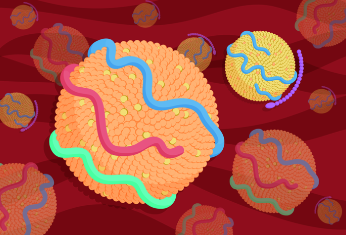
After myocardial infarction (MI), the heart secretes small extracellular vesicles (sEVs) — in particular, cardiac mesenchymal stromal cell (cMSC)-derived sEVs — with pro-neoplastic properties that can accelerate tumour growth when taken up by cancer cells. This finding provides new insights into the concept of reverse cardio-oncology.
The researchers generated a mouse model of heterotopic and orthotopic lung cancer that is characterized by post-MI left ventricular dysfunction (LVD). Consistent with previous findings, post-MI LVD accelerated tumour growth in these mice. Next, they assessed the role of cardiac sEVs in promoting tumour growth. The hearts of mice with MI were found to produce more than twofold the number of sEVs produced by hearts of control mice, and these cardiac sEVs had pro-neoplastic characteristics. In particular, cMSCs from infarcted hearts were a rich source of sEVs, and these cMSC-derived sEVs contained tumour-promoting microRNAs (miRs), such as miR-21 (which supports tumour cell survival and proliferation) and miR-24-1 (which is associated with the proliferation of ovarian cancer cells). Furthermore, cMSC-derived sEVs from infarcted hearts switched macrophages to a pro-tumorigenic state with pro-angiogenic, pro-inflammatory and immunomodulating traits. Finally, to establish whether inhibition of the renin–angiotensin–aldosterone system to treat post-MI LVD influences cMSC-derived sEVs and tumour growth, the researchers treated these mice with the aldosterone receptor antagonist spironolactone. Spironolactone reduced the number of cMSC-derived sEVs by 28% and suppressed tumour growth in mice with post-MI LVD but not in the sham mice.


留言 (0)