Patient population and design
Consecutive patients suspected of PMR were prospectively included between September 2020 and June 2022 from the Departments of Rheumatology at Aarhus University Hospital, Horsens Regional Hospital, and Silkeborg Regional Hospital in Denmark. To assess all potential patients with PMR, rapid referral clinics were established for all individuals suspected of PMR, and general practitioners were informed about this new service through information meetings and advertisements in newsletters dedicated to general practitioners. After referral, all patients were evaluated in the clinics preferably within 1 week. Inclusion criteria for the study were as follows: patients suspected of PMR, aged over 50, and experiencing proximal muscle pain. The exclusion criteria were as follows: any glucocorticoid treatment within the last 3 months; a medical history of PMR or giant cell arteritis (GCA); any other inflammatory rheumatic disease; symptoms of cranial GCA including headache, jaw claudication, scalp tenderness, or sight disturbances; and any active malignant cancers within the past 5 years (except basal cell carcinoma [26]).
The study design is graphically depicted in Supplementary Figure S1. At baseline, all patients had their medical history taken, lab work; were clinically examined, had vascular ultrasound as well as ultrasound of shoulders and hips performed; and were initially clinically diagnosed by the treating clinician. Subsequently, all patients had an FDG-PET/CT performed within days and before initiation of prednisolone treatment. Patients diagnosed with GCA subsequently to the FDG-PET/CT were excluded from the final analyses. Patients with a clinical diagnosis of solely PMR were administered 15 mg of prednisolone with a gradual taper (Supplementary Figure S1). If an adequate treatment response was not achieved within 2–4 weeks, based on lab work and telephone consultations at week 1 and week 4, the prednisolone dose was increased to 25 mg daily. Additionally, patients experiencing incomplete remission at week 1 underwent an additional telephone consultation along with lab work to assess whether an early increase in prednisolone dosage at week 2 was indicated. After 8 weeks of prednisolone treatment, a second clinical visit was performed consisting of a clinical examination, lab work, and a second FDG-PET/CT. Patients in clinical remission at week 8 were tapered to 5 mg prednisolone for 1 week followed by prednisolone discontinuation for a week. At week 10, during the temporary treatment discontinuation, a similar clinical visit was conducted including a third FDG-PET/CT. Prednisolone was subsequently restarted and tapered according to standard care [27]. Patients with incomplete remission at week 8 or relapse during prednisolone discontinuation continued or restarted prednisolone administration and were withdrawn from the week 10 visit, since this visit aimed at evaluating the effect of a short-term prednisolone discontinuation. At the 1-year follow-up, an experienced rheumatologist (KKK, ITH, BDN, CMS, SGK, JBN) determined the final diagnosis, including patients withdrawn from previous visits, considering the initial baseline diagnosis and the progression of the disease during the first year. This 1-year diagnosis served as the reference standard in this study. Patients initially diagnosed with other conditions, GCA or cancer, at the baseline visit received a telephone consultation and a medical chart review after 1 year to confirm their alternative diagnosis.
The study has been registered at the ClinicalTrials.gov database 17th of August 2020 (NCT04519580). Study data was collected and managed using REDCap electronic data capture tools hosted at Aarhus University [28, 29]
PMR activity
At each visit, the clinician independently assessed whether PMR patients were in remission or relapsed based on their clinical expertise, since a widely accepted scoring system for relapse is lacking. The PMR activity score (PMR-AS) was assessed: PMR-AS score < 7 indicating low PMR activity, 7–17 representing medium disease activity, and a score of more than 17 indicating high disease activity [30].
Blood samples
At baseline, laboratory tests were performed to investigate potential differential diagnoses. These tests included assessment of creatine kinase; p-25-hydroxy vitamin D2 + D3; thyroid-stimulating hormone; Ca2 + ; M-component; kappa and lambda chains; immunoglobulin A, G, and M; rheumatoid factor; anti-citrullinated-protein-antibody; and erythrocyte sedimentation rate (ESR). Routine tests were conducted at each visit including C-reactive protein (CRP), creatinine, alanine aminotransferase, alkaline phosphatase, platelet count, hemoglobin, white blood cell count, absolute neutrophil count, and absolute lymphocyte count.
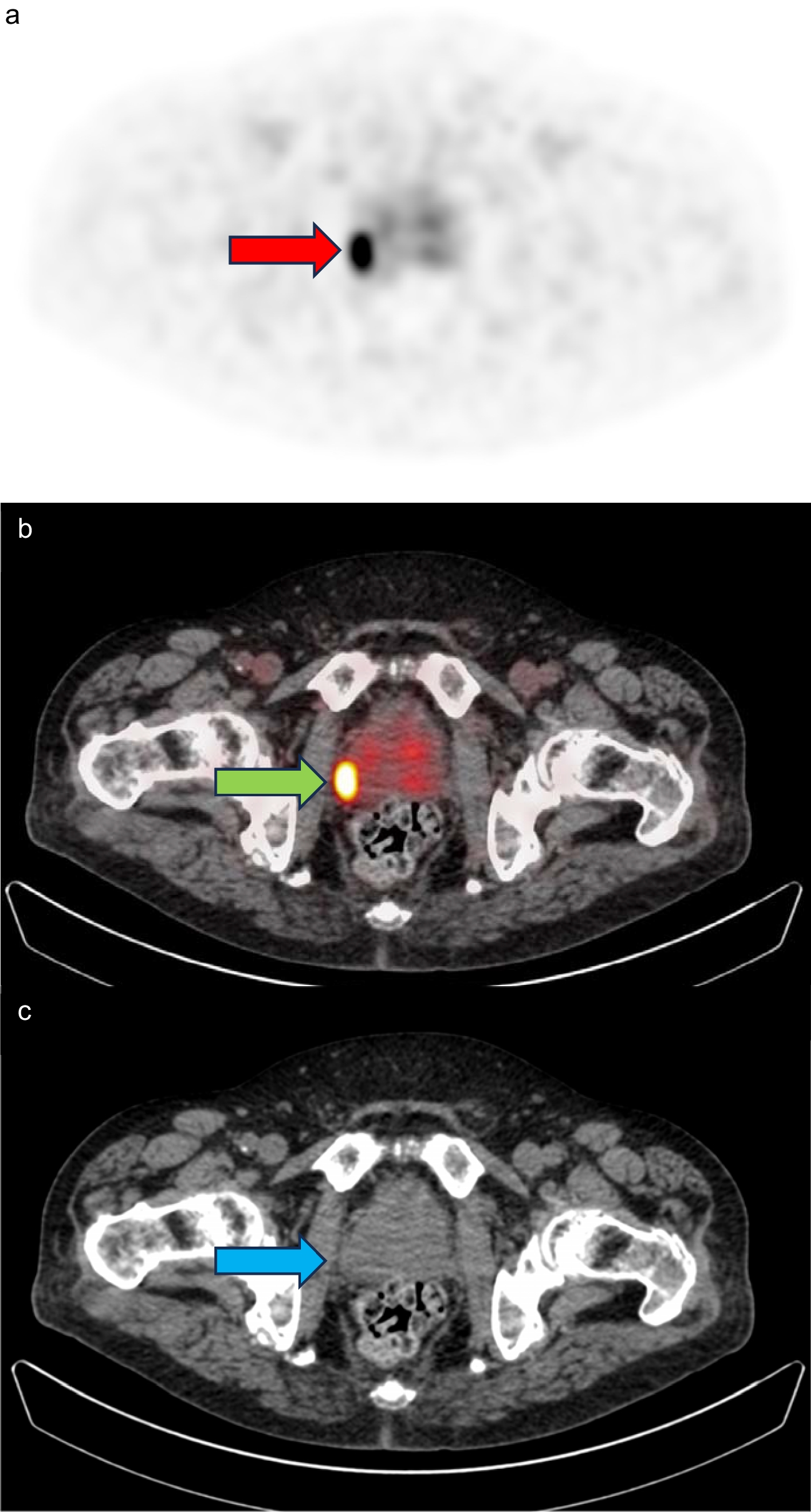
FDG-PET/CT scan procedure
Before the FDG-PET/CT scans, patients underwent a minimum fasting period of 6 h, except for diabetic patients who fasted for a minimum of 4 h. Patients received an intravenous infusion of 4 MBq FDG per kilogram body weight 60 min before undergoing a scan from the vertex of the skull to the mid-thigh using an integrated PET/CT scanner with continuous bed motion (Siemens Biograph Vision). Images were reconstructed using 4 iterations, 5 subsets, and 2-mm Gaussian post-processing filter, in matrix size 440 × 440. During the study, the FDG-PET/CT protocol was updated, and patients were subsequently instructed to have their arms down during the scan, aligning with the latest recommendation [31, 32]. An initial low-dose CT was performed for attenuation correction and anatomic mapping.
FDG-PET/CT evaluation
Two experienced nuclear medicine specialists (LCG and KR) and a trained medical doctor (AWN) independently evaluated the baseline, 8-week, and 10-week FDG-PET/CT scans blinded from the clinical diagnosis, clinical data, and scanning date. In accordance with standard daily procedures for FDG-PET/CT evaluation, each evaluator initially provided a binary classification of PMR (PMR yes/no) for each patient based on their regular practice, and any discrepancies were resolved through discussion between AWN, LCG, and KR. Furthermore, 12 different anatomic sites were evaluated for FDG uptake: shoulder joints, sternoclavicular joints, ischial tuberosities, hip joints, greater trochanters, and cervical/lumbar interspinous bursae. The FDG uptake was visually graded for each site and compared to liver uptake; 0, no uptake; 1, uptake lower than liver; 2, uptake equal to or higher than liver [11]. A summed PET-PMR score was calculated for each patient using the Leuven score and a cut-off of 16 or above was considered positive for PMR [11].
Sample size
Based on the only other prospective FDG-PET study in this research area, approximately 99 patients should be enrolled to obtain 95% confidence interval (95% CI) at baseline for a sensitivity of 85% (95% CI, 76–91) and a 95% CI for a specificity of 87% (95% CI, 79–93) [11].
Statistics
Clinical and FDG-PET/CT data between groups were compared with Student’s t-test or Mann–Whitney U test for continuous variables and chi-square statistics or a Fisher’s exact test for categorical variables. Sensitivity and specificity were calculated for a positive FDG-PET/CT using the Leuven score as well as the dichotomous diagnostic score. The interrater variability of the Leuven score between the three FDG-PET/CT raters was assessed with intraclass correlation coefficient (ICC) based on a mean-rating, absolute-agreement, two-way mixed-effects model [33]. An ICC estimate within the range of < 0.50, 0.50–0.75, 0.75–0.90, and > 0.90 corresponds to reliability levels categorized as poor, moderate, good, and excellent, respectively [34]. Repeated measurements within the same individuals were assessed using repeated measures ANOVA followed by a Bonferroni post hoc test. Statistical analyses were performed using STATA (version 17, StataCorp, USA). Two-tailed p-values less than 0.05 were considered statistically significant.


留言 (0)