Ethics statement
All samples used in this study were obtained from patients admitted to 108 Military Central Hospital (108-MCH), Hanoi, Vietnam. The study was approved by the institutional review board and an independent ethics committee of 108 Military Central Hospital Hanoi, Vietnam (108MCH-# 2723/HĐĐĐ- 25,062,020). Written informed consent was obtained from all study patients and/or their guardians. All samples used in this study were pseudo-anonymized prior to laboratory testing to de-identify the patients.
Bacterial strains
Positive control DNA was extracted from N. meningitidis serogroup B (MC58, #ATCC BAA-335). N. meningitidis serogroup C (M1628, #ATCC 13,102) and W135 (M-1574, #ATCC 43,744) were also used to confirm the diagnosis. Furthermore, non-meningococcal Neisseria species, including N. gonorrhoeae (F-18, #ATTC 49,226), and Escherichia coli DH5α (#18,258,012, Thermo Fisher Scientific Inc) were also tested for cross-reactivity.
Clinical CSF samples
All samples were collected from patients with suspected meningococcal disease (n = 139). Clinicians classified patients as having probable meningococcal disease based on clinical signs and symptoms, CSF parameters (such as white blood cell count, glucose and protein levels) and bacterial tests (such as CSF culture and/or latex agglutination test), and close contact with a meningococcal patient.
Detection of
N. meningitidis with Realtime-PCR
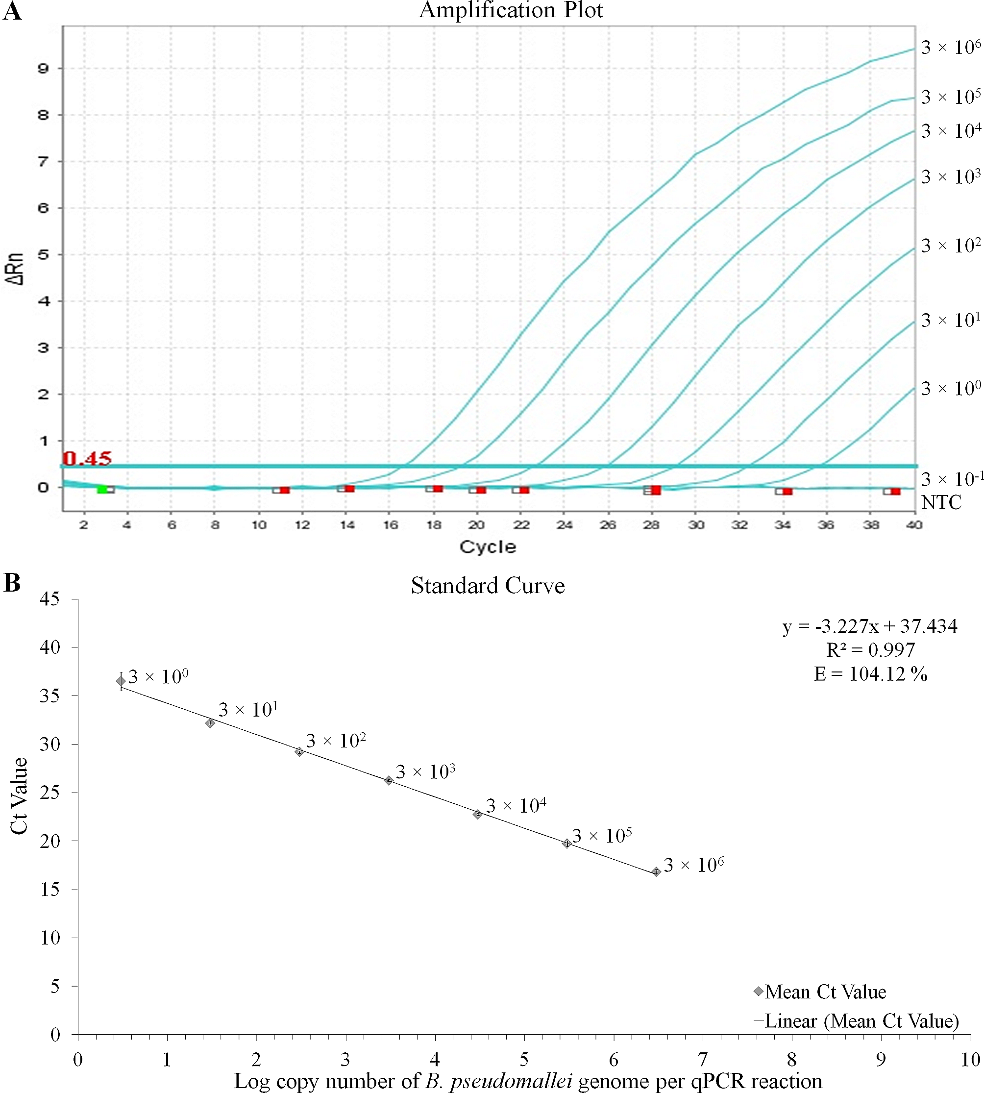
DNA was extracted from 200 μL saliva/oropharyngeal swabs/ CSF using the automated SACACE SYSTEM (Nucleic acid extraction laboratory workstation SaMag-12™, Sacace Biotechnologies S.r.l., Italy) with an elution volume of 100 μL. Real-time qualitative PCR targeting the capsule transporter gene ctrA of N. meningitidis was performed using the set of primers described in Table 1. Reactions were performed in volumes of 20 μL containing 1X QuantiTect Probe PCR (Qiagen, Hilden, Germany), 0.25 μM of each primer, 0.25 μM of probe and 5 μL of template DNA. Realtime PCR was performed in duplicates in AriaMx Real-Time PCR system (Agilent Technologies, Inc, Santa Clara, California, United States) as follows: initial denaturation at 95 °C for 15 min, followed by 45 cycles of denaturation at 95 °C for 15s, annealing at 60 °C for 60s when the fluorescence signal was measured. Samples were classified as negative if Ct values ≥ 40 or no amplification signal was detected.
Table 1 Primers and Probes employed for LAMP-CRISPR and Real-time PCR assayLoop-mediated isothermal amplification (LAMP) assays for
N. Meningitidis
LAMP-specific primer pairs for Neisseria meningitidis were designed. Details of the primer are described in Table 1. Reactions were performed in reaction volumes of 20 μL containing 1x isothermal buffer II (New England Biolabs, Ipswich, MA), 1.6 μM each FIP and BIP, 0.2 μM each F3 and B3, 0.4 μM each LF or LB, 0.25 μM dNTPs, 2.56 U of Bst3.0 DNA polymerase (New England Biolabs, Ipswich, MA), 0.4 M betaine (Sigma, St. Louis, MO), a truncated gene 2.5 protein (GP2.5-delta 21 C) and 5 μL of template DNA. LAMP reactions were performed in heat blocks (Block Thermostat BT200, Kleinfeld Labortechnik, Germany) at 63 °C for 20 min followed by an inactivation step for 5 min at 80 °C.
CRISPR/Cas12 cleavage reaction
Detection assays were performed using 1x r2.1 buffer, 25 nM LbCas12a (New England Biolabs, Ipswich, MA), 25 nM gRNA, 750nM custom synthesized homopolymer ssDNA FQ reporter or 125nM ssDNA FAM-biotin reporter (see Table 1). All these components were mixed within 20 μL and added directly into LAMP reaction tube (total volume 40 μL). It was incubated at 42 °C for 40 min. Finally, the entire volume was applied to lateral flow (PCRD, Abingdonhealth – FG-FD51673) for “naked-eye” detection. If the strip shows two bands (T2 and C lines), the result is negative. If it shows one band (C band line), the result is positive.
Neisseria meningitidis dilution series to determine the limit of detection (LOD)
The limit of detection (LOD) has been calculated as the lowest amount of analyte, which can be detected with more than a stated percentage of confidence, but not necessarily quantified as an exact value [15, 16]. In molecular assays, the LoD value is generally considered to be the lowest concentration of the target that can be detected in ≥ 95% of replicate measurements [17]. In this study, multiple aliquots of a specific matrix were spiked with serial dilutions of the target DNA and subjected to the entire process of LAMP-CRISPR/Cas testing. The LOD is then defined as the spike amount of target DNA in dilution that could be detected in 95% of replicates [18]. DNA extraction from colonies of N. meningitidis type B standard strains (MC58). DNA concentration was converted to copy number using a genome length for N. meningitidis type B standard strains (MC58) of 2,272,351 bp. LOD was calculated by probit regression analysis with 20 repeats for each level.
Statistical analysis
Sensitivity, specificity, positive predictive value (PPV), and negative predictive value (NPV) of real-time PCR tests were compared with those of LAMP-CRISPR/Cas (real-time PCR was considered the gold standard). Probit regression analysis was performed using SPSS software ver. 20 (IBM Armonk, New York United States).

留言 (0)