
記住我


Endothelial cell dysfunction, characterized by apoptosis, is observed in the lesion regions of the arterial vasculature during the early stages of AS. This dysfunction plays a significant role in plaque regression and plaque instability [34]. In these areas, there is an enlargement of the space between injured endothelial cells, which leads to the accumulation of lipids beneath the endothelium. This lipid accumulation is taken up by macrophages, resulting in the formation of foam cells and promoting the progression of AS.
To address these issues, we investigated the potential of GP and EM as therapeutic agents. GP has been shown to inhibit apoptosis in HUVECs, while EM has been found to reduce lipid accumulation caused by macrophages [9, 15]. We employed flow cytometry and confocal microscopy to evaluate the effects of GP and EM on apoptosis in HUVECs and the uptake of Dil-oxLDL by macrophages. Interestingly, we found that GP exhibited a stronger ability to inhibit apoptosis in HUVECs, while EM showed a superior capacity to reduce the uptake of Dil-oxLDL by macrophages. Motivated by these findings, we combined GP and EM to enhance the therapeutic potential for AS. As expected, the combination of GP and EM resulted in a more pronounced inhibition of HUVECs apoptosis and macrophage uptake of Dil-oxLDL (Fig. S1). Furthermore, to mitigate the hepatorenal toxicity of the drugs, we opted for a lower concentration of GP and EM [35, 36].
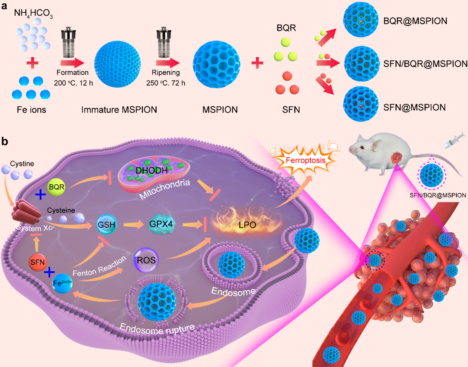
The liposomes were prepared using the thin film hydration method, as outlined in Fig. 1A. GP and EM were loaded into the LP NPs and modified with DSPE-TK-PEG2000. The fusion of Møm hybridization forms TK-MLP@(GP + EM) NPs. TEM images confirmed the spherical morphology of TK-MLP@(GP + EM) NPs, which were surrounded by a thin film coating resulting from the macrophage cell membrane hybridization (Fig. 1B). To accommodate the difference in solubility between GP and EM, LP NPs were loaded with a feed ratio of 1:20 (drug: lecithin), allowing EM to be incorporated into the phospholipid layer and GP into the core. The encapsulation efficiency percentages for GP and EM were determined to be 87.4% and 62.5%, respectively (Fig. 1C). In addition, the SDS-PAGE assay confirmed the preservation of membrane proteins in both Møm and TK-MLP@(GP + EM) NPs (Fig. 1D). Western blotting analysis further confirmed the existent of specific bands of CD11b in TK-MLP@(GP + EM) NPs, the corresponded protein marker for Møm (Fig. 1E). Furthermore, we adopted Förster resonance energy transfer (FRET) experiment to further verify the fusion of Møm and Liposome. Fig. 1F showed that compared with LP NPs alone, with the fusion of Liposome membrane and Møm, the trend of fluorescence intensity change at 565 nm is opposite to that at 663 nm as the fusion of the Liposome membrane and Møm leads to the separation of the DiI/DiD energy resonance transfer pair of the fluorophore, thus restoring the fluorescence signal of DiI (565 nm).
In the AS pathological environment, an H2O2 concentration of 1 mM is considered indicative of oxidative stress [28]. Therefore, the morphology changes of TK-MLP@(GP + EM) NPs were observed by TEM after incubation in a 1 mM H2O2 solution for 4 h (Fig. 1G). The bilayer and cavity structure vanished, the structure of NPs broke up into fragments, indicating the strong response of the nanoparticles to ROS. This suggests that ROS can serve as an intelligent switch to trigger the disintegration of TK-MLP@(GP + EM) NPs. Additionally, the average diameter of TK-MLP@(GP + EM) NPs, as measured by DLS, was approximately 184.6 nm (Fig. 1H), with a zeta potential of -46.93 mV (Fig. 1I). The cumulative drug release rates of GP and EM from TK-MLP@(GP + EM) NPs in PBS over 72 h were found to be 44.4% and 31.4%, respectively. In contrast, TK-MLP@(GP + EM) NPs disintegrated and dissociated in the presence of 1 mM H2O2 due to the robust ROS response mediated by TK. As a result, the cumulative drug release rates of GP and EM reached 86.5% and 64.2% after 72 h, respectively (Fig. 1J&K). Overall, these results demonstrate that TK-MLP@(GP + EM) NPs have the ability to dissolve and efficiently release drugs in the high ROS microenvironment at the AS site rapidly and efficiently.
Fig. 1
Characterization of TK-MLP@(GP + EM) NPs. (A) Schematic diagram of the synthetic process of TK-MLP@(GP + EM) NPs. (B) TEM image of TK-MLP@(GP + EM) NPs. (C) GP and EM entrapment efficiency of TK-MLP@(GP + EM) NPs. (D) SDS-PAGE analysis of retention protein bands of Møm and [LP + Mø]m NPs. (E) Western blot of Møm and [LP + Mø]m NPs for characteristic Møm marker CD11b. (F) The infusion efficiency investigation of Liposome NPs and [LP + Mø]m. The fluorescence recovery of DiI (565 nm) represented the fusion of LP NPs and [LP + Mø]m. (G) TEM image of TK-MLP@(GP + EM) NPs after being treated with 1 mM H2O2 solution for 4 h. (H&I) Particle size and zeta potential of TK-MLP@(GP + EM) NPs analyzed by DLS. (J&K) Release profile investigation of GP and EM in PBS and H2O2.
Cellular uptake of TK-MLP@(GP + EM) NPsMacrophages play a crucial role in the innate immune system. It was observed in Fig. S2 that the red fluorescence signal in macrophages treated with MLP NPs was significantly lower than that in the LP NPs group and TK-LP NPs group. This phenomenon was also visible in the red fluorescence signal of the TK-MLP NPs group, indicating that Møm possesses the ability to prevent macrophages from phagocytosing TK-MLPs. This can be attributed to the camouflage ability of Møm, which has been reported in previous study [37]. Additionally, we found that LPS-treated HUVECs exhibited a strong fluorescence signal after incubating with TK-MLP@Dil NPs, compared to intact HUVECs (Fig. S3). This result suggests that TK-MLP@(GP + EM) NPs can target injured endothelial cells, which is attributed to the interaction between the high ICAM-1 expression of activated endothelial cells and the CD11b surface molecule present on Møm [38].
As the “digestive organ” within cells, lysosomes play a role in the degradation and elimination of drugs, which can reduce their therapeutic effects [39]. Upon studying the interaction between TK-MLP NPs and lysosomes in activated HUVECs and macrophages, the uptake of TK-MLP NPs group by activated macrophages and HUVECs was highly efficient, surpassing that of the LP NPs group and TK-LP NPs group, while the green fluorescence of the lysosomal probe was observed. We discovered that the weak red fluorescence in the LP NPs and TK-LP NPs group was notably, this observation was consistent in both LPS-treated HUVECs and macrophages. Additionally, the yellow fluorescence signal (represents red fluorescence exhibited co-localization with the lysosomes) of the TK-LP NPs group was significantly stronger than that of the TK-MLP NPs group, suggesting that the hybridization of Møm facilitates the bypassing of lysosomes and internalization of NPs into activated endothelial cells and macrophages within the AS environment, provides a foundation for the precise and targeted therapeutic application of drugs (Fig. 2A&B).
To directly assess the cellular uptake of NPs within atherosclerotic plaques, a transwell system was employed, wherein LPS-treated HUVECs were incubated in the upper chamber, and activated macrophages were cultured in the lower chamber (Fig. 2C) [40]. The results displayed in Fig. 2D&E demonstrated that the red fluorescence within both activated macrophages and HUVECs was notably stronger in the TK-MLP group than in the LP group. These findings indicate that TK-MLP NPs can be effectively internalized by HUVECs and macrophages within the pathological environment characterized by heightened levels of ROS, attributed to the targeting and “homing effect” of Møm [25]. Moreover, these results establish a foundation for investigating the molecular mechanisms underlying the potential therapeutic applications of nanomaterials in atherosclerosis treatment.
Fig. 2
Cellular uptake of TK-MLP NPs. (A&B) Representative fluorescence images and quantitation of activated HUVECs and RAW264.7 cells 4 h after incubation with LP, MLP, TK-LP NPs, and TK-MLP NPs (red). Green represents the Lyso Tracker. Scale bar = 20 μm. (C)The schematic diagram showed that HUVECs were co-cultured with RAW264.7 cells in a transwell system for simulating plaque in vitro. (D&E) Phagocytosis and quantitation of LP NPsand TK-MLP NPs in activated HUVECs and RAW264.7 cells in transwell. BF indicates bright field. Scale bar = 20 μm. n = 3, ##P < 0.01, ###P < 0.001.
TK-MLP@(GP + EM) NPs can inhibit the apoptosis of HUVECsPrevious study has demonstrated that LPS can induce apoptosis in endothelial cells [41]. The apoptosis of endothelial cells leads to dysfunction, damage to the cell barrier, secretion of adhesion factors, and recruitment of monocytes to form macrophages, thereby contributing to the development and progression of atherosclerosis [42]. Thus, in this study, we utilized LPS-stimulated HUVECs as a model to investigate the effects of TK-MLP@(GP + EM) NPs on apoptosis. Flow cytometry analysis revealed that both early and late apoptosis of HUVECs in the Model group (∼ 21.4% and ∼ 22.85%, respectively). Compared with the Model group, TK-MLP@(GP + EM) NPs resulted in a significant ∼ 19.5% decrease in the early apoptosis rate and ∼ 14.9% decrease in the late apoptosis rate, respectively (Fig. 3A). Furthermore, western blot analysis demonstrated that the treatment with TK-MLP@(GP + EM) NPs up-regulated the expression of the antiapoptotic protein Bcl-2 and down-regulated the expression of the proapoptotic protein BAX. Additionally, Fig. 3B showed a 1.8-fold increase in Cleaved caspase-3 levels, a mediator of apoptosis, in HUVECs following LPS stimulation, consistent with previous reports [41]. However, treatment with TK-MLP@(GP + EM) NPs inhibited the expression of Cleaved cjaspase-3 (Fig. 3B). Collectively, these results further support the notion that TK-MLP@(GP + EM) NPs inhibit apoptosis in HUVECs.
LPS can change cell morphology and induce overexpression of intercellular cell adhesion molecule 1 (ICAM-1) in HUVECs by promoting apoptosis [43]. Phalloidin immunofluorescent staining revealed that LPS altered the morphology of HUVECs from cobblestone-like shapes to spindle shapes, which was attenuated by treatment with TK-MLP@(GP + EM) NPs. Moreover, TK-MLP@(GP + EM) NPs down-regulated the expression of ICAM-1 (Fig. S4). VE-cadherin, an endothelium-specific adhesion molecule, plays a crucial role in maintaining the integrity of endothelial cells and preventing AS [44]. We observed a significant 70% reduction in the expression of VE-cadherin (red fluorescence) in activated HUVECs. However, treatment with TK-MLP@(GP + EM) NPs resulted in a substantial 67% increase in the expression of VE-cadherin, compared to the Model group (Fig. 3C). These findings indicate that TK-MLP@(GP + EM) NPs are capable of reversing LPS-induced apoptosis and repairing the damage to HUVECs.
Endothelial cell apoptosis is primarily induced by excessive production of ROS resulting from mitochondrial damage [45]. Therefore, we utilized TEM to examine the ultrastructure of HUVECs mitochondria. In the Model group, we observed mitochondrial enlargement, swelling, hypodense matrix, and vacuolar degeneration of mitochondrial cristae. In contrast, the TK-MLP@(GP + EM) NPs group exhibited clear and well-preserved ultrastructure of endothelial mitochondria, characterized by dense mitochondrial cristae and a matrix with normal density (Fig. 3D). Subsequently, we investigated the impact of TK-MLP@(GP + EM) NPs on mitochondrial ROS in activated HUVECs by using fluorescent dyes MitoSOX. As demonstrated in Fig. 3E&F, the red fluorescence signal in activated HUVECs was significantly enhanced approximately 9.2 times compared to the Control group. However, the accumulation of mitochondrial ROS induced by LPS was mitigated by treatment with TK-MLP@(GP + EM) NPs, as evidenced by a remarkable 92% reduction in red fluorescence intensity. We attribute this reduction to the fact that the abundance of mitochondrial ROS triggered the breakdown of TK, consequently leading to the rapid release of the drugs. Additionally, mitochondrial dysfunction is characterized by a decrease in membrane potential [46]. Therefore, we employed JC-1 staining to evaluate the transmembrane potential (Δψm) of HUVECs. A higher red/green ratio of JC-1 fluorescence indicates a lower degree of mitochondrial dysfunction. LPS stimulation significantly lowered the Δψm of HUVECs. However, this impairment was significantly ameliorated after treatment with TK-MLP@(GP + EM) NPs, as demonstrated by a remarkable increase in the red/green ratio (Fig. 3G&H). These results suggest that TK-MLP@(GP + EM) NPs possess potent capability in maintaining the normal membrane potential of mitochondria. Hence, TK-MLP@(GP + EM) NPs exhibit the potential to inhibit HUVECs apoptosis resulting from mitochondrial damage and aid in the recovery of HUVECs functionality.
Fig. 3
TK-MLP@(GP + EM) NPs can inhibit the apoptosis of endothelial cells. (A) Apoptosis detection after various treatments by flow cytometry. (B) Western blotting assay of BAX, Bcl-2, and Cleaved caspase-3 levels in HUVECs with different treatments. (C) Confocal laser scanning microscopy images of VE-cadherin in HUVECs with different treatments. (D) The ultrastructure of mitochondria in HUVECs was observed using a transmission electron microscope (TEM). The arrows point to different states of mitochondrial structure. (E&F) Mitochondrial superoxide levels by MitoSOX Red fluorescent staining with MFI quantification. (G&H) Mitochondrial membrane potential by JC-1 staining with calculation of the ratio of red MFI (aggregated JC-1) to green MFI (monomer JC-1). 1: Control;2: Model; 3: GP + EM; 4: TK-MLP@(GP + EM). Scale bar = 20 μm. n = 3, ###P < 0.001 vs. the Control. ** P < 0.01, *** P < 0.001 vs. the Model.
TK-MLP@(GP + EM) NPs can inhibit lipid accumulation of macrophagesThe apoptosis of endothelial cells can have detrimental effects on the development of AS by causing further lipid accumulation and accelerating the progression of the lesion [47]. In this study, we also examined the impact of TK-MLP@(GP + EM) NPs on lipid deposition. We investigated the reduction of lipid uptake by activated macrophages by observing the change in fluorescence signal using Dil-oxLDL. The Model group exhibited a strong red fluorescence signal, indicating significant uptake of Dil-oxLDL by activated macrophages. However, in the TK-MLP@(GP + EM) NPs group, we observed a remarkable decrease in red fluorescence compared to the Model group, suggesting a better reduction in Dil-oxLDL uptake by activated macrophages (Fig. 4A). Furthermore, we assessed intracellular lipid droplets in activated macrophages using ORO staining. The results demonstrated a reduction in intracellular lipid droplets with TK-MLP@(GP + EM) NPs treatment, which can be attributed to the inhibition of ox-LDL internalization (Fig. 4B).
Fig. 4
TK-MLP@(GP + EM) NPs inhibits lipid accumulation of macrophages. (A) Confocal fluorescence images and semi-quantitative analysis of DiI-oxLDL internalization in RAW264.7 cells. (B) Optical microscopy images and semi-quantitative analysis of ORO staining. (C-E) Western blot analysis of ABCA1 and ABCG1 on activated macrophages with different treatment. (F&G) Confocal fluorescence images of ABCA1 and ABCG1 on activated macrophages with different treatment. 1: Control;2: Model; 3: GP + EM; 4: TK-MLP@(GP + EM). Scale bar = 20 μm. n = 3, ###P < 0.001 vs. the Control. *P < 0.05, ** P < 0.01, *** P < 0.001 vs. the Model
Considering that ABCA1 and ABCG1 play a crucial role in promoting cholesterol excretion during the development of atherosclerosis [48], we examined the effects of TK-MLP@(GP + EM) NPs on the expression of these transporters in activated macrophages. Compared to the Control group, LPS led to down-regulation of ABCA1 and ABCG1 levels in macrophages, which is consistent with our previous study [32]. However, treatment with TK-MLP@(GP + EM) NPs resulted in up-regulation of both ABCA1 and ABCG1 protein expression levels (Fig. 4C-E). Moreover, confocal microscopy images also demonstrated a significant increase in the expression of ABCA1 and ABCG1 with TK-MLP@(GP + EM) NPs treatment (Fig. 4F&G). These results indicate that TK-MLP@(GP + EM) NPs can effectively reduce lipid deposition by modulating lipid internalization and efflux. Previous study confirmed that re-polarizing macrophages from M1 to M2 type is beneficial for the treatment of AS [25], we subsequently evaluated the capability of TK-MLP@(GP + EM) NPs for reprogramming macrophages. In Fig. S5A&B, flow cytometry assay showed the signal decrease of CD80 while increase for CD206 in activated macrophages in the GP + EM group, because both GP and EM can induce M1-to-M2 repolarization of macrophages [20, 49]. What is more, the best therapeutic effect of TK-MLP@(GP + EM) NPs can attribute to the function of TK-MLP NPs on consuming high ROS levels in the Model group [50]. Therefore, these results indicate that TK-MLP@(GP + EM) NPs could efficiently reduce lipid deposition by inducing M1-to-M2 re-polarization of macrophages.
It has been established that the transformation of M1 macrophage is accompanied by mitochondrial dysfunction [51]. To assess mitochondrial integrity and condition among different groups, we observed it through TEM. As shown in Fig. S5C, mitochondria in the Control group appeared intact with clear mitochondrial crista, while the Model group displayed more swollen and damaged mitochondria. However, treatment with TK-MLP@(GP + EM) NPs resulted in the recovery of a clear and intact mitochondrial structure. Furthermore, we investigated the impact of TK-MLP@(GP + EM) NPs on mitochondrial ROS and Δψm in activated macrophages. As depicted in Fig. S5D&E, treatment with TK-MLP@(GP + EM) NPs attenuated the accumulation of mito-ROS caused by LPS by approximately 90% and restored mitochondrial Δψm. These results suggest that TK-MLP@(GP + EM) NPs can promote the repolarization of macrophages from M1 to M2 type, which is achieved by restoring mitochondrial function.
Overall, our findings provide further evidence that TK-MLP@(GP + EM) NPs possess the ability to reverse M1 macrophage polarization by preventing mitochondrial damage, eventually inhibit lipid accumulation and intervene atherosclerosis development.
Pharmacokinetics and targeting capability of TK-MLP@(GP + EM) NPsTo assess the prolonged circulation time of TK-MLP NPs, we conducted pharmacokinetic studies in male wild-type C57BL/6 mice. Ce6-labeled NPs were intravenously injected, and the residual content of nanoparticles was measured by analyzing the fluorescence intensity of Ce6 in blood samples collected at various time intervals. Compared to LP@Ce6 NPs with a circulation half-life (t1/2) of approximately 0.72 h, fluorescence imaging revealed that both MLP@Ce6 NPs and TK-MLP@Ce6 NPs exhibited extended blood circulation (t1/2 ≈ 1.13 h for MLP@Ce6 NPs; t1/2 ≈ 1.28 h for TK-MLP@Ce6 NPs), which is consistent with previous findings [23] (Fig. 5A). These results highlight the ability of membrane coating to prolong the circulation time of NPs, which is beneficial for enhancing drug accumulation in atherosclerotic plaques.
Fig. 5
Pharmacokinetics and targeting capability of TK-MLP NPs. (A) Representative photographs of blood samples collected from C57BL/6 mice after administration of different nano-materials at various time points. Pharmacokinetic curves of different nano-materials. n = 3. (B) Fluorescence photos and semi-quantitative show the fluorescent signals of Ce6 in aortas from ApoE−/− mice. ApoE−/− mice fed with HFD for 2 months were intravenously injected with different NPs. After administration of 12 h, the aortas of ApoE−/− mice were isolated for detection. n = 3. *P < 0.05, ***P < 0.001 vs. the LP@Ce6.
To assess the targeting capability of TK-MLP@(GP + EM) NPs, ApoE−/− atherosclerotic mice were injected intravenously with TK-MLP@Ce6 NPs to track its accumulation in the mice. As depicted in Fig. 5B, the fluorescence intensity of MLP@Ce6 NPs at the aortic site was observed to increase by approximately 1.40 times compared to LP@Ce6 NPs. This can be attributed to the high affinity between ICAM-1 on damaged endothelial cells and CD11b receptors on the macrophage cell membranes [52]. Furthermore, the signal intensity of the TK-MLP@Ce6 NPs treatment group increased by approximately 1.91 times. This enhanced targeting ability can attribute to the presence of abnormal ROS in atherosclerotic plaques, which effectively break the TK bonds of nanomaterials to trigger the release of Ce6. These results demonstrate that the Møm camouflaging and ROS responsiveness of TK significantly enhance the targeting and release abilities of TK-MLP@(GP + EM) NPs in ApoE−/− mice.
NPs are known to accumulate in the liver due to the first-pass effect, which involves the uptake of NPs by macrophages [53]. In our study, we observed that the fluorescent signal of the liver was higher in the LP@Ce6 NPs group compared to the MLP@Ce6 NPs and TK-MLP@Ce6 NPs groups (Fig. S6). This indicates that Møm coating is beneficial in preventing liver elimination and increasing the accumulation of TK-MLP@(GP + EM) NPs in plaques. Moreover, a strong fluorescence signal was observed in the kidneys of mice for all NPs, including TK-MLP@(GP + EM) NPs, due to the metabolism and excretion of NPs through the kidneys. Overall, these findings suggest that TK-MLP@(GP + EM) NPs have the ability to chronically localize and accumulate in atherosclerotic plaques.
The efficacy of TK-MLP@(GP + EM) NPs in ApoE-/- mice fed HFDBased on the promising results obtained thus far, we further investigated the therapeutic effect of TK-MLP@(GP + EM) NPs on atherosclerotic plaques in vivo. Following treatment, we isolated the aorta and observed a significant reduction in plaque size in the aortic arch region (area circled by the black dotted line) (Fig. 6A). Additionally, ORO staining revealed that the area of lipid deposition in the plaques was approximately 1.06% and 17.15% in the Control and Model groups, respectively, confirming successful construction of the atherosclerosis model. Notably, when compared to the Model group, the plaque area was significantly reduced in ApoE−/− mice treated with TK-MLP@(GP + EM) NPs. It is worth mentioning that in our animal experiments, we used MitoQ, a mitochondria-targeting antioxidant, as a positive group. The MitoQ group exhibited a significant reduction in plaque area (approximately 4.10%), indicating the potential of inhibiting mitochondrial oxidative stress for effective atherosclerosis treatment. Excitingly, TK-MLP@(GP + EM) NPs demonstrated a stronger inhibitory effect on aortic plaque formation compared to the free GP + EM group (plaque area approximately 9.64%). The plaque area with TK-MLP@(GP + EM) NPs treatment was approximately 3.50%, which was similar to the effect observed in the MitoQ group. This suggests that one of the mechanisms by which TK-MLP@(GP + EM) NPs reduce atherosclerotic plaques might be through the inhibition of mitochondrial oxidative stress (Fig. 6B&C). Furthermore, we investigated the impact of TK-MLP@(GP + EM) NPs on plaque formation in the high-occurrence region of the aortic roots. ORO staining of frozen sections revealed significant lipid deposition in the plaques of the Model group (approximately 22.68%), whereas TK-MLP@(GP + EM) NPs exhibited a significant anti-lipid deposition effect in all segments of the aortas (approximately 5.07%) (Fig. 6D&E).
Increased levels of ROS play a crucial role in the growth of the necrotic core, which is a key characteristic of atherosclerosis. In this study, the presence of large necrotic cores in the aorta root of the Model group was confirmed through H&E staining (Fig. 6F&G). However, treatment with TK-MLP@(GP + EM) NPs resulted in a significant reduction in necrotic cores by approximately 22.3%. Given that the extent of necrotic cores is positively associated with plaque vulnerability, Masson’s trichrome assay was performed to evaluate the stability of the plaques in ApoE−/− mice. Among the various groups investigated, TK-MLP@(GP + EM) NPs exhibited the highest collagen content and fibrous cap thickness surrounding the plaques (Fig. 6H&I).
Taken together, these findings demonstrate the remarkable therapeutic effects of TK-MLP@(GP + EM) NPs on atherosclerosis.
Fig. 6
The efficacy of TK-MLP@(GP + EM) NPs in ApoE−/− mice fed HFD. (A) Photographs of the aortic arch. (B&C) The representative images of en face ORO-stained aortas and semi-quantitative analysis. n = 4. (D&E) ORO-stained frozen sections and semi-quantitative analysis of the aortic roots. (F-I) Representative immunohistochemistry staining photographs and semi-quantitative analysis with H&E and Masson trichrome. Scale bar = 200 μm. n = 3, ###P < 0.001 vs. the Control. *** P < 0.001 vs. the Model.
TK-MLP@(GP + EM) NPs can restore endothelial function in ApoE-/- atherosclerosis micePrevious studies have highlighted the presence of elevated levels of ROS in atherosclerotic plaques in ApoE−/− mice [54]. In this study, DHE staining was performed on the freshly frozen aortic roots of ApoE−/− mice to assess the ROS content within the plaques (Fig. 7A&B). Notably, the Model group exhibited a strong red fluorescent signal, indicative of high ROS levels in the aortic root plaques compared to the Control group. In alignment with previous finding [54], treatment with MitoQ resulted in a ∼ 43% reduction in ROS levels. Similarly, the GP + EM group and the TK-MLP@(GP + EM) NPs group also demonstrated varying degrees of ROS reduction, consistent with the in vitro ROS level detection results shown in Figs. 3E and 4G. Importantly, it is worth noting that the TK-MLP@(GP + EM) NPs group exhibited superior ROS elimination capability and the weakest fluorescence intensity, exceedingly even that of the MitoQ group. These results further support the notion that TK-MLP@(GP + EM) NPs can be thought of as similar anti-mitochondrial oxidants. The abundant ROS levels within the plaques can trigger the breakdown of DSPE-TK-PEG2000, facilitating the release of the encapsulated drugs, which subsequently accumulate within the plaques. Overall, these findings provide evidence of the potent ability of TK-MLP@(GP + EM) NPs to effectively reduce ROS levels in atherosclerotic plaques.
The accumulation of ROS within atherosclerotic plaques has been shown to exacerbate endothelial cell injury and the expression of chemokines [55]. During the early stages of atherogenesis, dysfunctional endothelial cells secrete ICAM-1 [56], a factor implicated in the disease progression. In order to investigate whether TK-MLP@(GP + EM) NPs can inhibit this process in the treatment of AS, immunofluorescence staining was performed on a cross-section of the aortic root. As shown in Fig. 7C&D, the expression level of ICAM-1 in CD31-labeled endothelial cells was significantly increased by 7.4-fold in the Model group, displaying a strong yellow fluorescence signal compared to the Control group. However, treatment with TK-MLP@(GP + EM) NPs led to a notable 53% decrease in the expression of ICAM-1 in endothelial cells at the site of the lesion. These findings suggest that TK-MLP@(GP + EM) NPs may exert their therapeutic effects on atherosclerosis by inhibiting the upregulation of ICAM-1 in endothelial cells.
To investigate the impact of TK-MLP@(GP + EM) NPs on endothelial barrier integrity further, we analyzed the localization of VE-cadherin in the aortas. In the Model group, we observed separate fluorescence signals of VE-cadherin, indicating endothelial cell dysfunction. However, after treatment with TK-MLP@(GP + EM) NPs, we observed improved continuity of fluorescence signals in endothelial cells (Fig. 7E&F). This improvement may be attributed to the elevated concentration of ROS within the plaque region, which triggers the therapeutic effect of the drugs released by the nanomaterials on endothelial repair.
These findings suggest that TK-MLP@(GP + EM) NPs have the ability to restore endothelial cell function and suppress adhesion molecule expression by reducing ROS levels in the plaque area.
Fig. 7
TK-MLP@(GP + EM) NPs can restore endothelial function in ApoE−/− atherosclerosis mice. (A&B) Aortic root cross-sections were subjected to dihydroethidium staining for determination of the redox state with quantitative analysis of DHE MFI. (C&D) Aortic root cross-sections were conducted co-immunofluorescent staining with anti-CD31 (green) and ICAM-1 (red). (E&F) Immunofluorescence staining images of VE-cadherin in aortas from each group. Scale bar = 200 μm. n = 3, ###P < 0.001 vs. the Control. ** P < 0.01, *** P < 0.001 vs. the Model.
TK-MLP@(GP + EM) NPs can regulate cholesterol efflux in ApoE-/- atherosclerosis miceIt has been reported that macrophage infiltration is a characteristic of atherosclerotic plaques [57]. In accordance with this, Fig. 7C confirmed that TK-MLP@(GP + EM) NPs effectively inhibit the production of ICAM-1 by endothelial cells. Considering this, we further investigated whether TK-MLP@(GP + EM) NPs could reduce macrophage infiltration. As shown in Fig. 8A&B, the Model group exhibited a signific
留言 (0)