LONGITUDINAL ANALYSIS OF MICROVASCULAR CHANGES IN SICKLE CELL DISEASE USING SWEPT-SOURCE OPTICAL COHERENCE TOMOGRAPHY ANGIOGRAPHY
Purpose:
To analyze the changes in macular vascular densities (VDs) and foveal avascular zone (FAZ) over a 6-year period using swept-source optical coherence tomography angiography in patients with sickle cell disease compared with unaffected control subjects.
Methods:
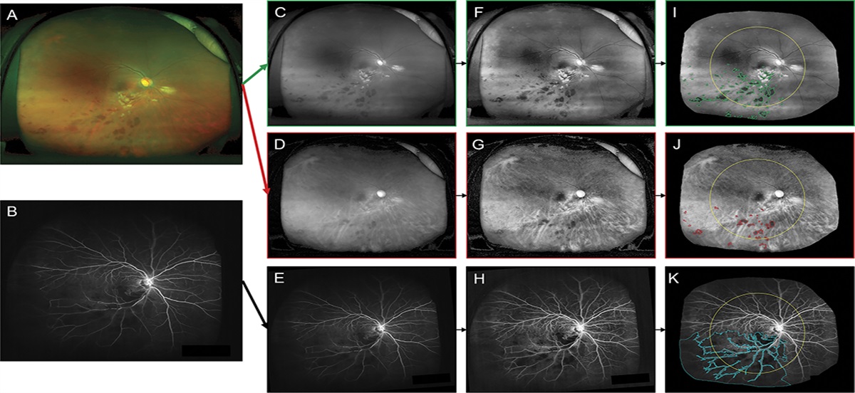
Thirty eyes of patients with sickle cell disease and 12 eyes of unaffected control subjects matched for age and ethnicity were examined at a 6-year interval using the same imaging protocol, including ultra-wide-field angiography, swept-source OCT, and 3 × 3-mm optical coherence tomography angiography. The macular VD and FAZ were measured on ImageJ software according to previously reported algorithms.
Results:
In sickle cell eyes, the mean FAZ significantly increased (P < 0.01), and the VD decreased in the foveal (within a circle of 1.5 mm in diameter around the foveal center) and temporal areas in both the superficial and the deep capillary plexuses (P < 0.01). The VD did not change over time in the parafoveal area (annulus between two circles of 1.5 and 3 mm in diameter) and in the superior, inferior, and nasal sectors. No worsening of peripheral retinopathy was observed in the cohort during the follow-up, except for one eye that developed sea-fan. In the control eyes, no microvascular change was observed over time in FAZ size and VD.
Conclusion:
These longitudinal optical coherence tomography angiography findings in patients with sickle cell disease showed an enlargement of the FAZ and a decrease in VD in the temporal and perifoveal regions despite the absence of progression of peripheral retinopathy, suggesting a worsening of the macular nonperfusion over time in sickle cell disease.


留言 (0)