Research object and organization
Patients with epithelial ovarian cancer treated with standard initial treatment (satisfactory cytoreductive surgery combined with chemotherapy) in the General Hospital of Ningxia Medical University from January 31, 2019, to June 30, 2021, were selected as sample sources. Inclusive criteria: (1) epithelial ovarian cancer diagnosed by surgery and pathology, (2) receiving standard initial treatment regimen (satisfactory cytoreductive surgery combined with chemotherapy), (3) the general data of the patients were complete, and the routine examination, serum CA 125, HE4 indexes and imaging examination were recorded regularly. Exclusion criteria:(1) combined with other malignant diseases, digestive tract diseases, acute and chronic inflammation; (2) not receiving platinum-containing chemotherapy, (3)follow-up data were incomplete.
According to the above criteria, 95 samples were collected for model construction, and 70 patients before December 31, 2020, as a training group, after 25 samples as a test set. All patients who participated in the study signed informed consent. All enrolled patients were divided into the platinum-resistant group(Res-group) and platinum-sensitive group(Sen-group) according to the following diagnostic criteria: patients in the platinum-resistant group include those who are platinum-resistant or platinum-refractory, platinum-resistant refers to those who respond to initial chemotherapy but progress or relapse within six months of completing chemotherapy, and platinum-refractory refers to those who do not respond to initial chemotherapy, such as stable tumors or tumors that progress, including those that progress within four weeks of chemotherapy. Platinum-sensitive refers to those who respond to initial chemotherapy but progress or relapse after six months of completing chemotherapy.
Clinical characteristics, tissue collection and patient follow-up
This study retrospectively collected the medical records of patients with epithelial ovarian cancer and collected the general conditions, examination results, treatment plans, and other data of patients through the electronic medical record system. Tumor tissue sections were obtained from the hospital pathology department for IHC.
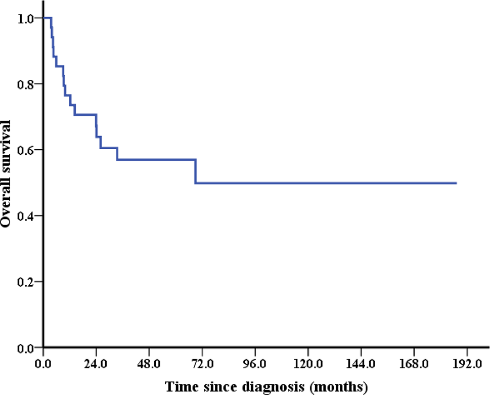
The enrolled patients were followed up by telephone and medical record data inquiry until February 2022. The median follow-up time was 27.0 (16.0\(\sim\)33.8) months. The follow-up interval of patients was every 2∼4 months in the first two years, every 4–6 months in the third five years, and every 6∼12 months after five years. Each follow-up included asking about symptoms, physical examination, tumor markers, and chest and abdomen CT or MRI examination.
Screening of platinum resistance genes
The GEO database (https://www.ncbi.nlm.nih.gov/geo/) was searched with the keywords “Ovarian cancer” and “Drug resistance”, and the screening conditions were: The species is “Homo sapiens”, the attribute is “Tissue”, and the study type is “Expression profiling by array”. Two datasets with significantly different genes (DEGs) are obtained: GSE45553 and GSE28739. The GSE45553 dataset is based on the GPL6244 data platform, including four platinum-resistant and four platinum-sensitive tissues. The GSE28739 dataset is based on the GPL7264 data platform, including 20 platinum-resistant and 30 platinum-sensitive tissues. The batch correction ratio is used to screen DEGs with “sva” and “limma” in R software, and the volcano diagram is drawn. The screening criteria of DEGs are set as follows: |log FC|> 1.0, p < 0.05. Heat map of DEGs was drawn by “heatmap” of R software, “clusterprofiler”,“org. db “,” richplot, “and” ggplot2 “were used to annotate the differential genes in Gene Ontology (GO), and Kyoto Encyclopedia of genes and genomes(KEGG) functional enrichment analysis was performed to p value < 0.05 was used as the inclusion criteria. Significant difference genes were analyzed by STRING online database (https://cn.string-db.org/) to analyze the protein-protein interaction (PPI) network of gene coding, and then the functional modules were constructed by Cytoscape software MCODE plug-in clustering, and the genes in the sub-network with the highest score were selected as hub genes.
In addition, this study screened nine classical platinum resistance-related genes to participate in the variable screening of the prediction model through the literature method, including the most critical intracellular transporter of platinum drugs, copper transporter 1 (CTR 1), whose expression level affects the chemotherapy effectiveness of platinum drugs Drug efflux-related genes: ABCC 1 transporter is also known as multidrug resistance-associated protein 1 (MRP 1), P-glycoprotein (P-gp), breast cancer resistance protein (BCRP), lung resistance-related protein (LRP), apoptosis-related genes such as p53, B-cell lymphoma-2 (Bcl-2), Survivin, DNA damage repair-related genes such as Resect and repair cross complementation one protein (ERCC 1) [12, 13].
Immunohistochemistry
The slices were washed with environment-friendly dewaxing solution I 10 min, environment-friendly dewaxing solution II 10 min, environment-friendly dewaxing solution III 10 min, absolute ethanol I 5 min, absolute ethanol II 5 min, absolute ethanol III 5 min and distilled water in turn. The antigen was recovered by heating. Endogenous peroxidase was blocked with 3% hydrogen peroxide. The slides were blocked with 3% BSA for 30 min at room temperature. The primary antibody prepared by PBS was dropped on the slides (SOCS 3 Polyclonal antibody, CEBPB Polyclonal antibody, IL-1 Beta Polyclonal antibody, CXCL 1 Polyclonal antibody, LIF Polyclonal antibody, P glycoprotein Polyclonal antibody, BCRP, ABCG 2 Polyclonal antibody, MVP/LRP Polyclonal antibody, human BCL 2 Polyclonal antibody, P53 Polyclonal antibody, SURVIVIN Polyclonal antibody, ERCC 1 Polyclonal antibody was from Wuhan Sanying Biotechnology, China, and SLC 31 A1/CTR 1 Antibody, ABCC 1 Antibody was from Affinity Biosciences, USA). The sections were placed in a wet box and incubated at 4 ° C for 12 h. Sections were rinsed three times with PBS and incubated with a secondary antibody (HRP-labeled rabbit anti-goat IgG, GB 23,204, Servicebio) for 30 min at room temperature, immunostained with 3∼30 diaminobenzidine tetrahydrochloride (DAB), and tissue samples were counterstained with hematoxylin. Sections were examined by light microscopy.
Image J software analyzed immunohistochemical pictures’ average optical density value (AOD). In addition, in the process of model establishment, IHC results were automatically scored by the IHC Profiler plug-in [14], and the average gray value of positive cells (staining intensity) and percentage of positive area (staining area) were used as IHC measurement indicators to give four scores: High positive (3+), Positive (2+), Low Positive (1+) and Negative (0).
Statistical method
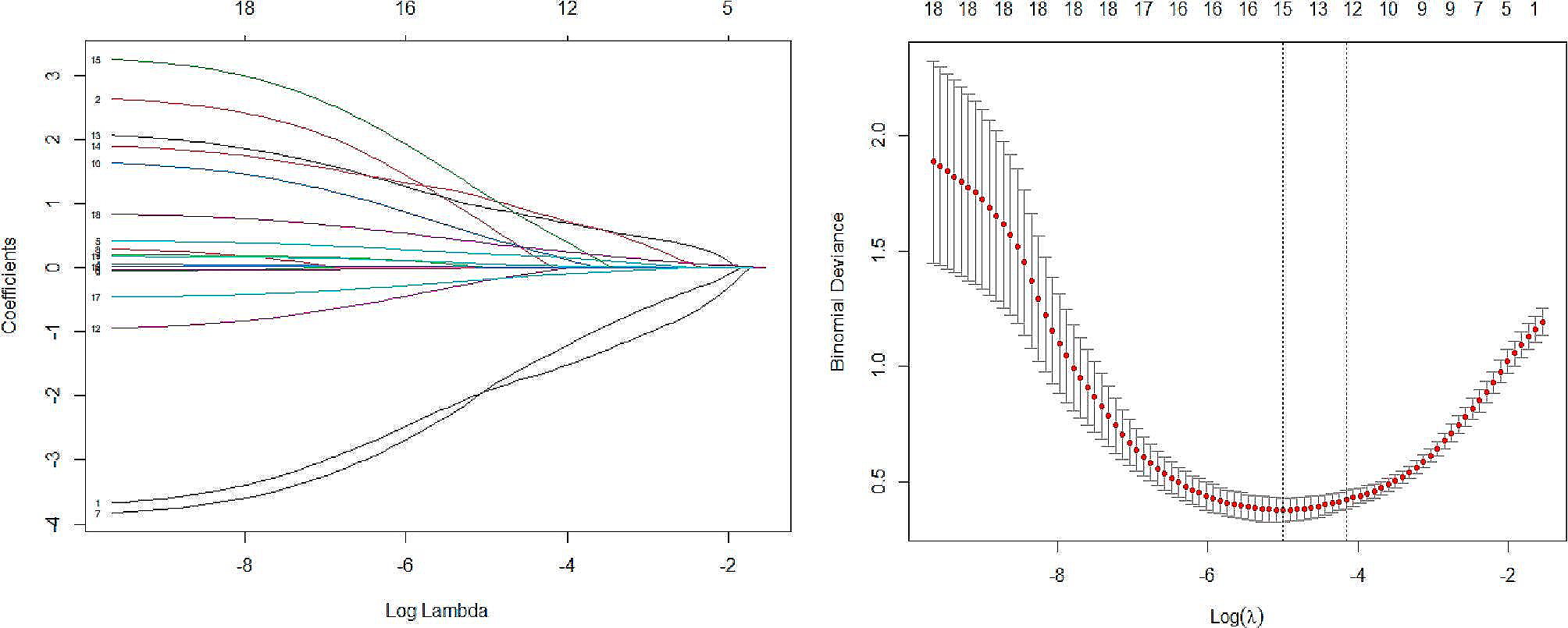
GraphPad Prism8.0 software was used to analyze the data. The measurement data were compared by independent sample t-test. The comparison of count data rates was tested and analyzed, and the IHC score was used as an ordered variable by the Mann-Whitney test. Using R® (version 4.1.1) to establish and verify the prediction model, using LASSO regression analysis to screen out the variables, based on the Logistics regression analysis to establish the prediction model, using nomogram function to draw the nomogram. Calculate the consistency index (C-index) to evaluate the model’s predictive value. Using 1000 times of enhanced Bootsrap to draw the calibration curve. Draw the Receiver operating curve(ROC) to calculate the model cut-off value and sensitivity specificity and the decision curve to analyze and evaluate the model performance. In the external verification process of the test set, draw ROC to calculate AUC, calculate the prediction model concordance rate, and evaluate the model’s accuracy by the kappa test.


留言 (0)