As endoparasitoids belonging to the family Braconidae oviposit in the host body cavity, they face the challenge of evading the host's immune system. In order to accomplish this, Braconidae parasitoids are known to inject venom (V) and polydnaviruses (PDV) into the host during oviposition, effectively suppressing the host's immune response (Teramoto and Tanaka, 2004). There are two types of serosal cells (Scs) in Braconidae parasitoid eggs (Beckage and de Buron, 1994, Dahlman and Vinson, 1993). Scs that develop in the head and tail of the embryo differentiate into teratocytes (Tcs) that are released into the host body cavity after hatching where they are then able to regulate the host's immunity and other physiological functions (Kato et al., 2016, Tanaka and Wago, 1990). Regarding host immunosuppression, Cotesia kariyai (Ck) Tcs suppresses the phenoloxidase activity involved in melanization of the host, Mythimna separata (host) larva. In addition, Tcs of Cotesia plutellae secrete Serpin and Rho GTPase-activating proteins to inhibit nodule formation in the host Plutella xylostella (Ali et al., 2015, Andrew et al., 2006).
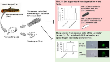
On the other hand, Scs surrounding the surface of the embryo are reported to be broken down after hatching (Dahlman and Vinson, 1993), but in some Braconidae parasitoids, these Scs continue to partially or completely surround the epidermis of the 1st instar larvae of the parasitoid after hatching (Grimaldi et al., 2006, Kitano, 1969, Lawrence, 1990, Pennacchio et al., 1994). The Scs of the 1st instar larvae are less well known than the Tcs, and have been reported in several papers to be involved in immunosuppression of the host (Grimaldi et al., 2006, Kitano, 1969). For example, proteoglycans, the main component of insect basement membranes, are present on the surface and outside of the Scs covering the epidermis of 1st instar larvae of Toxoneuron nigriceps, and if this component is similar to that found in the host basement membrane, it would be expected to be potentially protective against the encapsulation (Grimaldi et al., 2006). Furthermore, Kitano (1969) reported that when the Scs covering the epidermis of 1st instar larvae of Cotesia glomerata were physically destroyed and transplanted into Pieris rapae, the first instar larvae with the Scs disrupted were more encapsulated than those without the Scs disrupted.But little has been done on their physiological analysis.
Ck used in this study is a parasitoid belonging to the Braconidae that parasitizes host larva. Ck V and PDV are known to reduce the host's hemocytes and inhibit host encapsulation (Sawa et al., 2021, Teramoto and Tanaka, 2004), and the Tcs released during hatching are also involved in inhibiting host encapsulation (Hotta et al., 2001, Tanaka and Wago, 1990). In this study, we investigated the characteristics and functions of Scs surrounding Ck 1st instar larvae (1st Scs), and identify a novel mechanism of immunosuppression used to evade encapsulation by the host immune response.


留言 (0)