
記住我


Human thyroid cancer cell lines 8505 C and SW1736 harboring the BRAFV600E mutation were purchased from the European Collection of Cell Culture (Salisbury, United Kingdom) and Cell Lines Service GmbH (Eppelheim, Germany), respectively. BRAFWild Type (WT) cell line C643 was purchased from Cell Lines Service GmbH (Eppelheim, Germany) and THJ-16T derived from a patient with ATC was a kind gift from Dr. John A. Copland (Mayo Clinic, Jacksonville, FL). All cell lines were maintained in Dulbecco’s Eagle Medium (DMEM), supplemented with 10% fetal bovine serum (FBS), penicillin (10,000 U/mL), streptomycin (10,000 U/mL), and fungizone (250 ng/mL), in a standard humidified incubator at 37 °C in 5% CO2. All cell lines were authenticated by short tandem repeat profiling. Cell lines were tested for Mycoplasma from Idexx BioAnalytics (Columbia, MO, USA) and were negative for any contamination. S-8505 C = spheroid generated from 8505 C ATC cell line. S-C643 = spheroid generated from C643 ATC cell line.
Human specimensThyroid tissues were obtained from a patient who underwent surgery at Stanford Hospital. Information about the patient regarding sex, age, tumor size, and clinical stage was recorded. TNM staging was performed based on the eighth edition of the American Joint Committee on Cancer staging system. Approval for this study was obtained from the institution review board at Stanford University (approval number 50,782). The patient’s tumor tissue was procured after written informed consent had been obtained. The diagnosis was confirmed on hematoxylin-eosin (H&E)-stained slides by thyroid cancer surgical pathologists. Figure 1 summarizes the processing of the patient’s tumor samples to generate the 3D culture model.
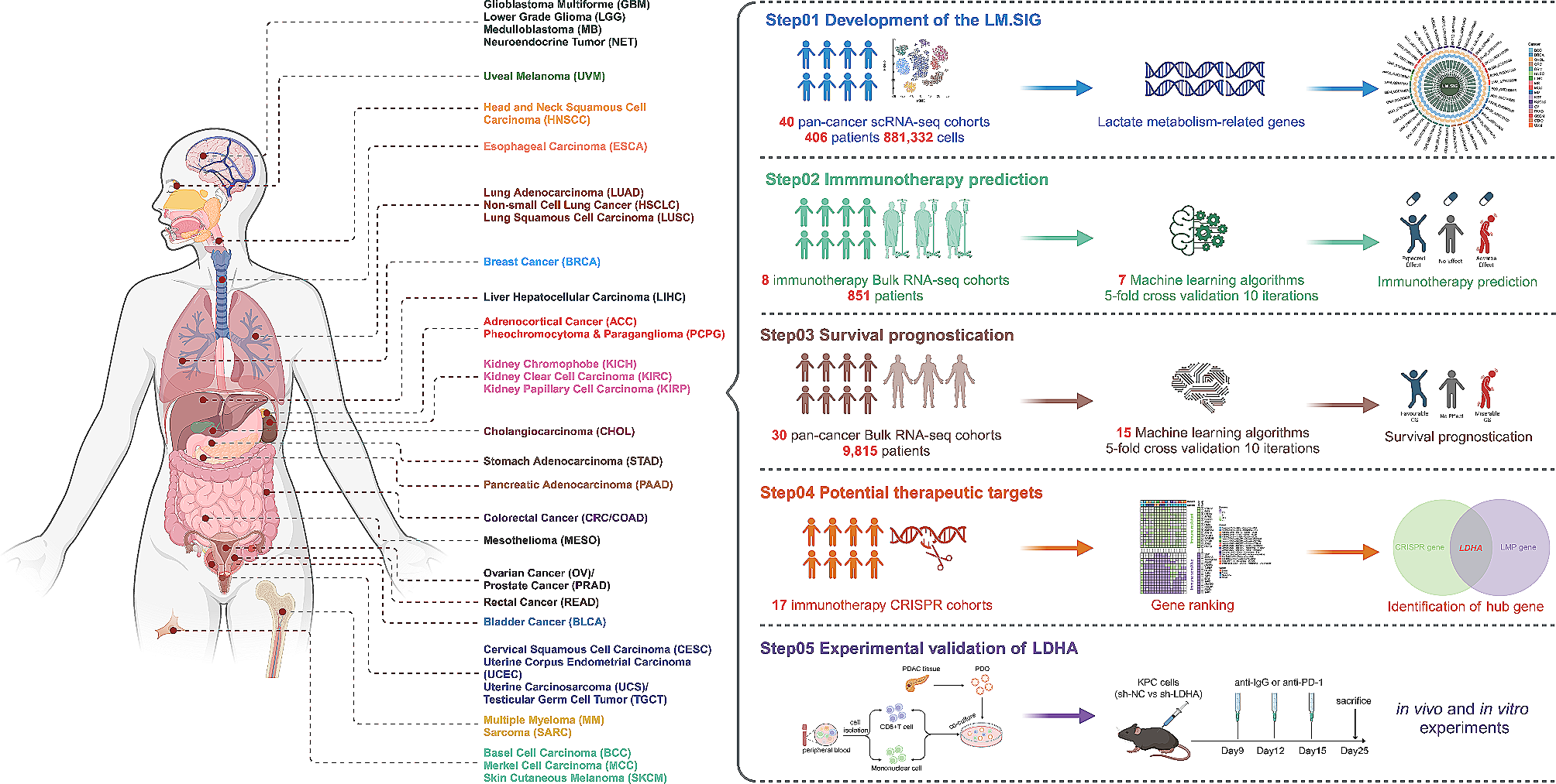
Fig. 1
Flow diagram of establishment and characterization of ATC organoids. Generation of ATC organoid lines from patients undergoing surgery using optimized medium components, as well as characterization, RNA-sequencing, and cell proliferation assay on ATC organoids (Figure Made in BioRender.com)
Tissue processingFresh tissue samples were immediately placed on ice, transported to the laboratory, and then washed and split into several smaller pieces. Two or three serial pieces of samples were snap-frozen and stored at -80 °C for DNA isolation, two pieces were fixed in formalin for histopathological analysis and immunohistochemistry, and the remaining tissues were dissociated and processed for spheroid derivation. Tumor tissue was finely sliced into 1–3 mm pieces with scissors. The minced tissues were digested with collagenase type II (5 mg/mL, Gibco, No. 17101-015) in the presence of the rho-associated protein kinase (ROCK) inhibitor Y-27,632 dihydrochloride (10 µM, AbMole Bioscience, No. M1817) for 20 min in a Thermo mixer at 37 °C with gentle shaking. Dissociated tissues were centrifuged at 350 g for five minutes, washed once with DMEM/F12 medium containing 15 mM HEPES and L-Glutamine (Gibco, No. 11330-032), 1% GlutaMAX (Gibco, No. 25030-081), and 1% antibiotic-Pen/strep (Gibco, No. 15140-122), and centrifuged again. The digested tissue suspension was resuspended with 5 mL of DMEM/F12 medium and filtered through a 70 μm cell strainer to remove large undigested fragments. The cell suspension was collected for primary culture. The patient sample was named ATC01. S-ATC01 = spheroid generated from ATC01 tumor sample.
Primary cell culture and viable cell selectionHarvested cell suspensions were placed into Matrigel-precoated plates for culturing. During the first three days, cells were cultured with Medium A (containing DMEM/F12 medium, 15% FBS, 1% GlutaMAX, 1% antibiotic-Pen/strep, and 5 µM ROCK inhibitor Y-27,632). After three days, the culture medium was removed and a new Medium B was added (containing DMEM/F12 medium, 10% FBS, and 1% GlutaMAX). For the first-round passage, centrifugation was not used due to the low number of cells. Instead, the cells were split into two wells by trypsinization, and the trypsin was neutralized with 20% FBS. Regular culture Medium B was applied once the cells had attached to the wells and continued in culture.
Spheroid generation using matrigel dropsThe cell pellets were resuspended in ice-cold Matrigel (Corning, No. 354,230), and five to six droplets of 50 µL Matrigel-cell suspension were placed onto a preheated Petri dish, which was then placed in an incubator at 37 °C, with 5% CO2, for 10 min to allow the Matrigel to solidify. The Matrigel droplets were then covered with warm complete Medium B and subsequently transferred into a low-attachment six-well plate (Thermo Scientific, No. 174,932) and cultured on an orbital shaker (120 rpm) at 37 ℃ incubator (5.0% CO2). The Medium B culture was changed every three days.
Spheroid generation using AggreWell and matrigel embeddingATC cells cultured in the flasks were dissociated into single cells and resuspended at a concentration of 2 × 10⁶ cells in 1 mL of Medium A per well in a 24-well AggreWell plate (STEMCELL Technologies, No. 34,415). Tumor aggregates formed at the bottom of the AggreWell after centrifugation (350 g, 5 min), and the plate was cultured in the incubator (37 °C, 5% CO2) overnight. On day two, an additional 500 µl of Medium A was added to provide a better proliferative environment. On day three, all aggregates from the AggreWell plates were collected and transferred to low-attachment six-well plates in Medium B (2 ml/well) and placed on an orbital shaker to provide a floating environment. On day 14 of culturing, tumor spheroids were individually embedded into the center of the Matrigel coat and transferred back to a low-attachment six-well plate for further culturing. Afterwards, Matrigel-coated ATC tumor spheroids continued to be cultured in Medium B. The medium was refreshed every three days.
Tumor spheroid passage and recoveryFor tumor spheroid passages, the spheroids were resuspended in 5 mL of 0.25% Trypsin-EDTA (Gibco, No. 25200-056) with incubation at 37 °C for approximately five minutes. Then they were manually shaken vigorously before the addition of DMEM/F12 (containing 10% FBS) and centrifuged at 350 g for five minutes. To maximize the development of tumor spheroids from dissociated cells, 5 µM of Y-27,632 was added during the first week of culture. Tumor spheroids were passed at a 1:2 dilution when necessary. To prepare frozen stocks, spheroids were dissociated and mixed with Recovery Cell Culture Freezing Medium (Gibco, No. 12648-010) and frozen following standard procedures [17]. When required, the tumor spheroids were thawed using standard thawing procedures and cultured as described earlier. The medium was refreshed every three days.
Histology and immunostainingTumor tissues and spheroids were immersed in 10% neutral-buffered formalin for more than 24 h, embedded in paraffin, and serially sectioned at a thickness of 5 μm. The slide sections were then deparaffinized and stained with H&E and immunohistochemical markers. For immunostaining, slides were treated with a 3% hydrogen peroxide buffer for 15 min to eliminate endogenous peroxidase activity. Then, the slides were boiled for 30 min in EDTA solution (pH 8.0) for antigen retrieval and blocked in a 5% bovine serum albumin blocking buffer for 20 min at room temperature to minimize nonspecific staining. Primary antibodies against Ki-67 (1:500, Cell Signaling Technology, No. 9449), cytokeratin 19 (CK19, 1:200, Maixin Biotech, No. Kit-0030), and thyroid transcription factor 1 (TTF-1, 1:100, Cell Signaling Technology, No. 12,373) were applied to the sections and incubated at 4 °C overnight. After washing with phosphate-buffered saline (PBS), the slides were incubated with a secondary antibody at room temperature for an hour. The secondary antibodies used in the assay are anti-rabbit IgG (Invitrogen, No. 31,460) and anti-mouse IgG (Invitrogen, No. 31,430). Then, the slides were developed using 3,3-diaminobenzidine (DAB) for a duration of 30 to 60 s, and counterstained with hematoxylin, mounted, and digitally photographed using a Keyence BZ-X710 microscope (Itasca, Illinois, USA).
RNA sequencingRNA was isolated from the cells using a RNeasy mini kit (Qiagen, No. 74,104). A Rapid Read RNA-Seq assay was used to prepare a poly-A-enriched mRNA library from the purified RNA on the Illumina HiSeq4000 platform. FastQC (version 0.11.7) from the Babraham Institute was used to perform quality control checks on raw sequence data (reads in fastq format). The December 2013 assembly of the human reference genome (GRCh38/hg38) was obtained from the UCSC Genome Browser download site. The corresponding reference annotations (GTF) were obtained from the iGenomes site hosted by Illumina. The splice-aware aligner STAR (version 2.7.0e) was used to align reads to the human hg38 reference genome, and SAMtools (version 1.9) was used to index the aligned and sorted BAM files. Cuffdiff (Cufflinks version 2.2.1) was used for gene expression quantification (in FPKM) and for the detection of differentially expressed genes based on the BAM alignments from STAR. MultiQC (version 1.7) was run to aggregate the results from STAR and FastQC analyses across all samples into a single report. The cummeRbund R package (version 2.24.0 with R version 3.5.0) was used for visualization and sample clustering. DESeq2 was used for differential expressed gene (DEG) analysis based on the negative binomial distribution [18]. The resulting P-values were adjusted using Benjamini and Hochberg’s approach to control the false discovery rate. Genes with an adjusted P-value < 0.05 as determined by DESeq2 were assigned as differentially expressed. DEGs were visualized by pheatmap in R. Enrichment analyses of DEGs, using ORA (overrepresentation analysis) and gene set enrichment analysis (GSEA), was conducted using WebGestalt [19] and clusterProfiler [20] and visualized by ggplot2 in R.
Drug panel and CellTiter-Glo® 3D cell viability assayATC patient-derived tumor spheroids were cultured in 24-well AggreWell plates and incubated (37 °C, 5% CO2) for three days before treatment. The medium contained DMEM/F12 and 10% FBS. On treatment day, individual tumor spheroids were transferred into low-attachment 96-well spheroid microplates (CORNING, No. 4520) under microscope visualization, with each well containing 50 µL of medium. The following compounds were applied in triplicate per treatment at 0 ∼ 40 µM dabrafenib (Selleckchem, No. S2807), 0 ∼ 40 µM, trametinib (Selleckchem, No. S2673), and control dimethyl sulfoxide (DMSO). The drugs were diluted in DMSO according to the manufacturer’s instructions to prepare stock solutions, and then stored at -80 °C. All stock solutions were diluted to the desired concentrations with prewarmed culture medium before use.
The plates were placed on an orbital shaker to provide a floating environment and ensure adequate distribution of the drugs. Redosing was performed after three days, and the viability of the spheroids was determined after five days of treatment with the CellTiter-Glo® 3D Cell Viability Assay (Promega, No. G9682) according to the manufacturer’s instructions. Luminescence was measured using a SpectraMax® i3x plate reader (Molecular Devices).
Statistical analysesAll data are expressed as mean ± SEM unless otherwise specified. Statistical analyses were performed using Prism GraphPad 7.0 (GraphPad Software). The significance in differences was determined by using a two-tailed Student’s t-test for means between two groups or by ANOVA with post hoc analysis among three more groups. Significance was set at a P-value of < 0.05.
留言 (0)