
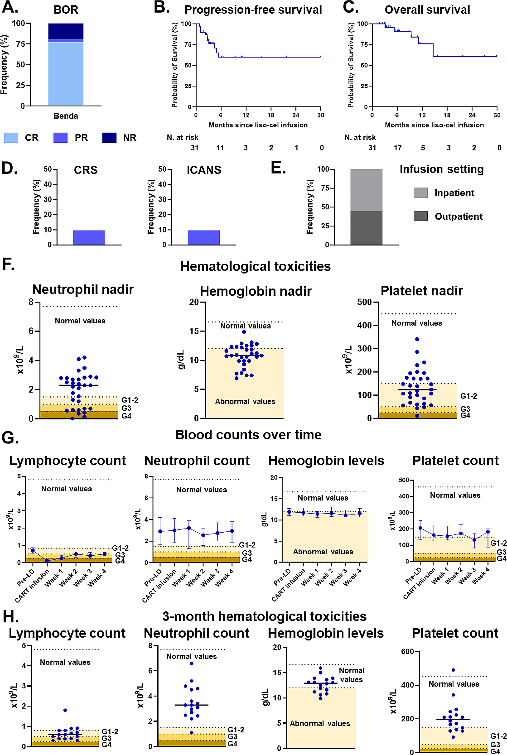
記住我

xCell analysis [7] found significantly higher platelets and CD8+ T cells level in NS and S group, respectively (Fig. 1B, C, Additional file 1: Fig. S1A, B and Table S2). Among all cell types, platelets exhibited the highest negative correlation with CD8 + T cells, and there was a potential direct physical interaction between platelets and CD8+ T cells (Fig. 1D, E and Additional file 1: Fig. S1C). NS group had significantly higher blood platelet count than S group (Additional file 1: Fig. S1D). Higher blood platelet count was related to shorter overall survival (OS) and progression free survival (PFS) in MSK-IMPACT ESCC immunotherapy cohort (log rank test P < 0.05) (Fig. 1F) [8]. Based on these findings, we speculated platelets might cause ESCC immunotherapy resistance by impairing CD8+ T cells function.
To further explore the connection of platelets and CD8+ T cells, we performed comparative analysis and found 298 significantly differential expression proteins (DEPs) between S and NS groups (Additional file 1: Fig. S1E). Pathway enrichment indicated that platelet activation and formation of fibrin clot pathway were enriched in NS group, and the upregulation of antigen processing and presentation, T cell receptor signaling pathway and fatty acid metabolism in S group, as well as molecules involved in these pathways at protein and phosphoprotein level (Additional file 1: Fig. S1F, G). GSEA analysis also showed platelet activation, aggregation pathway was significantly enriched in non-sensitive group (Additional file 1: Fig. S1H). The proteins involved in platelet activation such as F2, FGA, FGB and FGG were significantly upregulated in non-sensitive group, as well as phosphoprotein level (Fig. 1G and Additional file 1: Fig. S1I). Additionally, we also observed the similar expression of FGA, FGB and FGG in IMvigor210 metastatic urothelial carcinoma immunotherapy cohort (Fig. 1H) [9]. All of them showed significantly negative correlation with CD8+ T cells level (Fig. 1I). Among them, FGA expression exhibited significantly negative association with immunotherapy ORR across tumor types based on the TCGA pan-cancer mRNA expression datasets (Fig. 1J, K and Additional file 1: Fig. S1J) [10]. Overall, these results indicated that platelet activation attenuated immunotherapy response via inhibiting the immune effect of CD8+ T cells through a potential physical interaction (Fig. 1L).
We next set out to determine whether the DEPs between S and NS groups could distinguish sensitive patients from non-sensitive patients in response to immunotherapy (Fig. 2A). We randomized discovery cohort into a training set (80%, N = 42) and a testing set (20%, N = 11). Based on the DEPs, we finally screened 10 signatures (including ADD2, FGA, FGG, SPTB, ZC3H7B, LSR, NDUFB7, RNF214, WIPF2 and NCS1) with high accuracy (0.90), sensitivity (92%) and specificity (88%) on training set, and 1, 100% and 100% on testing set (Additional file 1: Supplementary methods). The receiver operating characteristic (ROC) curves showed high predictive power of the model with area under curve (AUC) of 0.93 and 1 on training and testing sets, respectively. Furthermore, the model was also validated in an independent validation cohort (N = 20), including 6 S patients and 14 NS patients. Notably, the model also achieved high accuracy (1), sensitivity (100%) and specificity (100%) with AUC of 1 (Fig. 2B–E and Additional file 1: Fig. S2A–G).
Fig. 2
The construction and validation of predictive model for immunotherapy response. A Diagram describing a construction and validation of the predictive model for sensitive (S) and non-sensitive (NS) groups. B The heatmap displaying the 10 signatures that discriminate S and NS for ESCC immunotherapy in the discovery cohort. C Classification error matrix using logistic regression classifier of 80% training set and 20% testing set in the discovery cohort based on the 10 signatures combination. The number of samples identified is noted in each box. D ROC curves showing the predictive effect of this model in the 80% training set and 20% testing set of the discovery cohort. E Classification error matrix and ROC curve showing high sensitivity and specificity of the 10 signatures in the independent ESCC immunotherapy validation cohort
Overall, the comprehensive proteomic analysis described an atlas of immunotherapy in ESCC. The activation of platelets in ESCC tumor microenvironment could decrease the anti-tumor efficacy of CD8+ T cells through a potential direct physical interaction, causing resistance to immunotherapy. Finally, we screened 10 biomarkers and constructed predictive model for predicting ESCC immunotherapy response, which could distinguish S patients from NS patients and contributed to personalized immunotherapy of ESCC patients.
留言 (0)