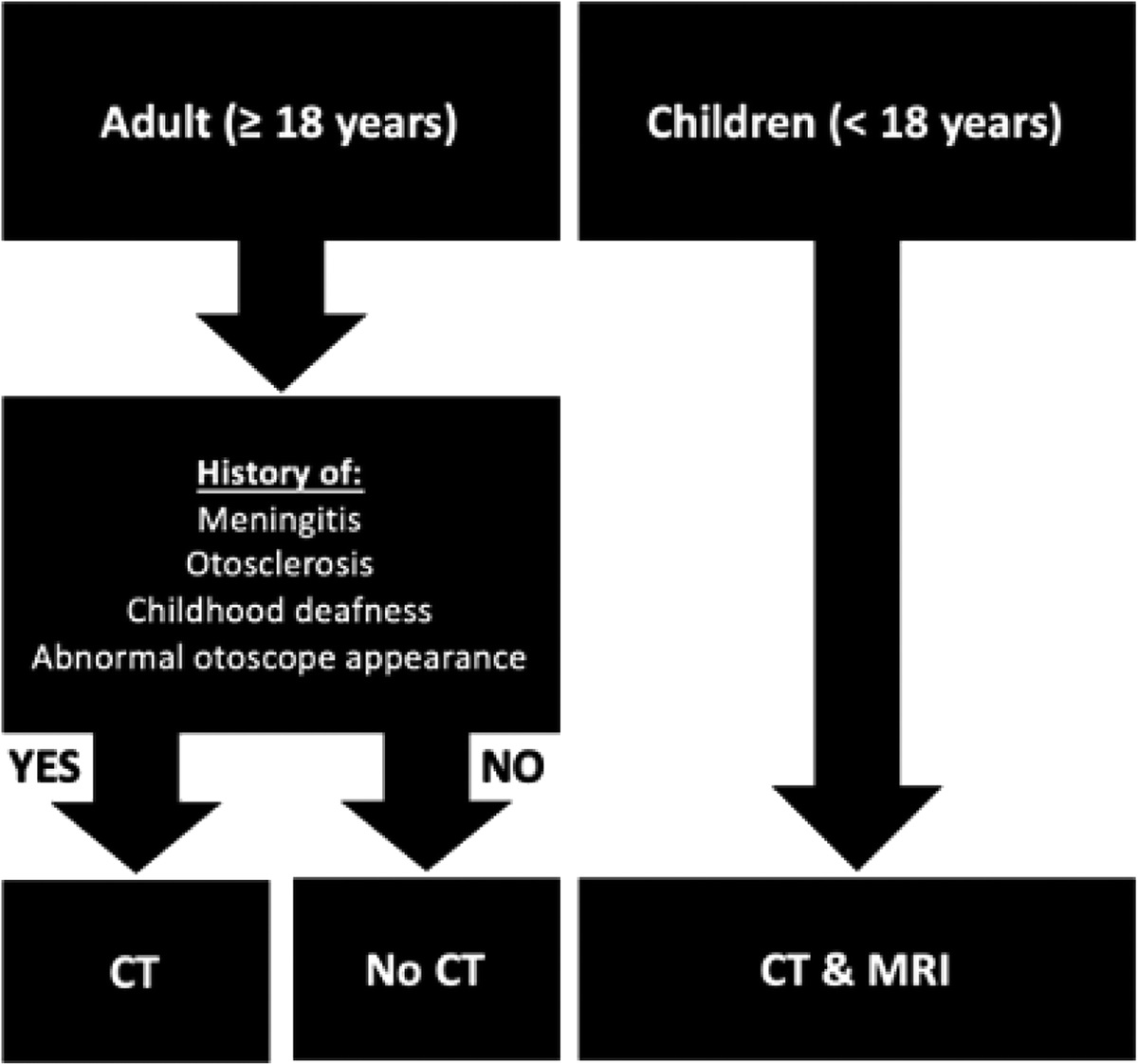
Preoperative Imaging in Cochlear Implants
Objective
To determine the utility of computed tomography (CT) and magnetic resonance imaging (MRI) in cochlear implant candidates.
Study Design
Retrospective case review.
Setting
Tertiary referral hospital.
Patients
A total of 207 cochlear implanted patients with CT and/or MRI
Intervention(s)
N/A.
Main Outcome Measure(s)
Age versus abnormal radiologic findings, imaging abnormality versus postoperative outcomes, postoperative outcomes versus electrode design, Cambridge Cochlear Implant Protocol (CCIP) status for imaging abnormalities, sensitivity and specificity of CT and MRI for round-window/cochlear occlusion, and MRI for incomplete partitions.
Results
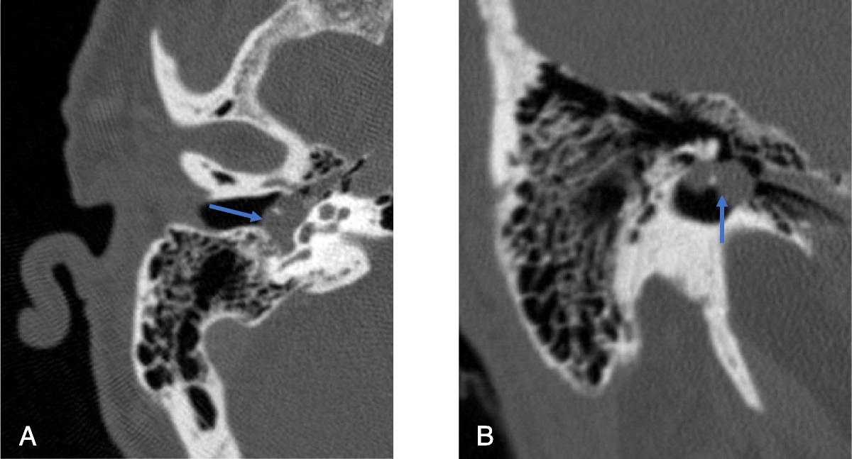
A total of 207 patients with CT, MRI, or both were reviewed retrospectively. Less than half (15.5%) of CT scans had findings that might affect surgical intervention compared with 5.9% of MRI. No significant difference was found between children and adults for relevant imaging abnormalities (grade 4 or higher) with either CT (p = 0.931) or MRI (p = 0.606). CCIP status correlated with cochlear abnormalities (p = 0.040); however, only 46.2% of radiographic abnormalities on CT would be identified by these criteria. For detecting cochlear occlusion requiring surgical intervention, the sensitivity and specificity for CT were 40% (4 of 10; 95% confidence interval [CI], 12.16–73.76) and 95.73% (95% CI, 91.40–98.27), respectively. For MRI, the sensitivity and specificity were 33.33% (1 of 3; 95% CI, 0.84–90.57) and 96.97% (63 of 65; 95% CI, 89.32–99.63), respectively. There was no difference for postoperative AzBio scores for higher-grade imaging abnormalities (p = 0.6012) or for electrode designs (p = 0.3699).
Conclusions
Significant radiographic abnormalities were relatively uncommon in cochlear implant patients on either CT or MRI at our single-center institution. If present, abnormal imaging findings rarely translated to management changes. CCIP status does not reliably predict which patients are likely to have abnormalities. Both MRI and CT have low sensitivity for round-window or cochlear occlusion, but detection likely leads to changes in surgical management.

留言 (0)