Mice
Six female 12-month-old C57/Bl6J mice (JAX stock #:000664) were single housed on a 12 h light/dark at 22–24 °C with ad libitum access to food and water. The present study was carried out in accordance with the European Communities Council Directive of 2010 (2010/63/EU) for care of laboratory animals and approved by a local ethics committee (Bezirksamt Arnsberg) and the animal care committee of North Rhine-Westphalia, Germany. The study was supervised by the Animal Welfare Commission of the Ruhr-University Bochum.
AAV vector production
The AAV2/8-CMV-mCherry was produced using HEK293T cells (Sigma Aldrich 12022001; no recent mycoplasma screening) utilizing the three-plasmid system with polyethylenimine as previously described [11]. We performed a qPCR dilution series with pAAV-CMV-mCherry DNA (100–109 calculated copies) for titer determination (Fig. S1A). The quantitation limit of the reaction was determined at ten DNA copies. The precision was adequate (R2 = 0.9936), resulting in the standard curve equation: y = −3.137x + 37.452. A virus titer of 2.1 × 10⁹ genomes/µl was calculated.
Intracranial injections
A sagittal incision along the cranial midline was performed. The craniotomy was performed 5.9 mm from bregma and 2 mm towards the left hemisphere. The AAV2/8-CMV-mCherry (8.4 × 10⁸ copies) was pressure injected into the cerebellum from 1.8 to 1.4 mm depth in 100 µm steps with 2 min incubations between injections [11].
Sample collection
The sample collection was performed as described by the workgroup of Le Guiner under sterile conditions [12]. Mice were individually placed in sterilized cages without bedding and one fresh feces pellet, a saliva swab, and 120 µl of urine were collected per mouse and resuspended in growth medium followed by vortexing, centrifugation (900 × g, 5 min, RT), filtering (0.22 µm) and storage at −80 °C. The DNA extraction was performed using the QIAamp Viral RNA Mini Kit (Qiagen, Hilden, Germany) according to manufacturer’s protocol. To increase DNA yield the elution step was repeated resulting in 80 µl eluate. One sample was excluded from the analysis due to contamination during the sample collection (saliva: M2, 72 h).
Infection assay
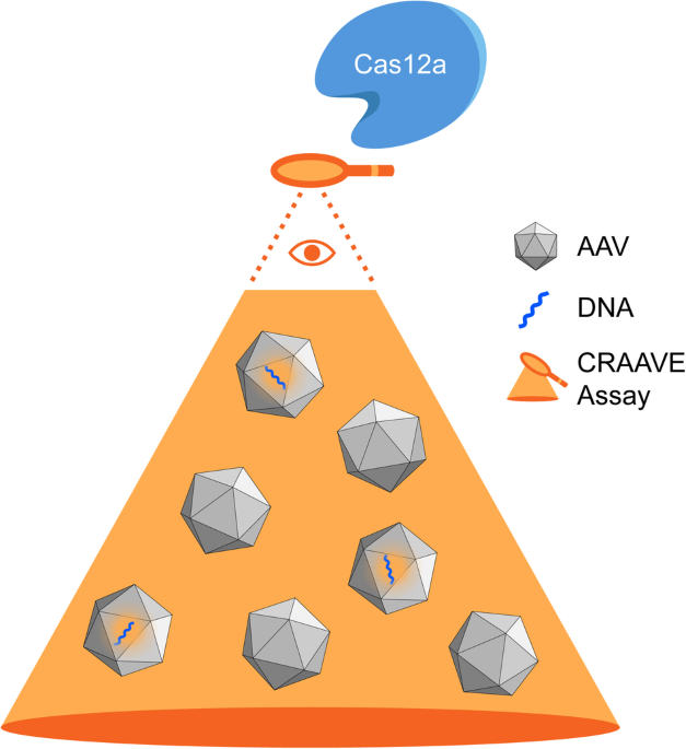
To test the infectivity of shed AAV DNA from collected biological samples, we designed a helper virus-free, BSL-1 compatible version of commonly used infection assays, based on the three-plasmid system [13,14,15,16] allowing AAVs to replicate in HEK293T cells in presence of helper plasmids to increase their abundance prior to qPCR detection. In brief, 7.5 × 10⁴ low passage HEK293T cells were seeded in 24-well plates. After 18 h of incubation, cells were transfected with pRep2/Cap8-plasmids (68 ng/well) and p-helper-plasmids (82 ng/well) using PEI. The collected samples were added 6 h after transfection and incubated for 72 h. The DNA of the supernatant (140 µl) was extracted with the QIAamp Viral RNA Mini Kit and from cells with the DNeasy Blood and Tissue Kit (Qiagen, Hilden, Germany) and quantified with a NanoDropTM2000. The sensitivity of the assay was determined using a dilution series of functional AAV particles ranging from 2 × 10⁵ to 0 as input/well followed by qPCR quantification.
Quantitative real-time PCR
The used primer and probe sequences were validated against the vector expression cassette: 5′-forward-primer: GTCCAAGCTAGGCCCTTTTG, 3′-reverse-primer: GCTTCAAGGTGCACATGGA and Taqman-Probe: 5′-FAM-CGAGGAGGATAACATGGCCA-TAMRA-3′. All reactions were set up with a volume of 20 µl: 1xGo Taq Probe qPCR Mastermix (Promega, Madison, Wisconsin, USA) 0.9 µM per primer, 2 µM probe, 5 µL template (supernatant samples) or 100 ng DNA (cellular samples). The qPCR reactions consisted of denaturation at 95 °C for 2 min and 40 cycles of denaturing for 15 s at 95 °C combined with annealing and extension for 60 s at 60 °C. The cycle threshold values were generated with the Rotor GeneQ series Software 2.3.5 (Qiagen, Hilden, Germany). A standard curve was generated from a dilution series (108–0.1 copies) and an efficiency between 0.95–1.1 and vector copies/reaction using the formula: 10^((CT−37.452)/−3.137) were calculated. Every qPCR sample set included non-template controls and samples with known AAV copy numbers.
Biodistribution analysis of the liver was performed with the BRYT green® Dye (Promega, Madison, Wisconsin, USA) and normalized with amplicons of genomic DNA (Chr. 10, product size: 270 bp) with the primers 5′-TTGTTATGTGGGTCCTGCGG-3′ and 3′-GTAGAAGCCCTCAGTCCTCG-5′. Specificity of obtained signals was determined with gel electrophoresis. One sample displaying unspecific qPCR signal was removed from the analysis (saliva 72 h).
Nested polymerase chain reaction
A nested polymerase chain reaction (PCR) was performed to detect minuscule amounts of AAV DNA using the GoTaq Hot Start Mastermix Green (Promega, Madison, Wisconsin, USA). 0.5 µM per primer and 100 ng of DNA from the biological samples were applied. The first PCR used the primer 5′-TCACTAGGGGTTCCTGCGG-3′ located in the inverted terminal repeat region of the AAV genome. The Robocycler96s program consisted of initial denaturation at 94 °C for 3 min and 40 cycles of denaturing for 30 s at 94 °C, annealing for 30 s at 55 °C and extension at 72 °C for 3 min. A final extension step was performed at 72 °C for 3 min. Subsequently, the samples were diluted 1:20 and subjected to additional 35 cycles of PCR with the same parameters using 0.5 µM of the primers 5′-ATTACGGGGTCATTAGTTCA-3′ and 3′-GCACGTGGTTACCTACAAA-5′ located at the edges of the mCherry insert before visualization by gel-electrophoresis. Two samples (feces 24 h, saliva 24 h) were used up before the final analysis and are therefore not included in the reported data set reducing the sample size for those data points from six to five.
Statistical analysis
The statistical analyses were performed with SigmaPlot (14.0) and GPower (3.1.9.4). A sample size of six was calculated a priori assuming a moderate effect size of 0.4 leading to a power prediction of 0.96. The error bars display mean ± SEM. Statistical significance is reported as follows: *p < 0.05; **p < 0.01; ***p ≤ 0.001. The generated data were tested for normal distribution and equal variance to apply the appropriate statistical tests. Animals were not randomized since only one experimental group was present, but the samples for the subsequent molecular analysis were randomized by encoded labeling.


留言 (0)