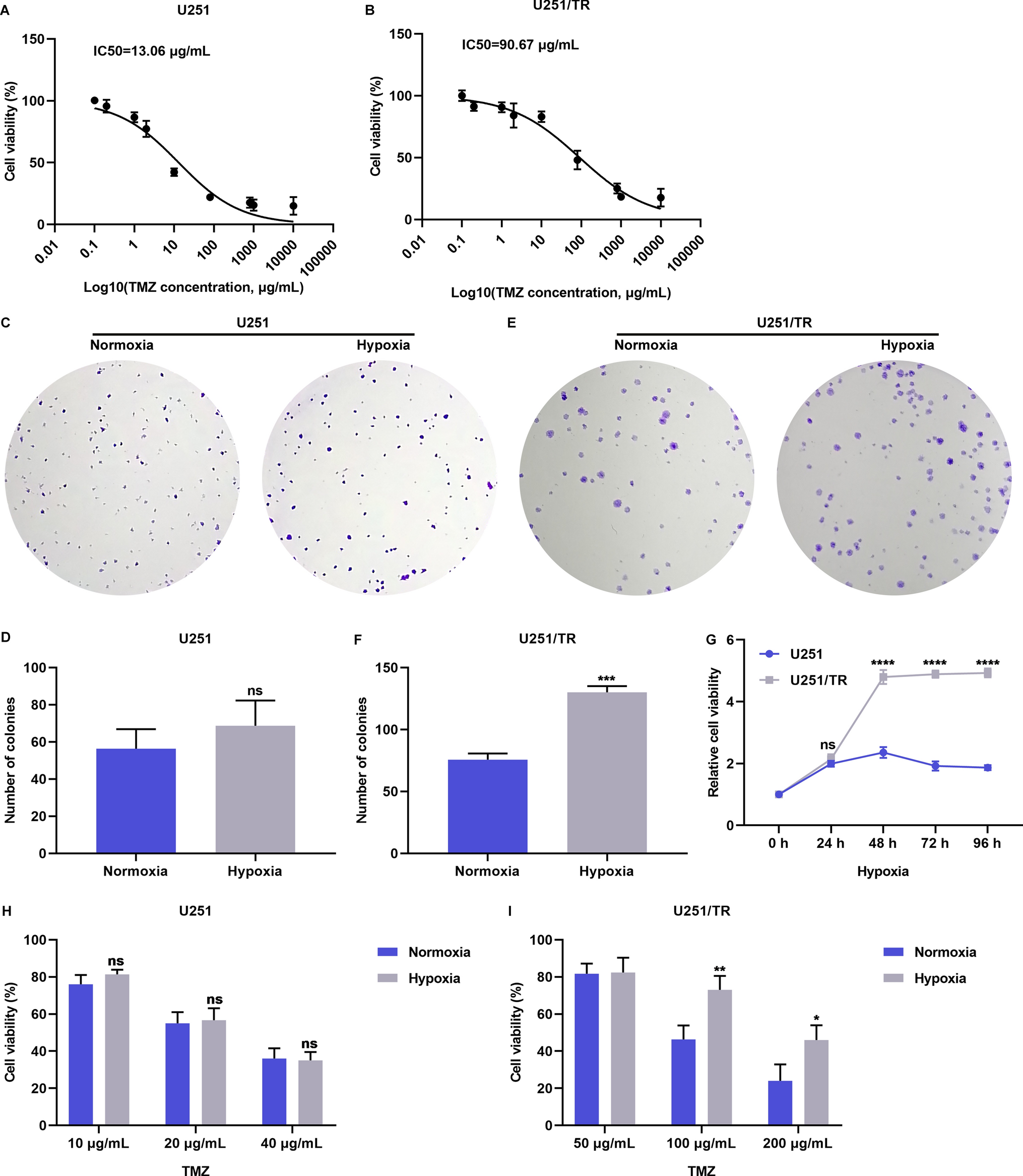
2.1 Patients
After the approval of the institutional review board (no. 2022GR0196), we retrospectively reviewed the medical records of 83 patients, who received systemic therapy followed by consolidative local radiotherapy at Korea University Guro Hospital between March 2017 and June 2022. The inclusion criteria were as follows: (1) having histologically confirmed NSCLC; (2) having stage IV disease with one to three metastatic sites; (3) showing partial response or stable disease or oligo-progressive disease on tumor response evaluation after systemic treatment; (4) having an Eastern Cooperative Oncology Group performance status of 0–2; and (5) having physiologic suitability to undergo radiotherapy. Therapeutic decisions were made on an individual patient basis at a multidisciplinary lung cancer conference. In the current study, we defined localized metastatic disease as the presence of one to three metastatic sites at the time of diagnosis [10,11,12,13]. And patients who showed favorable tumor response after systemic treatment, including oligo-progressive disease at the thoracic site which was amenable to curative high-dose local radiotherapy, were included.
2.2 Treatment scheme
Consolidative radiotherapy was administered to up to three sites that remained active on the positron emission tomography-computed tomography (PET-CT) data. The planned total dose and fraction size depended on the location of lesions. With respect to the target delineation of lung lesion, the gross tumor volume (GTV) was delineated under the lung window setting. The internal target volume (ITV) was delineated following four-dimensional CT with special regard to the patient’s respiratory motion. The clinical target volume (CTV) was generated with a 5-mm expansion of the GTV-ITV in all directions and then modified according to the adjacent normal anatomic structures. The planning target volume (PTV) was generated with a 5-mm expansion of the CTV. Regarding treatment planning, a hypofractionated regimen was administered, whenever possible, to patients with NSCLC. Stereotactic ablative radiation therapy (SABR) with a total dose of 60 Gy in four fractions was administered to patients with NSCLC of small-sized (≤ 4 cm) and peripherally located tumors. For patients who received intensity-modulated radiation therapy (IMRT), different dose-fractionation schedules were planned to deliver 60 Gy in 20 fractions over 4 weeks or 50–70 Gy in 5–10 fractions over 1–2 weeks, respectively. When the shortest distance between the CTV margin and the esophagus was ≥ 1.5 cm, 5–10 fractions were preferred to 20 fractions.
With respect to the target delineation of bone lesion, the GTV were delineated under the bone window setting. The CTV were generated with 5 mm expansion of the GTV in all directions, which were then modified according to the adjacent anatomic structures. The simultaneous integrated boost (SIB) technique was applied to deliver different dose levels to GTV and CTV within a single treatment fraction. SABR was conducted for spine lesions with total dose of 32/24 Gy in four fractions. In cases of IMRT, two different dose-fractionation schedules were used for the delivery of 35/30 Gy in 5 fractions or 30/25 Gy in 5 fractions, respectively, depending on whether the lesion contains joint areas. In patients with other lesions, IMRT was administered with a total dose of 35–45 Gy in 5–15 fractions, depending on the distance between the CTV margin and normal organ, with reference to the PET-CT data.
The prescription guideline was to deliver at least 97% of the prescribed dose to 95% of the PTV. The minimum and maximum doses to 1 cc of PTV were 95% and 107%, respectively. The percentage lung volume that received ≥ 20 Gy was to be kept ≤ 35%, and the mean lung dose was ≤ 20 Gy. The maximum doses to the spinal cord and esophagus were not to exceed 45 Gy and 60 Gy, respectively, satisfying the dose-volume constraints of normal organ.
2.3 Surveillance
We assessed tumor responses using contrast-enhanced chest/abdomen/pelvis computed tomography (CT) scans for every two cycles of systemic therapy and at completion of therapy to assess disease progression during follow-up. The revised Response Evaluation Criteria in Solid Tumors guidelines (version 1.1) was used to evaluate tumor responses. Treatment-related complications were evaluated using the Common Terminology Criteria for Adverse Events (version 4.03).
2.4 Statistical analysis
PFS was defined as the time from the initiation of systemic therapy to the date of the first documentation of disease progression or the latest documented follow-up visit after receiving consolidative radiotherapy. Overall survival (OS) was defined as the time from the initiation of systemic therapy to the date of death from any cause or the latest documented follow-up visit. The 2-year PFS and OS rates were calculated using the Kaplan–Meier method and compared using the log-rank test. Statistical significance was set at p < 0.05. Statistical analyses were performed using IBM SPSS Statistics for Windows (version 24.0; IBM Corporation, Armonk, NY, USA).


留言 (0)