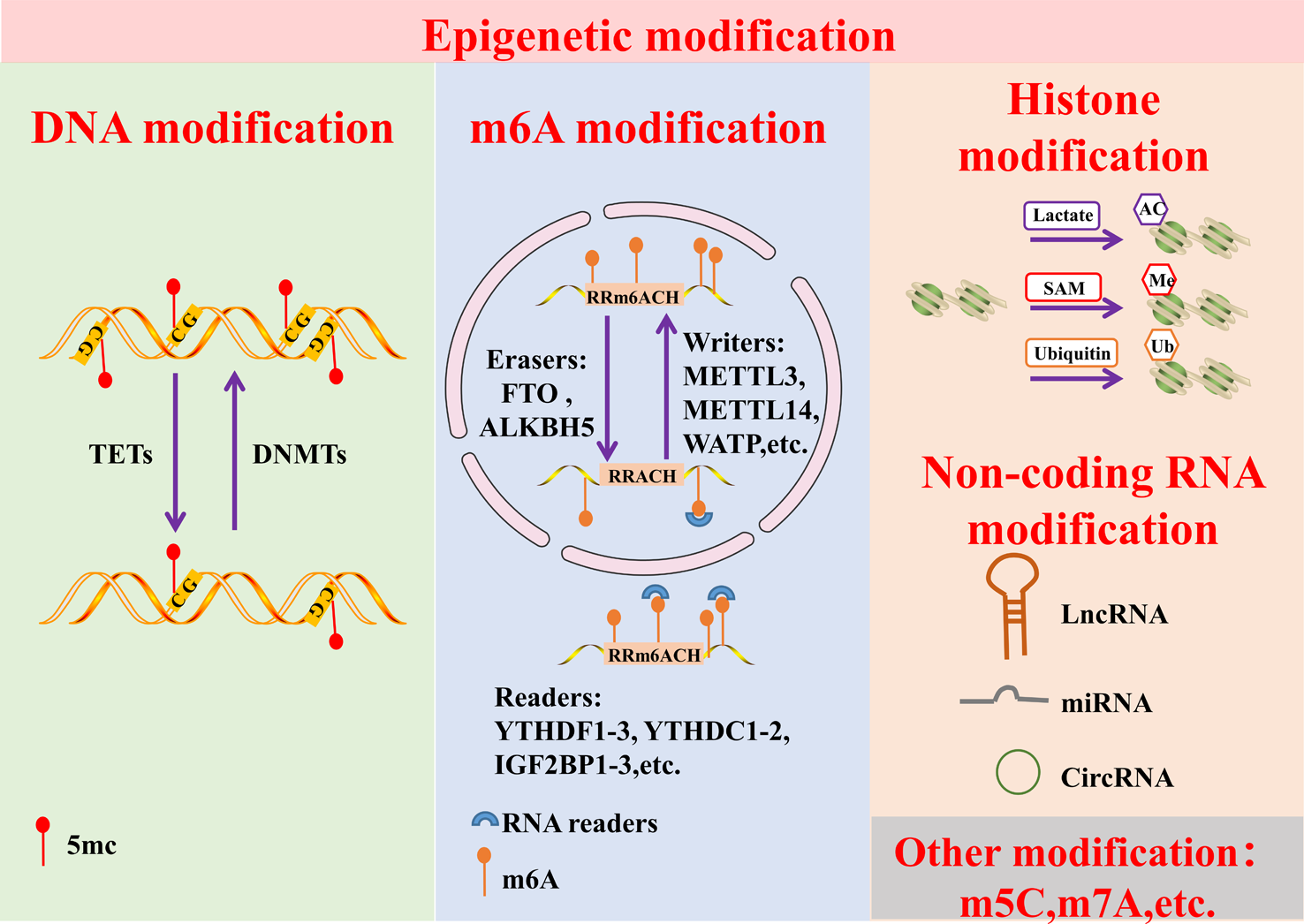
CD70 RNA-seq analysis
Pan-cancer bulk RNA-seq data from 28 different cancer types from the PCAWG Firehose cohort included in The Cancer Genome Atlas (TCGA) were downloaded from the cBioPortal for Cancer Genomics platform (https://www.cbioportal.org) using the package “cgdsr” in R (R Core Team) [25]. RNA-seq by Expectation–Maximization (RSEM) values were converted into integers, and genes with counts greater than 10 in at least 10% of the tumor samples per cancer type were filtered in for further analysis. Resulting expression profiles were scaled by the number of counts per million reads, log2-transformed, and quantile normalized. Single sample gene set enrichment analysis (ssGSEA–BioC-package “GSVA”) was performed on colon adenocarcinoma (COAD), rectal adenocarcinoma (READ) and pancreatic adenocarcinoma (PAAD) data sets for two published gene sets related to general CAFs and CAFs associated with resistance to immune checkpoint inhibition (ICB; [26]). Associations between CD70 expression and CAF (general CAFs and CAFs associated with ICB) abundance scores were tested using Spearman’s correlation coefficients or linear regression models using CD70 as the dependent variable. Comparison of different linear models was done using log ratio tests. In each case, p-values inferior to 0.05 were considered significant.
Patient and healthy donor samples
Formalin-fixed paraffin embedded (FFPE) tissue blocks of 23 PDAC patients and nine historical PDAC FFPE sections previously involved in a clinical study [1] were provided by the Antwerp Biobank (Antwerp, Belgium; ID: BE71030031000) and approved by the Ethics Committee of the Antwerp University Hospital-University of Antwerp (UZA-UAntwerp) under study reference EC14/47/480 and EC13/47/469, respectively. Generation and usage of patient-derived PDAC organoids was approved by patients via written informed consent and by the UZA-UAntwerp Ethics Committee under study reference EC14/47/480. Buffy coats from four healthy donors were purchased from the Blood Transfusion Center of the Red Cross-Flanders (Mechelen, Belgium) and usage was approved by the UZA-UAntwerp Ethics Committee under study reference EC5488.
Immunohistochemistry
CD70 expression in the TME of PDAC patients was evaluated on 23 tumor resections by manual staining for hematoxylin and eosin (H&E), alpha-smooth muscle actin (α-SMA), and CD70 on consecutive sections from FFPE tissue blocks. Five µm-thick sections were baked for 2 h at 60°C and exposed to heat-induced epitope retrieval by incubation in Envision FLEX+ antigen retrieval solution (DAKO; 20 min) at 97°C (PT-Link instrument, DAKO). Subsequently, endogenous peroxidase activity was quenched by incubating the slides in peroxidase blocking buffer (DAKO; 5 min). Immunohistochemical staining of CD70 using an anti-CD70 polyclonal antibody (#PA5-102557, 1:750, Thermo Fisher) was performed with the protocol previously described [27]. After blocking with normal goat serum (Jackson Immuno Research; 30 min), the anti-CD70 antibody was incubated at room temperature (35 min), followed by rabbit enhanced polymer-based linker incubation (DAKO; 15 min), and visualized using the Envision FLEX+ detection kit (DAKO; 25 min) according to the instructions of the manufacturer. Immunohistochemical staining of α-SMA using an anti-α-SMA monoclonal antibody (mAB; clone 1A4, 1:100, Agilent) was performed in an analogous manner to CD70 without blocking with normal goat serum or addition of a rabbit enhanced polymer-based linker. All sections were counterstained with hematoxylin (Merck; 2 min), dehydrated and mounted with Expert mounting medium (Cellpath). Tonsil and placenta tissues were used as positive controls for CD70 and α-SMA staining, respectively.
Scoring for CD70 positivity was restricted to the stromal compartment and performed by pathologists experienced in immunohistochemical staining patterns of CD70 [1, 19, 28, 29]. Positive staining was assigned when CAFs displayed strong membrane or cytoplasmic intensity and was expressed as a percentage of positive CAFs of the total stromal fibroblasts. To rule out non-specific binding of the CD70 antibody in the stromal compartment, nine historical PDAC tissue sections that were stained with a clinically approved anti-CD70 mAb (clone 301731, 1:40, R&D Systems, not commercially available anymore) were included [1].
Dissected tumors from Raji cell line-bearing NOD-Prkdcscid-IL2rgTm1/Rj (NSG) mice were fixed in 4% formaldehyde, processed, and paraffin embedded. Five µm-thick sections from the FFPE tissue blocks were stained for H&E and CD70 as described above.
Cell lines and culture conditions
The NK-92 and Raji cell lines were purchased from the German Collection of Microorganisms and Cell Cultures, the LIM2099 cell line from Sigma-Aldrich, and the PANC-1 cell line from the American Type Culture Collection. The CT5.3hTERT, RLT-PSC, and hPSC21 CAF cell lines were kindly provided by Prof. O. De Wever (Ghent University, Ghent, Belgium) [30], Prof. R. Jesenofsky (University of Heidelberg, Mannheim, Germany) [31], and Prof. A. Masamune (Tohoku University Graduate School of Medicine, Sendai, Miyagi Prefecture, Japan) [32], respectively.
NK-92 cells were cultured in GlutaMAX alpha Minimum Essential Medium (α-MEM; Life Technologies) supplemented with 12.5% Fetal Bovine Serum (FBS; Life Technologies), 12.5% horse serum (Life Technologies), 2mM L-glutamine (Life Technologies), 1% Penicillin/Streptomycin (P/S; Life Technologies) and 150 U/mL recombinant IL-2 (ImmunoTools), as described before [33]. Raji and LIM2099 cells were cultured in Roswell Park Memorial Institute medium (Life Technologies) supplemented with 10% FBS, 2mM L-glutamine, and 1% P/S. CT5.3hTERT and PANC-1 cells were cultivated in Dulbecco-Modified Eagle Medium (DMEM; Life Technologies) supplemented with 10% FBS, 2mM L-glutamine, and 1% P/S. Lastly, RLT-PSC, and hPSC21 cell lines were cultured in DMEM/F12 (Life Technologies) supplemented with 10% FBS, 2mM L-glutamine, and 1% P/S. Cell cultures were maintained in exponential growth in 5% CO2 in a humidified incubator at 37°C, confirmed Mycoplasma free through regular testing with the Mycoalert Mycoplasma detection kit (Lonza), and their identity validated by short tandem repeat profiling.
Generation of CD70-CAR NK cells
CD70-CAR and CD70-CAR-IL-15 constructs were designed via Creative Biolabs using the CellRapeutics™ CAR Technology platform and inserted into a pST1 vector (kindly provided by Prof. Uğur Şahin, University Medical Center Mainz, Mainz, Germany) behind the T7 promoter site. The pST1-CD70-CAR and pST1-CD70-CAR-IL-15 plasmids were transformed in Escherichia coli (SoloPack Gold Supercompetent Cells, Agilent) using a heat shock of 42°C for 30 s, selected on Luria–Bertani broth agar (Miller; Sigma-Aldrich) plates containing Kanamycin (Sigma-Aldrich), amplified in Luria–Bertani broth medium (Miller; Sigma-Aldrich) containing Kanamycin, purified with the NucleoBond Xtra Midi Plus EF kit (Macherey Nagel), and linearized with the PmeI restriction enzyme (Life Technologies). In vitro transcription of CD70-CAR- and CD70-CAR-IL-15-encoding messenger RNA (mRNA) was performed using the mMESSAGE mMACHINE T7 transcription kit (Life Technologies) following manufacturer’s instructions. NK-92 cells were electroporated as described elsewhere [33]. In short, cells were pulsed using a Gene Pulser Xcell (Bio-Rad) with time constant protocol (300V, 12 ms) in 4 mm cuvette (ImmunoSource), dissolved in 200 μL Opti-MEM (Life Technologies) at a concentration of 25–125 × 106 cells/mL in the presence of 100 μg/mL CD70-CAR- or CD70-CAR-IL-15-encoding mRNA. NK-92 cells electroporated without CAR-encoding mRNA (MOCK) were used as control cells. Electroporated cells were recovered in NK-92 medium without IL-2 for at least 4 h until further use in downstream applications. IL-2 was never supplemented in downstream experiments unless otherwise stated.
Flow cytometric phenotyping
Expression of CD70 was assessed on peripheral blood mononuclear cells (PBMCs) from healthy donors by flow cytometry. PBMCs were isolated by Lymphoprep (Stemcell technologies) density gradient centrifugation from buffy coats of four healthy donors, purchased from the Blood Transfusion Center of the Red Cross-Flanders. Prior to antibody staining, PBMCs were incubated 30 min at 4°C with human serum (Sigma-Aldrich) to prevent aspecific binding to Fc receptors. Subsequently, PBMCs were stained for 30 min at 4°C with a multicolor panel containing CD3-FITC (clone SK7, Biolegend), CD4-BB700 (Clone SK3, Biolegend), CD8-BV570 (Clone RPA-T8, Biolegend), CD56-PE-Cy7 (NCAM16.2, BD Biosciences), CD19-BV421 (Clone HIB19, BD Biosciences), CD14-BV785 (Clone 63D3, Biolegend), and CD70-PE (Clone ki-24, BD Biosciences). Live-Dead Fixable Near-IR (Thermo Fisher) was included to exclude non-viable cells from the analysis. Acquisition was performed on a NovoCyte Quanteon (Agilent Technologies).
Surface expression of the CD70-CAR and CD70-CAR-IL-15 constructs on NK-92 cells was evaluated via flow cytometry by staining with a CD27-PE mAb (Clone O323, Cell Signaling Technology) or corresponding isotype control IgG1κ mAb (Clone MOPC-21, Cell Signaling Technology) for 30 min at 4°C. Expression of CD70 on tumor and CAF cell lines was determined by staining with a CD70-PE mAb or corresponding isotype control IgG3κ mAb (Clone J606, BD Biosciences) for 30 min at 4°C. CD27 and CD70 expression were measured on a CytoFLEX (Beckman Coulter) after 15 min incubation with the fluorescent intercalating viability dye 7-amino-actinomycin D (7-AAD; Biolegend) to exclude non-viable cells.
Expression of immune checkpoint molecules DNAM-1, TACTILE, TIGIT, PD-1, and LAG-3 was determined on MOCK, CD70-CAR, and CD70-CAR-IL-15 NK cells 24 h post-electroporation by pretreating the cells with human serum for 30 min at 4°C, staining the cells with a multicolor antibody panel for 30 min at 4°C, and measuring the samples using a NovoCyte Quanteon. The multicolor antibody panel included DNAM-1-FITC (Clone11A8, Biolegend), CD96-BV421 (Clone NK92.39, Biolegend), TIGIT-PE-Dazzle (Clone A15153G, Biolegend), PD-1-BV650 (Clone EH12.2H7, Biolegend), and LAG3-BV785 (Clone 11C3C65, Biolegend). Live-Dead Fixable Near-IR marker was used to exclude non-viable cells from the analysis.
Analysis of IL-15, IFN-γ and TNF-α secretion
In order to analyze secretion of the IL-15, IFN-γ and TNF-α pro-inflammatory cytokines by CD70-CAR, CD70-CAR-IL-15, or MOCK control NK cells into the supernatant, cells were seeded after electroporation in flat-bottom 96-well plates (Greiner bio-one) at a concentration of 1 × 10⁶ cells/mL. Supernatant was taken 4 h, 24 h, 48 h and 72 h after electroporation and stored at -80°C until downstream analysis. Detection was performed by electrochemiluminescence (Meso Scale Discovery Inc.) using a U-PLEX detection kit (IL-15) or V-PLEX detection kits (IFN-γ and TNF-α) according to the manufacturer’s protocol.
In vitro CD70-CAR NK cell-mediated cytotoxicity of CD70+ tumor and CAF target cell lines
Cytotoxic activity toward CD70+ tumor cell lines (Raji, PANC-1, and LIM2099) was assessed by co-culturing CD70-CAR, CD70-CAR-IL-15 and MOCK NK cells 24 h after electroporation with PKH67 (Sigma-Aldrich)-labeled CD70+ tumor cell lines (Raji, LIM2099, PANC-1) in a 5:1 effector:target ratio in sterile FACS tubes (VWR). After 4 h, co-cultures were stained with 7-AAD and Annexin V-PE (BD Biosciences). Target cell survival was measured on a CytoFLEX flow cytometer and calculated as previously described [34]. To investigate if the observed killing was CAR-mediated, CD70-CAR, CD70-CAR-IL-15, and MOCK NK cells were incubated overnight and during the co-culture with PKH67+ Raji cells with 10 μg/mL or 100 μg/mL anti-CD27 neutralizing mAb (Clone MAB382, R&D Systems) or isotype control IgG1κ mAb (clone MAB002, R&D Systems). Co-culture and target cell survival analysis was performed as described above. To evaluate the efficacy of CD70-CAR NK cell-mediated killing after cytokine stimulation, CD70-CAR NK cells were incubated overnight prior to the co-culture with the effector dose 50 (ED50) of IL-2 (0.25 ng/mL), IL-7 (0.50 ng/mL), IL-12 (0.05 ng/mL), IL-15 (2.60 ng/mL) and IL-18 (9.00 ng/mL) cytokines, purchased from R&D Systems. Subsequently, stimulated NK cells were co-cultured with PKH67+ Raji, LIM2099 and PANC-1 target cells and analyzed as described above. To exclude if the improved cytotoxic capacity was only due to IL-15 stimulation, CD70-CAR NK cells and MOCK control cells were incubated overnight with IL-15 (ED50: 2.60 ng/mL) and co-cultured with PKH67+ LIM2099 cells. Co-culture and target cell survival analysis was performed as described above. Lastly, to investigate the mode of action of IL-15 stimulation, CD70-CAR NK cells were stimulated overnight with the ED50 (2.60 ng/mL) and 20 pg/mL IL-15 (i.e., rounded number of the highest amount of IL-15 measured in the supernatant of CD70-CAR-IL-15 NK cells via electrochemiluminescence), and CD70-CAR expression and cytotoxic capacity were measured 24 h post-electroporation in the same manner as described above.
Longitudinal cytotoxic activity of CD70-CAR, CD70-CAR-IL-15, and MOCK NK cells toward CD70+ CAF cell lines (CT5.3hTERT, RLT-PSC, and hPSC21) was analyzed using the xCELLigence Real-Time Cell Analysis (RTCA; Agilent) that records cell viability and growth by impedance measurements. Performance of the NK cell-mediated killing assay with the xCELLigence RTCA was executed as explained previously [35]. In short, seeding density was optimized for each CAF cell line to ensure continuous growth until the end of the assay. CAF cells were seeded in gold-coated 16-well plates and background impedance of the plates was measured before seeding of the target cells. After a 24 h incubation to allow proper adhesion to the plates, CAF cell lines were treated with CD70-CAR, CD70-CAR-IL-15, or MOCK NK cells in a 1:1 effector:target ratio (based on the amount of CAFs seeded at day 0), or left untreated. The impedance signal was monitored by automated measurements every 15 min starting from cell seeding and ending 48 h after treatment. Measurement of the impedance was expressed as Cell Index (CI) and normalized to 1 after starting the co-culture.
CD70+ Raji xenograft mouse model
In vivo efficacy of CD70-CAR, CD70-CAR-IL-15, and MOCK NK cells was evaluated in a subcutaneous Raji xenograft mouse model. Female NSG mice of six weeks old, were obtained from Janvier Labs and maintained at the Animal Core Facilities of the UAntwerp under specific pathogen free conditions in individually ventilated cages enriched with nesting material. All animal procedures were conducted in accordance with, and approval of, the Animal Ethics Committee of the UAntwerp under registration number 2021–55. Mice were injected subcutaneously with 1.0 × 10⁶ Raji cells suspended in 100 μL sterile PBS (Life Technologies) containing 12 mg/mL Geltrex (Life Technologies) at the left abdominal flank. Tumor growth was measured using a digital caliper and tumor area was assessed by measuring length and width, and calculated using the formula ‘tumor area = length x width’. When tumors reached 50 mm2, mice were randomized based on tumor size into four different treatment groups (day 0): (1) untreated, (2) MOCK NK cells, (3) CD70-CAR NK cells and (4) CD70-CAR-IL-15 NK cells. All mice, except for the untreated group, were treated on day 0 and day 4 intravenously (i.v.) via the tail vein with 1.0 × 10⁷ CD70-CAR, CD70-CAR-IL-15, or MOCK NK cells suspended in 200 μL sterile PBS. Prior to injection, CD70-CAR, CD70-CAR-IL-15, and MOCK NK cells were irradiated 4 h post-electroporation with a sublethal radiation dose of 10 Gy using the X-RAD 320 irradiation device (Precision X-ray Inc.) at a concentration of 4.0 × 10⁶ cells/mL in sterile culture flasks. Dosimetry was performed using Gafchromic EBT3 films, irradiations were performed from the top of the culture flasks and the dose was prescribed at the exit. Mice were sacrificed when tumor size reached 150 mm2.
CD70-CAR NK cell-mediated cytotoxicity of CD70+ microtumors
Longitudinal cytotoxic activity of (CAR) NK cells toward CD70+ CAFs in an advanced in vitro model was evaluated using 3D patient-derived PDAC microtumors containing PDAC tumor organoids with RLT-PSC CAF cells. Patient-derived PDAC organoids (P002, P044 and P87) from our in-house organoid bank, were cultured as previously described [36, 37]. In short, for passaging organoids were digested into single cell suspension using TrypLE (Life Technologies) and plated in 100% cultrex (Bio-Techne Ltd) drops on flat bottom culture plates (Greiner bio-one). The drops were covered with Advanced DMEM/F12 medium (Life Technologies) supplemented with 1% GlutaMAX (Life Technologies), 1% HEPES (Life Technologies), 1% P/S, 4% Noggin-Fc Fusion Protein (ImmunoPrecise Antibodies), 4% R-Spondin-Fc Fusion Protein (ImmunoPrecise Antibodies), 1 × B27 supplement (Life Technologies), 1.25 N-Acetylcysteine (Sigma-Aldrich), 10 mM Nicotinamine (Sigma-Aldrich), 500 nM A83 (Bio-Techne), and 5 nM Wnt Surrogate-Fc Fusion Protein (ImmunoPrecise Antibodies).
For downstream microtumor generation, organoids were mixed with red fluorescent-transduced RLT-PSC cells, seeded in Advanced DMEM/F12 medium containing 3% Cultrex, supplemented with 1% GlutaMAX, 1% HEPES, and 1% P/S in 384-well ultra-low attachment microplates (Corning), and incubated at 37°C for two days to allow assembling of microtumors. On day three, microtumors were treated with CD70-CAR, CD70-CAR-IL-15, or MOCK NK cells in a 1:1 effector:target ratio (based on the amount of RLT-PSC cells seeded on day 0) or left untreated and followed up by the Tecan Spark Cyto live-cell imager every 2 h for 36 h. Brightfield and fluorescent images were analyzed using our in-house developed analysis software Orbits [38] and growth rate of RLT-PSC cells in the microtumors was determined by normalizing the fluorescent red signal (sum red area) to the first measurement (T0) or the control (untreated) at the respective timepoint (12 h, 24 h, 36 h).
Data analysis
FlowJo v10.8.2 Software (BD Life Sciences) and ImageJ v1.53t (U. S. National Institutes of Health) were used for flow cytometry data and microtumor image analysis, respectively. Prism version 9.1.2 (GraphPad) was used for graphical data representation. Statistical computations were performed in JMP Pro v16.0.0 (SAS Institute Inc.). Linear Mixed Models were used to compare means between two or more groups. In the case of comparing the means of more than two groups and the null hypothesis was rejected by the Fixed Effects test, a multiple comparison post hoc analysis was applied. Dunnett’s correction for multiple comparison was used when comparing to a control sample. For all pairwise comparisons, Tukey’s multiple comparison correction was applied. A nonlinear Spearman’s correlation with a simple linear regression was done to analyze the correlation between CD70-CAR and CD70-CAR-IL-15 NK-mediated cytotoxicity and CD70 target expression. Differences in probability of survival between treatments groups in in vivo experiments were analyzed using the Log-Rank (Mantel-Cox) test with the SPSS Statistics v28.0.1.1. software (IBM). To analyze differences in tumor kinetics over time, we used R with the “afex” and “emmeans” [39] packages to perform mixed model ANOVAs. Differences were considered to be significantly different if p-value < 0.05 (*p < 0.05; **p < 0.01; ***p < 0.001; and ****p < 0.0001). Error bars represent mean values ± standard deviation (SD) unless stated otherwise.


留言 (0)