
記住我


Baseline information, including age, gender, main lesion side (left or right), presence of diabetes mellitus, thyroid disease, rotator cuff disease, numeric rating scale (NRS), and shoulder ROM, were retrospectively collected between January 2022 and May 2022 from the selected 141 patients who were diagnosed with AC, obtained significant pain reduction (50% or more reduction compared with pretreatment) after 5 weeks of conservative treatments and could be followed up at 6–8 months after completion of the treatment. The diagnosis of AC was determined by painful and limited ROM of the shoulder joint for at least 1 month [7]. Limited shoulder ROM was described as a 25% or more reduction in ROM in abduction, flexion, and external rotation measured by a goniometer for the diagnosis of AC [2]. This study did not include the data of patients with traumatic shoulder conditions affecting shoulder ROM, inflammatory joint disease, full-thickness rotator cuff tendon tear using ultrasonography, or history of surgery [8]. This study was approved by the Investigational Review Board (IRB) of Wooridul Spine Hospital (2022-03-WSH-002), which waived the requirement for written informed consent owing to the retrospective nature of the study. The Helsinki Declaration was adhered to in this study.
TreatmentThe treatment lasted 5 weeks. Passive ROM exercise, with physical therapies, including heat and electrical therapy, was performed for 30 min twice weekly. Each patient was taught to perform active ROM exercise with ten repetitions thrice daily.2
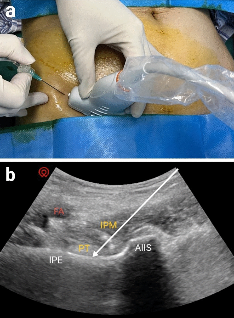
The intraarticular injection was administered while the patient was in supine position, with the arm adducted and internally rotated. First, the skin was anesthetized with lidocaine, and then a 21-gauge spinal needle, 2.5–3 inches long, was directed into the shoulder joint space via an anterior approach under fluoroscopic guidance. Approximately 1 cc of contrast was injected to confirm the proper location of the needle inside the joint, followed by an injection of 40 mg triamcinolone (1 cc) and 0.5% lidocaine (5 cc) [9] (Fig. 1). The repeated intraarticular injections were administered at 1 to 2-week intervals under C-arm fluoroscopy until a conceivable pain reduction was achieved [9, 10].
Fig. 1
Contrast spreading and needle position during glenohumeral intraarticular injection under fluoroscopic guidance
Clinical EvaluationPain score was calculated using NRS, ranging from 0 (no pain) to 10 (worst possible pain) based on the degree of pain experienced in the previous week.
Passive ROM was measured with a goniometer while the patient was sitting upright on a chair. Abduction, flexion, and external rotation angle were measured, with the patients asked to relax as much as possible and the examiner pressing down on the clavicle and scapula using one hand to eliminate scapular movement during ROM measurement. For flexion and abduction angle measurement, the examiner moved the patient’s arm in sagittal and coronal planes from arm adduction posture with the elbow joint extended [10, 11]. The external rotation angle was measured with the shoulder fully adducted, 90° elbow flexion, and neutral position of the forearm. Internal rotation angle could not be measured with a goniometer because most patients could not accomplish a 90° abduction, which is mandatory for an accurate internal rotation angle measurement [12].
NRS and passive ROM were assessed at pretreatment (baseline, T0), at the end of 5 weeks of the treatment sessions (T1), and 6–8 months after the completion of treatment (T2).
Patients were divided into successful (n = 96) and unsuccessful (n = 45) groups according to the degree of pain reduction at the T2 timepoint. Pain reduction of ≥ 50% compared to the initial pain was considered successful. We compared the clinical and demographic data of the two groups at the T0 and T1 to identify the related factor that might affect the significant pain reduction maintenance at the T2 timepoint.
Statistical AnalysisStatistical analysis was performed using the SPSS Version 14.0 statistical package (SPSS Inc., Chicago, IL, USA). The paired t test was used to evaluate the significance of NRS reduction and ROM improvement at the T1 and T2 compared to the T0 in the total population and the successful and unsuccessful pain reduction groups. The proportions of gender, main lesion side, number of injections, and association of diabetes, thyroid disease, and rotator cuff disease were compared between the successful and unsuccessful groups using the Chi-square test. Comparison of age, pain duration, and NRS and shoulder ROM at the T1 and T2 between the successful and unsuccessful groups were conducted with the student t test. p < 0.05 was considered to be statistically significant.
留言 (0)