
記住我


We present a report on the surgical treatment of peripartum pubic symphysis rupture in five patients. In this cohort, four out of five women (75%) underwent revision surgery due to symphyseal construct failure. Postnatally, the symptoms manifested at an average of 6.75 months (ranging from 3 to 14 months).
During the one-year postoperative follow-up of the patient initially treated with external fixation (Case 2), who subsequently underwent plate fixation, it was observed that the screws had become loose, and the symphysis had recurrently widened, leading to the removal of the plate. The remaining three patients experienced failure of plate fixation, necessitating revision surgery at an average of 10.6 weeks (ranging from 6 to 12).
In this series, one patient (Case 3) underwent pubic symphysis fixation using a 4-hole plate, following the recommendation by Rommens et al. [8]. However, she experienced complete fixation failure, necessitating the removal of the initial fixation and subsequent fixation of the pubic symphysis using a 6-hole plate. Notably, Sagi and Papp’s [34] retrospective analysis suggests that the two-hole symphyseal plating technique group had a higher rate of implant failure and a significantly increased rate of pelvic malunion. Based on these findings, they recommend using multi-hole plating for unstable pubic symphyseal disruptions.
Two patients (Cases 4 and 5) underwent revision surgery with two orthogonal anterior plates. Ultimately, the distance between the pubic bones was 5-8 mm, and the Lindahl score assessment was determined to be good or excellent. It is noteworthy that the patient (Case 1), who initially had a 70 mm pubic symphysis diastasis, underwent external fixation 1 month after giving birth. This procedure was successful, although a 12 mm pubic symphysis diastasis remained.
It is essential to highlight that our analysis has revealed a notably higher rate of fixation failure than previously reported by Najibi et al. [9], who described 50% complications in subacute and chronic groups, and van Zwienen et al., who observed 47% complications within their cohort [35].
Literature reviewThe literature review indicates that performing surgical treatment 2 weeks after delivery results in fewer complications compared to cases that are subacute or chronic (P = 0.041). Various factors may contribute to the clinical context. The pubic symphysis allows for small-magnitude movement of pubic bones during everyday activities. As humans ambulate, torsion of the sacrum (nutation) affects the front part of the pelvic ring [36, 37]. Walheim et al. found that the magnitude of anteroposterior sagittal movements when standing on alternate legs is 1.3 mm in nulliparous women and 2.1 mm in multiparous women [38]. Additionally, Garras et al. reported significant differences between the pelvic translations of nulliparous women (1.6 ± 0.8 mm) and multiparous women (3.1 ± 1.5 mm) [39].
In an early radiographic study from 1934, Abramson et al. [40] observed an average width of 7.7 mm at the symphysis in the last 2 months of pregnancy, indicating a mean increase of 3 mm compared to non-pregnant multiparous individuals serving as controls. The data mentioned suggest that the range of motion of pelvic joints during and after pregnancy increases. Therefore, if the patient begins walking before the pelvic ligaments have completely healed, it may lead to a failure of the construct [41, 42].
Previous biomechanical cadaveric studies have demonstrated that when symphyseal diastasis widens beyond 25 mm, the posterior iliosacral ligaments (sacrotuberous, sacrospinous, and the anterior iliosacral) become compromised in a sequential order [43]. This is classified as an APC II injury. In more severe cases, the posterior iliosacral ligaments may also be affected, resulting in a completely unstable APC III injury [2]. According to Matta [44], a fixation technique involving a single plate for pubic symphysis rupture is a reliable method. However, several subsequent studies have reported complications in treating APC II injuries with anterior fixation alone [41, 42].
Furthermore, Sagi et al. asserted that ligamentous damages to the iliosacral joint can be more substantial than what is seen on static imaging [45]. They conducted a stress examination under anesthesia with dynamic fluoroscopy, revealing occult instability of presumed APC I and APC II injuries. They concluded that inadequate treatment of wrongly identified trauma and chronic instability could contribute to unfavorable functional outcomes related to pelvic fractures.
Additionally, a retrospective study by Frank et al. revealed that using an anterior plate and an additional posterior screw for APC II pelvic ring injuries significantly reduces the incidence of anterior plate failure and malunion compared to using an anterior plate alone [46].
The ability of soft tissue to heal in the subacute and chronic group is poorer than that of the acute group, as mentioned by several authors [9, 35, 47]. When soft tissues such as ligaments, tendons, and muscles are torn, they often heal with contraction and shortening. Consequently, it can be challenging to reduce and maintain the reduction of chronic pelvic ring injuries [35, 39, 48]. Lybrand et al. [49] found that symphyseal cartilage removal in acute injury cases resulted in closer apposition of the pubic bones. This was associated with significantly reduced rates of implant failure and the need for revision surgery.
Najibi et al. suggested that the incapability to walk normally may lead to disuse osteopenia, making it difficult to achieve sound fixation [9]. Another specific factor for this group of patients is the excessive loss of bone during the third trimester of pregnancy, particularly during lactation [50, 51]. According to Athonvarangkul and Wysolmerski [52], a dramatic and reversible physiological response alters bone and mineral metabolism to accommodate the increased calcium demands for milk production throughout lactation. Research indicates that women may experience a decrease of up to 10% in their bone mineral content over 3-6 months of exclusive breastfeeding. However, full restoration of bone mineral content is achievable within 6-12 months of weaning [51, 52].
Our patients have reported difficulties in adhering to the postoperative weight-bearing protocol. As young mothers, they attended to their and their infants’ needs. Previous research has shown the challenge of enforcing partial weight-bearing restrictions on post-surgery patients. Vasarhelyi et al. demonstrated that most of their patients and healthy experimental controls exceeded their weight-bearing limitations following surgery [53]. Early loading increases the risk of implant breakdown since the osseous and soft tissue structures have insufficient time to heal.
Najibi et al. recommended using 6-hole plates and larger caliber screws (4.5 mm instead of 3.5 mm) to prevent failure in fixing the pubic symphysis. Other solutions, which include undertapping the screw tract [54, 55] or using K-wire pilot hole preparation, may improve screw pullout strength [56].
Furthermore, in subacute and chronic cases, fusing the symphysis was considered a better option than performing open reduction and internal fixation [9]. Weil et al. described a series of 19 patients who experienced persistent postpartum pelvic pain [57]. Most patients experienced pain relief with nonoperative treatment.
Nevertheless, four patients in this group underwent fusion of the symphysis pubis. In two cases, the iliosacral joint was also fixed with screws, and one patient had fusion of the iliosacral joint using 2 iliosacral screws. Three out of four patients experienced the resolution of their symptoms. One patient had symptoms of L5 nerve root irritation, and it was found that the iliosacral screw was malpositioned, necessitating its removal and the placement of a new screw. They recommended the use of percutaneous iliosacral fixation in cases where there are symptoms in the posterior area, corresponding changes in the X-rays, and a positive response to CT-guided injection into the iliosacral joint. Van Zwienen et al. demonstrated satisfactory results in severe pregnancy-related low back pain treated surgically using triple pelvic ring fixation, which includes symphysiodesis and bilateral percutaneous iliosacral screw fixation [35]. In the initial stage of the study, plates were used to fix the symphysis. However, after six cases of nonunion, a bone graft was added for further stabilization.
Two-plate fixation for peri-partum pubic symphysis rupture in acute cases with good outcomes was described by Hou et al. [4] and Yoo et al. [15]. Simonian et al. demonstrated on a cadaveric double-leg stance model that a “box plate fixation” using two-hole, 4.5 mm dynamic compression plates (DCP) positioned parallel to each other on the pubic symphysis results in the least amount of symphysis motion [58]. Yao et al. in their study using finite element analysis reported that the most effective pelvic fixation in both the anterior and posterior regions was achieved through the use of dual implants [59]. The data mentioned above, along with the positive mid-term results of the two revision cases described (Cases 4 and 5), indicate that utilizing two parallel plates could offer stable support for the pubic symphysis and serve as an alternative to symphysiodesis.
Analysis of literature cases indicates that the application of closed reduction and external fixation for acute cases is associated with more complications compared to anterior plate fixation. However, specific clinical scenarios may warrant the use of external fixators. For instance, Klotz et al. documented a case involving a complete longitudinal urethral rupture, pubic symphysis rupture, and pelvic fracture during spontaneous vaginal delivery [16]. Successful adaptation of the urethra was achieved after external skeletal fixation was applied to stabilize the pelvic fracture. Using an external fixator might be preferred over plate fixation in situations with a high risk of soft tissue infection.
Limited data exist regarding deliveries following the fixation of the pubic symphysis with a plate or symphysiodesis. We present the case of a patient (Case 1) who initially had the pubic symphysis fixed using an external fixator, which was later removed. Four years later, she underwent an uneventful pregnancy and cesarean delivery. According to Najibi et al., vaginal delivery after plate fixation is considered “not contraindicated but not recommended” [9]. However, symphysis fusion suggests a preference for cesarean section. Osterhoff et al. reported an incident-free vaginal delivery 15 months after the surgical fixation of postpartum symphyseal rupture [10].
Out of the five patients in our case series, only two (Cases 1 and 2) were referred to our department by obstetricians. The remaining three independently sought treatment, contributing to the delayed initiation of surgical intervention. This underscores the importance of a close collaboration between obstetricians and orthopedic surgeons. Recognizing the critical role of the time elapsed since delivery to surgery, early diagnosis by obstetricians can facilitate prompt intervention by the orthopedic team if required.
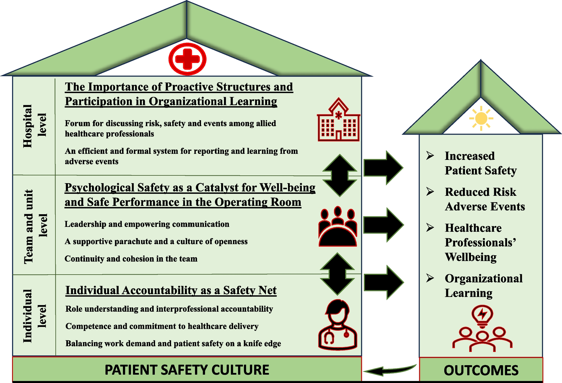
Several treatment algorithms have been proposed for acute peri-partum pubic symphysis rupture by Herren et al. [60] and Osterhoff et al. [10], as well as for chronic pelvic postpartum pain by Weil et al. [57]. We present an algorithm that takes into account the duration since childbirth (Fig. 7).
Fig. 7
Proposed Algorithm for Treating Pubic Symphysis Rupture during the Peripartum Period based on Time Elapsed since Delivery
The study has some limitations. It was conducted retrospectively and lacked a control group. The primary constraint is the small sample size and the application of various treatment approaches to a limited cohort, possibly introducing bias. Given the rarity of this complication, assembling sizable and comparable patient groups is challenging. To provide a more comprehensive view, a literature review was conducted, resulting in a heterogeneous study group. Nonetheless, a protocol for managing such clinical conditions should be established as part of a prospective multicenter study.
留言 (0)