
記住我


’Not everything that counts can be counted and not everything that can be counted counts’
Albert Einstein
Healthy kidneys increase the rate of glomerular filtration in response to a hemodynamic and/or a metabolic stimulus. Bosch termed the increase between baseline glomerular filtration rate (GFR) and stimulated GFR, measured 2 h after a protein meal, the renal functional reserve (RFR) [1]. This change from unstimulated to stimulated or stressed GFR is also called renal reserve capacity or renal functional response [2].
RFR=GFRStimulated−GFRBaseline
The induced increase from baseline GFR is termed hyperfiltration, either generated in a kidney with normal numbers of nephrons by an elevation of GFR in all single nephrons, or by a larger increase in single nephron GFR in the remaining functioning nephrons in a kidney with reduced nephron numbers (congenital or acquired) [2–5]. Hyperfiltration can therefore be a sign of a kidney under stress, but is not easy to detect in humans especially early in the process (this may later manifest with albuminuria) [6▪▪]. Static measurement of GFR does not capture RFR. Hence, a ‘normal’ serum creatinine or Cystatin C can mask a significant loss of RFR and compensation via hyperfiltration. Despite its plausible physiologic relevance; however, RFR is not routinely measured in clinical practice [2,7,8,9▪].
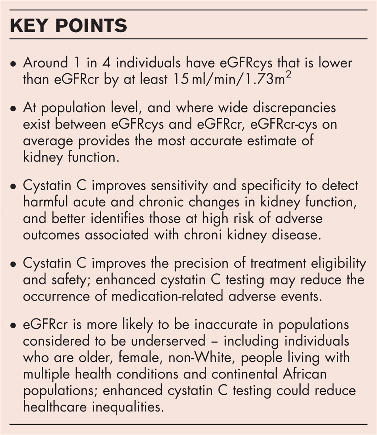
Poor accuracy of baseline GFR estimates remains a major challenge for all kidney care, and hampers easy measurement of RFR [10–12]. Measurement of GFR is cumbersome [13]; therefore, estimations of GFR (eGFR) using derived formulas are most commonly used. In routine clinical care, however, the determination of baseline eGFR is of low quality. For example, the 2009 CKD-Epi formula has a P30 value of only 84%, meaning that only 84% of GFR estimates fall within 30% of measured GFR values [10,14▪▪]. This wide range can have important clinical consequences. Newer eGFR equations including Cystatin C have improved somewhat, with a P30 above 90% [15].
 Box 1:
 no caption available
WHY IS THE RENAL FUNCTIONAL RESERVE IMPORTANT?Renal functional reserve and hyperfiltration are key paradigms in kidney physiology and pathophysiology [4,16]. They closely link nephron mass, hemodynamics, response to injury, progression of kidney disease, and therapeutic approaches and treatment effects. Given the close glomerulo-tubular balance, and the importance of tubulo-interstitial function and fibrosis in prognosis of CKD, the concept of RFR has now been extended to include tubular capacity [5,17,18].
In other organ systems, assessment of functional reserve capacity has been integrated into routine diagnostics. In nephrology, we currently diagnose and treat patients without knowing the true function, capacity and vulnerability of the kidneys. This is comparable to treating hypertension without measuring 24-h BP, assessing pulmonary function without assessing residual lung function, evaluating cardiac function without stress tests, or not being able to diagnose impaired glucose tolerance in pregnancy.
The prediction of loss of kidney function and time-point of need for kidney replacement therapy are major questions in nephrology and are most frequently asked by patients. Since the early work by Mitch and Walser [19] a relatively steady, predictable course of renal functional decline in the individual patient, reflecting the natural progression of kidney disease has been proposed, using previous creatinine measurements to model the future progression [20]. This extrapolated time course has been used to time fistula placement, wait-listing for transplantation, timing of dialysis start and/or transplantation [11,19]. The predictability of the ongoing functional decline reflects a certain fatalism in the approach to kidney disease: continuous progression of kidney disease is inevitable and there is a point of no return in the steady loss of nephrons.
Downward or upward deviations from the ‘expected’ Mitch curve indicate either accelerated deterioration, likely due to additional injury, or an improvement in function potentially due to a therapeutic intervention or quiescence of disease. However, in daily clinical practice, the actual vs. the predicted course and the observed vs. the expected time-points frequently differ significantly, which may have negative consequences for the individual patient. These inaccuracies, in particular of the linear prediction of GFR progression, question the value of this approach, and alternate risk calculators have been proposed which use variables such as actual GFR, age, or blood pressure rather than slopes of previous decline over time [21,22].
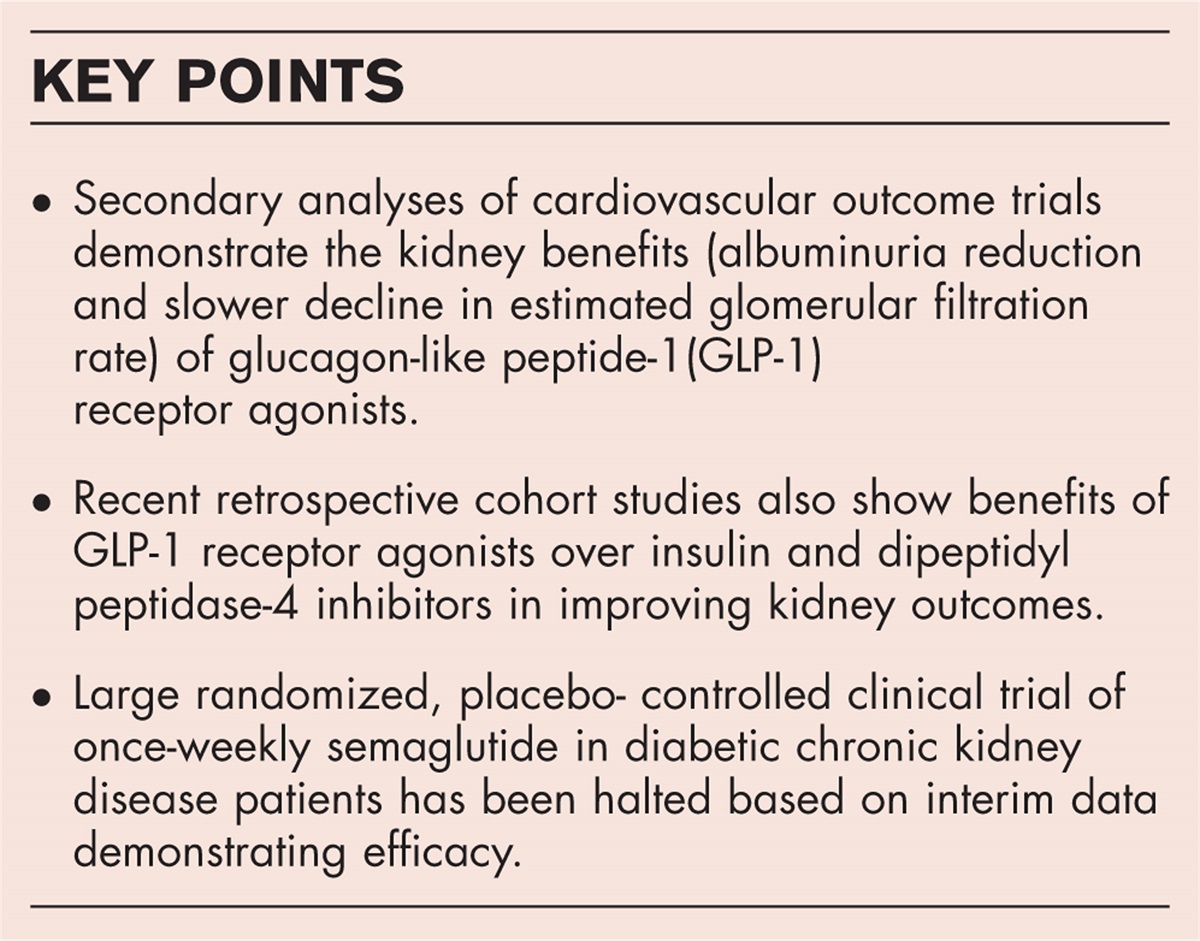
However, slope analyses of progression over time have recently garnered renewed interest, largely due to availability of large and longitudinal data sets and recognition of the need for better surrogate endpoints in studies of chronic kidney disease (CKD) progression [23,24]. In particular, the large trials of SGLT2-inhibitors have changed the outlook in nephrology [25]. We should no longer be fatalistic bystanders but should become active drivers of therapeutic approaches. The possibility of effectively targeting renal hemodynamics and metabolism and the commercial interest in marketing these therapies are pushing not only to develop better benchmarks, but also to improve the understanding of progression of kidney disease [26]. The need to individualize therapies with pathophysiology-based use of, for example, renin-angiotensin system, and SGLT2-inhibitors, GLP1 and mineralocorticoid receptor antagonists requires accurate measurement of GFR and understanding of the dynamics in response to these therapies, hence the RFR [27].
CHALLENGES WITH MEASUREMENT OF RENAL FUNCTIONAL RESERVEDespite the plausibility of its key role in renal physiology, RFR testing has not been integrated into the clinical routine. Reasons for this include that RFR testing is labor and time intensive and costly, and that robust studies evaluating the impact on outcomes are lacking [2]. More importantly, however, the lack of an accurate and practical method to measure RFR has hindered progress, as expertly discussed by de Moor et al.[2].
Measurement of RFR relies not only on measurement of GFR, but the method should also be able to accurately and reliably detect acute dynamic changes in GFR. Measurement of RFR therefore requires a precise measure of GFR and a robust and reproducible method to stimulate GFR.
Various stimulants have been used to dynamically increase GFR (Table 1). In general, intravenous dopamine is the most commonly used hemodynamic stimulant, and intravenous amino acids or oral protein load are the most commonly used metabolic (and likely also hemodynamic) stimulants [2,9▪,28]. These stimulants variably increase GFR through increasing effective renal plasma flow and/or filtration fraction. As shown in Table 1, the effect of the hemodynamic (immediate) vs. metabolic (after 1–3 h) stimuli differ [9▪,29,30]. Furthermore, the type of protein used for oral loading has a differential impact, with meat-based protein having a greater effect on the GFR than vegetable-based protein [2,30]. The optimum dose of oral protein has not been well defined [2,31], and it is not clear whether there is maximum enteral absorptive capacity, which may limit the maximal potential effect of oral protein loading [31]. In addition, postprandial hypotension due to splanchnic pooling might have a paradoxical lowering effect on poststimulation GFR [8,32]. Therefore, many variables must be considered in the selection of GFR stimulation method. One certainty is that these methods cannot be compared against each other and should be standardized within any study.
Table 1 - Stimulation methods for renal functional reserve testing and their physiological importance Stimulating agent Effect on GFR Effect on ERPF Effect on FF Effect on RFR (GFR) Dopamine, intravenous ↑ ↑↑ ↓ ↑ AA, intravenous ↑↑ ↑ ↑/ ↔ ↑↑ Dopamine + AA, intravenous ↑↑↑ ↑↑↑ ↔ ↑↑↑ Oral protein ↑↑ ↑↑ ↑/ ↔ ↑↑/ ↑↑↑ ↑ - increase; ↓ - decrease; ↔ - no change.ERPF, effective renal plasma flow; FF, filtration fraction; GFR, glomerular filtration rate; RFR, renal functional reserve.Adapted with permission from [9▪].Multiple methods to measure GFR are highlighted in Table 2, again each having strengths and limitations [14▪▪,33]. The gold standard for GFR measurement is the constant intravenous infusion technique using inulin and urinary inulin clearance. Inulin is only filtered and not secreted, and therefore its excretion reflects glomerular clearance better than molecules such as creatinine, which are also secreted [14▪▪]. Because of inconvenience and expense, inulin clearances are rarely performed [14▪▪]. As an alternative, Zitta et al.[8] investigated the use of sinistrin to capture GFR kinetics. Sinistrin is an inulin-like polyfructosan with higher solubility and easier handling than inulin [34]. On the basis of their protocol for RFR testing, sinistrin bolus injection and subsequent plasma levels over time appeared to yield robust results for RFR [35,36]. However, sinistrin was subsequently withdrawn from the market due to hypersensitivity reactions.
Table 2 - Methods to measure glomerular filtration rate Marker Advantages Disadvantages Inulin Gold standard for GFR measurement, usually measured together with ERPF Costs, need for infusion, more blood sampling in short period, bladder catheter, possible allergic reactions Iothalamate Reliable GFR measurement, usually measured together with ERPF Continuous infusion, often need for two venous lines, urine collection, possible allergic reactions Iohexol Plasma clearance reliable, nonionic, cheap, stable compound, easy measurement, low toxicity Intravenous administration, more blood sampling required if reduced renal function 99mTc-DTPA Plasma clearance reliable, split renal function possible Radioactive, logistics, cost, protein binding (underestimates GFR), accuracy affected by renal depth, injection dose. Camera methods not recommended. RFR requires 2 days 51Cr-EDTA Plasma clearance reliable Radioactive, logistics, cost availabilityOther alternatives for measuring RFR are the use of nonradiopharmaceutical markers such as iohexol or isotopes such as Cr-EDTA or Tc-DTPA [14▪▪]. For the isotopes, usually serial plasma measurements are analyzed with area under the curve, slope-intercept, or single-sample methods to calculate the plasma clearance curve [14▪▪]. However, the measurements of the isotope decays after a bolus injection usually require two separate days of analysis, one day for the baseline GFR test and one day for the stimulated GFR test [13]. Two days of measurements are cumbersome for the patients and costly [2]. In addition, the models of calculating GFR from plasma curves all rely on assumptions according to the kinetics of biomarker distribution and filtration, impact of protein binding, preanalytic and analytic problems. Not clear whether they are ready for widespread clinical use [14▪▪].
Given the logistical challenges with all of these methods to measure GFR, Ronco and his team developed a practical, clinically useful method for creatinine-based RFR measurement [28,37]. In principle, after standardized prehydration and then drinking to replace with urine output, creatinine clearances (CrCl) are measured in 1-h urine collections 2 h before and 3 h after oral protein load. The lower of the two values of CrCl before stimulation were used as baseline GFR, the peak CrCl after protein load was taken as stress GFR, and RFR was calculated as the difference between stress and baseline GFR [37]. The strength of this method is its standardization and feasibility. It can be done within 8 h in one day and costs are acceptable [37]. However, it is unclear whether 1-h urine collections and creatinine determinations can capture the true kinetics of GFR changes, given that creatinine is secreted by the renal tubules, and has unclear kinetics and distribution over such a short time. Hourly urine collection without an indwelling bladder catheter may be inaccurate, and selection of the baseline GFR as the lower of the two CrCl before stimulation, and the peak GFR as the highest CrCl within 3 h post stimulation, needs further validation [5]. Replacement of creatinine with the more accurate filtration marker cystatin C did show conflicting results, questioning whether changes in plasma levels of creatinine and cystatin C reflect the stimulation-induced filtration dynamics fast enough to be detected within an hour [35,38].
More work is therefore required to standardize stimulation and GFR and to optimize the detection of dynamic changes in GFR to improve robustness of RFR measurement. In addition, further open questions relevant to RFR evaluation include the need to measure renal blood flow to assess filtration fraction, the duration of poststimulation measurements required to account for variable factors such as delayed gastrointestinal emptying or impaired kidney function, the optimal route of application of the stimulant (intravenous or oral), and the need to adjust GFR measurement to body surface area. These uncertainties and the weaknesses of the different methods have hampered better understanding of the clinical relevance of hyperfiltration and the RFR.
POTENTIAL UTILITY OF RENAL FUNCTIONAL RESERVE MEASUREMENTSIn the current paradigm, progression of kidney disease and loss of RFR is seen as a biphasic process [39]. In the early phase of stress and nephron loss, RFR compensates without a measurable increase in serum creatinine or fall in baseline GFR. Once RFR is lost, further functional deterioration will result in a clinically manifest reduction in GFR and increase in creatinine [39]. It is still unclear whether this rather rigid model really reflects progression of kidney disease [6▪▪]. Some studies have demonstrated loss of RFR in older and obese kidney donors, which may be consistent with a proportion of the RFR being ‘used’ at baseline to maintain GFR, and therefore dampening the response to further stimulation (various scenarios reviewed in ref [44] and [12]). Given the observation, however, that in living donors, postnephrectomy, the remaining single kidney does not increase its GFR to 100% of the predonation value, and that most donors do retain some RFR, albeit lower than the 2-kidney value, it is likely that even kidneys functioning under some degree of ‘stress’ maintain the capacity to increase GFR in response to a stimulus, possibly until advanced stages of kidney injury and failure [40]. Inter-individual variability is however high in these studies, suggesting that better knowledge about the RFR in individual patients may be useful to inform treatment decisions and interpret response to treatment.
RFR measurements would be useful in evaluating risk associated with hyperfiltration (for in detail reviews see [2,4]. The safety of living kidney donation, success of renal transplantation, life-long good function in most children born with a single kidney, and the ‘benign’ hyperfiltration associated with normal pregnancy have all questioned a universal pathological impact of hyperfiltration on long-term kidney function [2,4]. In the setting of transplantation, however, a sufficient RFR is essential to ensure long-term safety after living kidney donation for the donor [9▪,41] and the donor kidney's capacity to meet the metabolic demand of the recipient [40,42]. Various studies have shown the relevance for living organ donation, indicating a significant loss of RFR in obese and old donors [37,40,43]. Detection of reduced or absent RFR may inform decisions about a potential donor's eligibility to donate. Similarly, measurements of RFR may be also helpful to understand the molecular changes and mechanisms associated with hyperfiltration [44,45] and to study the impact of hyperfiltration in children born with a single kidney [46,47], with low birth weight/low nephron number [48,49], or in pregnancy [50].
Monitoring of hyperfiltration and RFR could help in the assessment and treatment of patients with diabetic nephropathy [51], obesity [52], autosomal dominant polycystic kidney disease [53], presurgery and in follow up of patients requiring tumor nephrectomy [47,54], in intensive care [7], as well as cardio, hepato, onco-renal, or metabolic syndromes [55–57], and as a prognostic indicator after acute kidney injury (AKI) [21,39,57]. Furthermore, measurement of RFR may create opportunities for more evidence-based individualization of therapies in diverse settings [6▪▪]. Given the game-changing potential of the sodium-glucose-cotransporter 2 (SGLT2)-inhibitors, in patients whose creatinine levels rise significantly after treatment initiation for example, knowledge of an individual's RFR over time may enhance personalization of therapy, and may provide confidence to continue therapy in patients in whom this may otherwise have been stopped out of ‘caution’ [6▪▪,26].
In populations and in individuals, better understanding of RFR may permit detection of renal stress/injury at an earlier time-point, highly relevant now that disease-modifying therapies are becoming a reality, and to understand individual variability in CKD progression [58,59]. Longitudinal measurements over time could provide insights on possible breakpoints in progressive loss of renal function, permit detection of when benign hyperfiltration may switch to pathologic hyperfiltration, the existence of a point of no return, identify time windows for intervention before the reversible becomes irreversible, and guide the use of modulators of intra-glomerular pressures and/or tubular reabsorption [6▪▪,21,24,39,42,45,60].
CONCLUSIONThe precise assessment of both renal baseline function and renal reserve function would elevate nephrology care on another level. RFR measurement would be useful to precisely diagnose impaired renal function [61], predict future disease course [19,24], differentiate between benign vs. pathologic hyperfiltration [42,62,63], analyze links to mitochondrial stress and inflammation [64], and to individualize therapies [6▪▪,25]. More robust methodology is required to standardize GFR stimulation and GFR measurement, and more evidence is required to understand the true impact of RFR on kidney function and outcomes. We suggest therefore that what can be counted should be counted.
AcknowledgementsNone.
Financial support and sponsorshipNone.
Conflicts of interestThe authors have no conflicts of interest to declare.
REFERENCES AND RECOMMENDED READINGPapers of particular interest, published within the annual period of review, have been highlighted as:
▪ of special interest
▪▪ of outstanding interest
REFERENCES 1. Bosch JP, Saccaggi A, Lauer A, et al. Renal functional reserve in humans. Effect of protein intake on glomerular filtration rate. Am J Med 1983; 75:943–950. 2. De Moor B, Vanwalleghem JF, Swennen Q, et al. Haemodynamic or metabolic stimulation tests to reveal the renal functional response: requiem or revival? Clin Kidney J 2018; 11:623–654. 3. Helal I, Fick-Brosnahan GM, Reed-Gitomer B, Schrier RW. Glomerular hyperfiltration: definitions, mechanisms and clinical implications. Nat Rev Nephrol 2012; 8:293–300. 4. Cortinovis M, Perico N, Ruggenenti P, et al. Glomerular hyperfiltration. Nat Rev Nephrol 2022; 18:435–451. 5. Palsson R, Waikar SS. Renal functional reserve revisited. Adv Chronic Kidney Dis 2018; 25:e1–e8. 6▪▪. Luyckx VA, Rule AD, Tuttle KR, et al. Nephron overload as a therapeutic target to maximize kidney lifespan. Nat Rev Nephrol 2022; 18:171–183. 7. Jufar AH, Lankadeva YR, May CN, et al. Renal functional reserve: from physiological phenomenon to clinical biomarker and beyond. Am J Physiol Regul Integr Comp Physiol 2020; 319:R690–R702. 8. Zitta S, Stoschitzky K, Zweiker R, et al. Dynamic renal function testing by compartmental analysis: assessment of renal functional reserve in essential hypertension. Nephrol Dial Transplant 2000; 15:1162–1169. 9▪. Figurek A, Luyckx VA, Mueller TF. A systematic review of renal functional reserve in adult living kidney donors. Kidney Int Rep 2020; 5:448–458. 10. Levey AS, Stevens LA, Schmid CH, et al. A new equation to estimate glomerular filtration rate. Ann Intern Med 2009; 150:604–612. 11. Steiner RW. How many disparate measurements of kidney function unfairly allow selected candidates to accumulate time on the deceased donor kidney transplant waiting list? J Am Soc Nephrol 2021; 32:1539–1540. 12. Shafi T, Zhu X, Lirette ST, et al. Quantifying individual-level inaccuracy in glomerular filtration rate estimation: a cross-sectional study. Ann Intern Med 2022; 175:1073–1082. 13. Delanaye P, Ebert N, Melsom T, et al. Iohexol plasma clearance for measuring glomerular filtration rate in clinical practice and research: a review. Part 1: How to measure glomerular filtration rate with iohexol? Clin Kidney J 2016; 9:682–699. 14▪▪. Warwick J, Holness J. Measurement of glomerular filtration rate. Semin Nucl Med 2022; 52:453–466. 15. Inker LA, Eneanya ND, Coresh J, et al. New creatinine- and cystatin C-based equations to estimate GFR without race. N Engl J Med 2021; 385:1737–1749. 16. Brenner BM, Hostetter TH, Olson JL, et al. The role of glomerular hyperfiltration in the initiation and progression of diabetic nephropathy. Acta Endocrinol Suppl (Copenh) 1981; 242:7–10. 17. Chawla LS, Ronco C. Renal stress testing in the assessment of kidney disease. Kidney Int Rep 2016; 1:57–63. 18. Ichikawa I, Hoyer JR, Seiler MW, Brenner BM. Mechanism of glomerulotubular balance in the setting of heterogeneous glomerular injury. Preservation of a close functional linkage between individual nephrons and surrounding microvasculature. J Clin Invest 1982; 69:185–198. 19. Mitch WE, Walser M, Buffington GA, Lemann J Jr. A simple method of estimating progression of chronic renal failure. Lancet 1976; 2:1326–1328. 20. Kovesdy CP, Coresh J, Ballew SH, et al. Past decline versus current eGFR and subsequent ESRD risk. J Am Soc Nephrol 2016; 27:2447–2455. 21. Tangri N, Inker LA, Hiebert B, et al. A dynamic predictive model for progression of CKD. Am J Kidney Dis 2017; 69:514–520. 22. Li L, Astor BC, Lewis J, et al. Longitudinal progression trajectory of GFR among patients with CKD. Am J Kidney Dis 2012; 59:504–512. 23. Raynaud M, Aubert O, Reese PP, et al. Trajectories of glomerular filtration rate and progression to end stage kidney disease after kidney transplantation. Kidney Int 2021; 99:186–197. 24. Inker LA, Collier W, Greene T, et al. A meta-analysis of GFR slope as a surrogate endpoint for kidney failure. Nat Med 2023; 29:1867–1876. 25. Heerspink HJ, Perkins BA, Fitchett DH, et al. Sodium glucose cotransporter 2 inhibitors in the treatment of diabetes mellitus: cardiovascular and kidney effects, potential mechanisms, and clinical applications. Circulation 2016; 134:752–772. 26. Savedchuk S, Phachu D, Shankar M, et al. Targeting glomerular hemodynamics for kidney protection. Adv Kidney Dis Health 2023; 30:71–84. 27. Heerspink HJL, Vart P, Jongs N, et al. Estimated lifetime benefit of novel pharmacological therapies in patients with type 2 diabetes and chronic kidney disease: a joint analysis of randomized controlled clinical trials. Diabetes Obes Metab 2023; doi: 10.1111/dom.15232. 28. Sharma A, Zaragoza JJ, Villa G, et al. Optimizing a kidney stress test to evaluate renal functional reserve. Clin Nephrol 2016; 86:18–26. 29. Anastasio P, Viggiano D, Zacchia M, et al. Delay in renal hemodynamic response to a meat meal in severe obesity. Nephron 2017; 136:151–157. 30. Premen AJ. Potential mechanisms mediating postprandial renal hyperemia and hyperfiltration. FASEB J 1988; 2:131–137. 31. Schoenfeld BJ, Aragon AA. How much protein can the body use in a single meal for muscle-building? Implications for daily protein distribution. J Int Soc Sports Nutr 2018; 15:10. 32. Borg MJ, Xie C, Rayner CK, et al. Potential for gut peptide-based therapy in postprandial hypotension. Nutrients 2021; 13:806–923. 33. White CA, Akbari A, Allen C, et al. Simultaneous glomerular filtration rate determination using inulin, iohexol, and (99m)Tc-DTPA demonstrates the need for customized measurement protocols. Kidney Int 2021; 99:957–966. 34. Oettl K, Payerl D, Zitta S, et al. Quantitative analysis of sinistrin in serum with high-performance liquid chromatography for renal function testing. Anal Biochem 2004; 331:183–188. 35. Mueller TF, Raeder J, Oettl K, et al. Cystatin C does not detect acute changes in glomerular filtration rate in early diabetic nephropathy. Ren Fail 2008; 30:21–29. 36. Zitta S, Schaffellner S, Gutschi J, et al. The effect of mammalian target of rapamycin versus calcineurin inhibitor-based immunosuppression on measured versus estimated glomerular filtration rate after orthotopic liver transplantation. Transplantation 2015; 99:1250–1256. 37. Spinelli A, Sharma A, Villa G, et al. Rationale for the evaluation of renal functional reserve in living kidney donors and recipients: a pilot study. Nephron 2017; 135:268–276. 38. Christiadi D, Simpson C, O’Brien K, et al. Cystatin C kidney functional reserve: a simple method to predict outcome in chronic kidney disease. Nephrol Dial Transplant 2022; 37:1118–1124. 39. Sharma A, Mucino MJ, Ronco C. Renal functional reserve and renal recovery after acute kidney injury. Nephron Clin Pract 2014; 127:94–100. 40. van Londen M, Kasper N, Hessels NR, et al. Renal functional reserve capacity before and after living kidney donation. Am J Physiol Renal Physiol 2018; 315:F1550–F1554. 41. Mueller TF, Luyckx VA. The natural history of residual renal function in transplant donors. J Am Soc Nephrol 2012; 23:1462–1466. 42. Giral M, Foucher Y, Karam G, et al. Kidney and recipient weight incompatibility reduces long-term graft survival. J Am Soc Nephrol 2010; 21:1022–1029. 43. van Londen M, Schaeffers A, de Borst MH, et al. Overweight young female kidney donors have low renal functional reserve postdonation. Am J Physiol Renal Physiol 2018; 315:F454–F459. 44. Stefansson VTN, Nair V, Melsom T, et al. Molecular programs associated with glomerular hyperfiltration in early diabetic kidney disease. Kidney Int 2022; 102:1345–1358. 45. Sharma M, Sharma R, McCarthy ET, et al. Hyperfiltration-associated biomechanical forces in glomerular injury and response: Potential role for eicosanoids. Prostaglandins Other Lipid Mediat 2017; 132:59–68. 46. Groen In ’t Woud S, Roeleveld N, Westland R, et al. Uncovering risk factors for kidney injury in children with a solitary functioning kidney. Kidney Int 2023; 103:156–165. 47. Cochat P, Febvey O, Bacchetta J, et al. Towards adulthood with a solitary kidney. Pediatr Nephrol 2019; 34:2311–2323. 48. Lillas BS, Tondel C, Melsom T, et al. Renal functional response-association with birth weight and kidney volume. Kidney Int Rep 2023; 8:1034–1042. 49. Keijzer-Veen MG, Kleinveld HA, Lequin MH, et al. Renal function and size at young adult age after intrauterine growth restriction and very premature birth. Am J Kidney Dis 2007; 50:542–551. 50. Odutayo A, Hladunewich M. Obstetric nephrology: renal hemodynamic and metabolic physiology in normal pregnancy. Clin J Am Soc Nephrol 2012; 7:2073–2080. 51. Tonneijck L, Muskiet MH, Smits MM, et al. Glomerular hyperfiltration in diabetes: mechanisms, clinical significance, and treatment. J Am Soc Nephrol 2017; 28:1023–1039. 52. Denic A, Glassock RJ. Obesity-related glomerulopathy and single-nephron GFR. Kidney Int Rep 2020; 5:1126–1128. 53. Messchendorp AL, van Londen M, Taylor JM, et al. Consortium D: kidney function reserve capacity in early and later stage autosomal dominant polycystic kidney disease. Clin J Am Soc Nephrol 2018; 13:1680–1692. 54. Imam N, Burjonrappa S. Nephron sparing surgery outcomes in Wilms’ tumor: is it ready for primetime? Pediatr Surg Int 2022; 39:5. 55. Bystad EO, Stefansson VTN, Eriksen BO, Melsom T. The association between metabolic syndrome, hyperfiltration, and long-term GFR decline in the general population. KI Rep 2023. 56. Reboldi G, Verdecchia P, Fiorucci G, et al. Glomerular hyperfiltration is a predictor of adverse cardiovascular outcomes. Kidney Int 2018; 93:195–203. 57. Mancianti N, Guarnieri A, Lenoci M, et al. Rationale for the evaluation of renal functional reserve in allogeneic stem cell transplantation candidates: a pilot study. Clin Kidney J 2023; 16:996–1004. 58. Rowe C, Sitch AJ, Barratt J, et al. Biological variation of measured and estimated glomerular filtration rate in patients with chronic kidney disease. Kidney Int 2019; 96:429–435. 59. Cantaluppi V, Guglielmetti G, Dellepiane S, et al. A call to action to evaluate renal functional reserve in patients with COVID-19. Am J Physiol Renal Physiol 2020; 319:F792–F795. 60. Denic A, Mathew J, Lerman LO, et al. Single-nephron glomerular filtration rate in healthy adults. N Engl J Med 2017; 376:2349–2357. 61. Levin A, Okpechi IG, Caskey FJ, et al. Perspectives on early detection of chronic kidney disease: the facts, the questions, and a proposed framework for 2023 and beyond. Kidney Int 2023; 103:1004–1008. 62. Hostetter TH, Olson JL, Rennke HG, et al. Hyperfiltration in remnant nephrons: a potentially adverse response to renal ablation. Am J Physiol 1981; 241:F85–93. 63. Lenihan CR, Busque S, Derby G, et al. Longitudinal study of living kidney donor glomerular dynamics after nephrectomy. J Clin Invest 2015; 125:1311–1318. 64. Tang C, Dong Z. Mitochondria in kidney injury: when the power plant fails. J Am Soc Nephrol 2016; 27:1869–1872.
留言 (0)