
記住我

This retrospective cohort study adhered to principles of the Declaration of Helsinki and its subsequent amendments. It also conformed to guidelines of the Institutional Review Board (IRB) of the Friedrich-Alexander-University, Erlangen/Nuremberg, Germany under auspices of the Bavarian Hospital Act (Bayerisches Krankenhausgesetz Art. 27 [4]). All subjects granted general permission for scientific use of their clinical data, supplying written informed consent for anonymous data publication. Ethics committee approval was waived since this was a retrospective study.
We enrolled 148 patients (men, 43; women, 105) surgically treated for thyroid nodules between 2010 and 2021, categorized (n = 37, each) as follows: group 1, MTC (men, 14; women, 23); group 2, PTC (men, 6; women, 31); group 3, FTC (men, 13; women, 24); or group 4, FTA (men, 10; women, 27). All patient data were acquired from our institutional database. Within the designated time period, there were 37 patients with MTCs, 290 with PTCs, 63 with FTCs, and 911 with FTAs. Equivalent patient samplings were achieved for groups 2–4 using a random number generator. Biographic data of all qualifying patients are presented in Table 1. Recruitment of study subjects is shown in Fig. 1.
Fig. 1
Recruitment algorithm of the study subjects. MTC: medullary thyroid carcinoma, PTC: papillary thyroid carcinoma, FTC: follicular thyroid carcinoma, FTA: follicular thyroid adenoma
Table 1 Age and sex distribution of the patients in the four subsetsEach enrollee underwent thyroidectomy in one of two surgical departments. All diagnoses of thyroid carcinoma or thyroid adenoma were confirmed histologically by board-certified pathologists with expertise in thyroid neoplasms.
In all patients, US devices used for preoperative examinations were equipped with high-resolution longitudinal probes transmitting at 10.0 MHz (LOGIQ P6 Pro; GE Healthcare, Chicago, IL, USA). The entire body of imaging data was stored in a picture archiving and communication system (PACS) for later analysis by two nuclear medicine specialists, each with > 10 years of experience reviewing > 2000 thyroid US examinations annually.
Patients were examined in supine positions, necks slightly extended. In doing so, both anterior and lateral neck areas were freely accessible by US probe. First, the entire right lobe was examined in transverse and longitudinal orientations. Then, the same procedure was applied step-wise to left lobe and to isthmus.
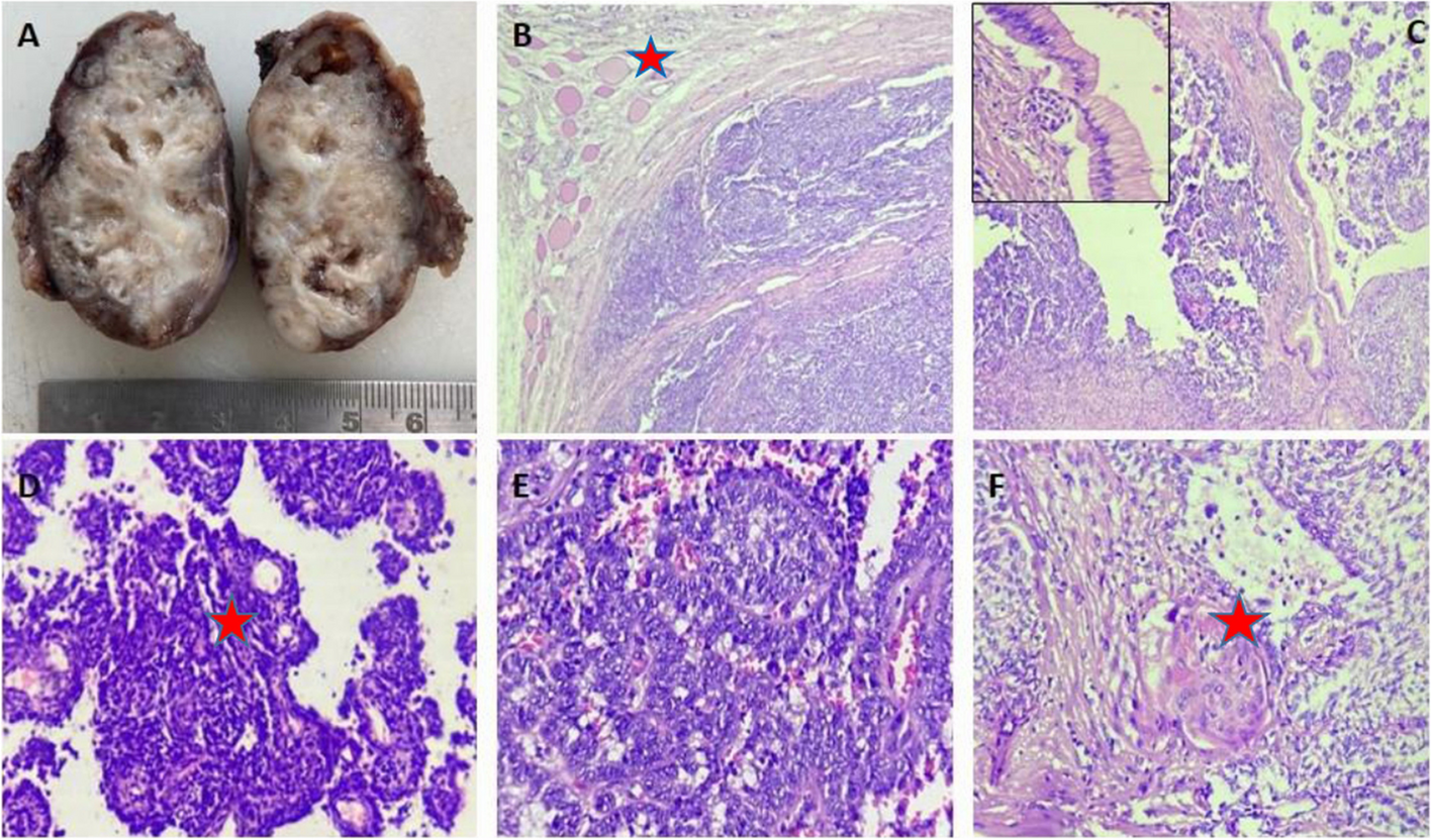
Focal thyroidal lesions were recorded in two dimensions and stored as above. Examiners assessed the following seven morphologic tumor criteria: (1) Volume (mL), calculated as v = 0.5 * (dx * dy * dz) using maximal lateral (dx), anteroposterior (dy), and craniocaudal (dz) axial diameters; (2) Shape, whether round (dx = dy = dz [± 10%]), oval (> 10% disparity in axial diameters, except TTW), irregular (undulating or complex shape), or TTW (anteroposterior diameter > lateral diameter, craniocaudal diameter discounted); (3) Contour (smooth vs. ill-defined); (4) Internal structure (homogeneous vs. non-homogeneous); (5) Echogenicity, whether hypoechogenic (below adjacent tissue level but not anechoic), hypoechogenic with cysts (anechoic components), hyperechogenic (beyond adjacent tissue level), or hyperechogenic with cysts; (6) Calcification (+/−); and (7) Focality (one or multiple sites).
Criteria (2) through (7) for focal lesions corresponded to those defined by Russ et al. [9].
Artificial neural network architectureThis study was conducted to compare tumors by groups (i.e., MTC vs. PTC, MTC vs. FTC, MTC vs. FTA, PTC vs. FTC, PTC vs. FTA, and FTC vs. FTA) and with respect to the EU TI-RADS system, based on neural network processing of ultrasonographic traits. Only three hidden layers were involved, given the relative paucity of data. Demographic (age and sex) and morphologic characteristics (volume, shape, contour, structure, echogenicity, calcifications, and focality) served as input.
The network architecture is illustrated in Fig. 2. There are seven input neurons fully connected to three hidden layers of 50, 250, and 100 neurons respectively. Output is shown as a vector reflecting respective tumor probabilities. In the hidden layer, a rectified linear unit is invoked as activation function, the output layer assuming a sigmoidal function and the mean squared error a loss function. To evaluate the network, a leave-one-out cross-validation is carried out. Of all available datasets, 75% were used initially for training, 20% for validation, and 5% for testing. Features of the implemented artificial neural network are listed in Table 2. The neural network was implemented in Python, Tensorflow and Keras.
Fig. 2
Architecture of the neural network with seven input neurons to discriminate among the classes of thyroid nodules
Table 2 Features of the implemented neural network Statistical analysisDepending on the nature of data distribution, analysis of variance (ANOVA), Fisher’s exact test, or chi-square test was applied to assess differences among groups. Significance in linear relations was gauged via Pearson’s correlation coefficient, using multinomial logistic regression for multivariate relations. By default, confidence intervals of binary variables involved binomial distributions. In neural network performance analysis, the following metrics were generated: accuracy, sensitivity, specificity, positive predictive value, negative predictive value, Fleiss’ κ, Cohen’s κ, and F-score. All computations were driven by standard software (MATLAB vR2012b; The MathWorks Inc, Natick, MA, USA), setting significance at p < 0.05.
留言 (0)