
記住我

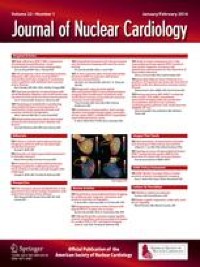


Twenty-five years ago, a seminal event led to my strong view that myocardial perfusion imaging (MPI) and coronary artery calcium (CAC) scanning should be combined whenever possible. In 1998, we began introducing CAC scanning to our imaging repertoire at Cedars-Sinai Medical Center. A 72-year-old asymptomatic cardiologist with moderate hypercholesterolemia (LDL 152) and no other coronary artery disease (CAD) risk factors was referred for a SPECT MPI scan. He was not taking any medications at the time. Both his stress ECG and SPECT scan were completely normal, and nothing changed in his lifestyle or medications after the test. However, an “introductory” CAC scan was soon offered. His CAC score was 2,946 (Figure 1). After the scan, he started on a statin and aspirin, reduced his animal fat intake, lost weight, and increased the intensity of his exercise routine. He maintained his healthy lifestyle from then on and never developed symptoms despite his very high CAC score. He went on to live for another eighteen years, never having had a coronary event. This initial experience seared in my mind the value that CAC scanning could have for improving patient management after MPI imaging and marked the beginning of my advocacy of combining CAC with MPI.
Figure 1
Images from 72-year-old asymptomatic male with CAC scan after normal SPECT
Similar experiences with a large cohort of patients undergoing CAC scanning led our group to create a large prospective randomized trial to assess the impact of a cardiac imaging test on clinical outcomes. Our approach was quite novel at the time. Specifically, in what came to be known as the “EISNER” (Early Identification of Subclinical Atherosclerosis by Noninvasive Imaging Research) trial, Alan Rozanski and I assigned 2,137 volunteers with CAD risk factors to either CAC scanning or a control group.1 The subjects were followed for 4 years. The CAC group manifested better risk factor control at the end of the trial. Significantly, the study demonstrated that this was accomplished without increasing medical costs, which was an important concern at that time. The reason? Physicians “de-risked” patients with zero CAC scores, resulting in less downstream testing or use of medical resources in the patients without CAC abnormality.
In the mid-2000s, the NHLBI decided to sponsor a large clinical trial that would compare the downstream clinical impact of coronary CT angiography (CTA) versus stress testing in symptomatic patients with suspected CAD. At the time, based on our extensive clinical experience with CAC, I suggested that the trial should include a comparison of the clinical impact of combined SPECT MPI/CAC versus coronary CTA. Ultimately, the trial was awarded to a group of Duke investigators in what came to be known as the PROMISE trial.2 For this trial, 10,003 symptomatic candidates for noninvasive testing were randomized to either coronary CTA or stress testing, which was SPECT MPI in two-thirds. Clinical outcomes were comparable in the two groups over a median follow-up of 25 months. However, in the coronary CTA arm there was greater statin, aspirin, and beta blocker use, and the patients adopted a healthier diet and experienced more weight loss than those in the stress testing arm. These changes parallel the changes we previously observed in the EISNER Trial studying CAC. Ultimately, the PROMISE trial did not include a subgroup who would undergo MPI plus CAC scanning and unfortunately no subsequent study has evaluated the clinical impact of this combined imaging approach.
Thus, a fair comparison among today’s imaging modalities was lost. A fair comparison would acknowledge potent ways in which the addition of CAC score can improve the clinical utility of stress MPI. As listed in Table 1, the benefits of combining CAC with MPI are many, including improvements in diagnosis, prognosis, and the guiding therapeutic management of patients with suspected CAD.
Table 1 Clinical benefits of combining coronary artery calcium scanning with stress/rest myocardial perfusion imaging (MPI).Regarding diagnosis, there are multiple ways in which the combination of CAC with MPI provides benefit in the diagnosis of CAD. First, MPI has both a notable strength and flaw. Its strength is its ability to relate cardiac symptoms to inducible myocardial ischemia and to guide which patients may or may not be candidates for coronary revascularization procedures for either symptomatic relief or mortality benefit.3,4 On the other hand, the intrinsic limitation of stress MPI is that it cannot detect CAD that is not hemodynamically significant—nonobstructive disease that is prognostically important and present in most patients with normal MPI studies.
In contrast, CAC is a direct marker of coronary artery atherosclerosis, establishing the diagnosis of its presence. Its extent provides a measure of a patient’s overall coronary atherosclerotic burden, reflecting the integrated lifetime effect of all factors producing atherosclerosis in an individual patient. In an early study of 1,195 patients who underwent both SPECT MPI and CAC scanning at our Institution5 we found that CAC was common among the patients with normal perfusion studies: 78% had CAC and 31% had extensive CAC (Agatston score ≥400) (Figure 2).
Figure 2
Distribution of coronary artery calcium (CAC) scores among 1193 patients undergoing SPECT MPI and CAC scans. Left: normal SPECT scans; Right: ischemic SPECT scans. 31% of the patients with normal SPECT had CAC ≥400 (5)
A second diagnostic benefit of combining CAC and MPI relates to the likelihood of obstructive CAD. The main intent of the seminal article on the likelihood of CAD by my colleagues George Diamond and Jim Forrester was to propose the application of serial Bayesian analysis of multiple tests.6 Accurate pre-test likelihood assessment is key to determining the overall post-test likelihood of obstructive CAD at the conclusion of an MPI report, which is the bottom line that a clinician needs from an MPI study in patients with no known CAD. The amount of CAC provides the most accurate assessment of the likelihood of obstructive CAD, being superior to assessment based on age, sex, risk factors, and symptoms.7
A third diagnostic benefit of combined MPI and CAC analysis is that the CAC score can help improve reader confidence in MPI test interpretation. The final SPECT/PET MPI interpretation—whether using subjective visual analysis or standard quantitative analysis of perfusion defects—is often complicated by lack of observer certainty regarding the reporting of a scan as either normal or abnormal. In a patient with minimal visual or quantitative perfusion abnormalities, this uncertainty is often expressed by use of the term “equivocal” or “borderline.” Knowledge of the degree of underlying atherosclerosis is effective in reducing the proportion of patients in whom the interpreter is “on the fence” among patients with subtle perfusion findings.8
This relates to a fourth diagnostic benefit. Stress-only SPECT MPI studies are advocated in ASNC guidelines and are becoming increasingly common. In our laboratory, over 30% of our studies are stress only, increasing to nearly 50% among exercise patients (unpublished observations). When minimal stress perfusion defects are suspected to be due to artifact, the knowledge of a zero CAC score increases the use of stress-only imaging.
With respect to prognosis, concomitant MPI and CAC imaging corresponds to a growing interest in utilizing cardiac tests for evaluating both long-term as well as short-term clinical risk. A large, consistent body of literature from registries and population-based studies have demonstrated a progressive increase in the rate of all-cause and cardiovascular mortality over time as the amount of CAC increases.9,10 Among patients with a normal SPECT MPI scan, short-term risk has been shown to be low regardless of patients’ concomitant CAC score, but after 5 to 6 years of follow-up, risk becomes increasingly heterogeneous, varying according to the magnitude of CAC abnormality.11 Multiple reports of patients having both CAC and SPECT or PET MPI have shown that the combination of the two studies is stronger prognostically than either test alone.12,13,14
In addition, the dual application of MPI and CAC can reduce the concern for missing high-risk CAD. Assessment of regional perfusion defects depends on there being a greater reduction in perfusion to a region compared to a normal or less abnormal zone. This phenomenon can lead to an underestimation of ischemic extent. While not common, patients with high risk CAD may manifest “balanced ischemia,” with no perfusion defect.15,16 This is of particular concern to the scan reader in highly symptomatic patients or those with abnormal stress ECG and clinical responses to exercise. Knowledge of the presence and extent of CAC provides the opportunity to express caution in the integrative final summary of studies manifesting normal or mildly abnormal perfusion scans.
Perhaps most importantly, as illustrated by our patient, combining CAC scoring with MPI results in a particularly tangible clinical benefit: the opportunity to optimize preventive management recommendations and healthy patient behaviors after the MPI scan. In the EISNER trial, we observed improvement in blood pressure, triglycerides, weight, and Framingham Risk Score in patients randomized to receiving a CAC scan.1 Knowledge of the presence and extent of CAC can guide therapy not only in primary prevention of asymptomatic patients but also in the risk reduction of symptomatic patients undergoing diagnostic SPECT or PET. This includes guidance about both the need for and intensity of LDL therapy (e.g., use of statins alone, or with ezetimibe, or the need for PCSK9 inhibitors). The CAC score can also influence decisions regarding the initiation of aspirin and the aggressiveness of medically managing other CAD risk factors, including hypertension, insulin resistance, and obesity. Advocating for lifestyle management (e.g., healthy diet, exercise) is also important but challenging. Two meta-analyses in this regard indicate that use of CAC scanning leads to improved patient adherence to medications and changes in health behaviors.17,18
In prior work, we observed that the frequency of abnormal SPECT MPI among diagnostic patients referred for scanning decreased dramatically over time, from 41% in 1991 to 9% in 2009.19 In the 91% with normal scans, the MPI test result is unlikely to have impacted the implementation of beneficial preventive treatments, patient adherence, or lifestyle—changes that affect long-term patient outcomes. Such impact may be achieved in patients with normal MPI studies if they are combined with CAC assessment.
There are many ways in which CAC can be assessed, without requiring a dedicated CAC scan. With hybrid SPECT/CT and PET/CT systems, assessment of CAC should be routine.20 Its presence and extent of CAC can be qualitatively assessed visually19 and quantified21 from CT attenuation correction (CTAC) scans, making it available on all SPECT/CT and PET/CT systems, without separate CAC scanning. Many patients referred for SPECT or PET MPI have already had standard chest CT, from which CAC can be qualitatively or quantitatively analyzed.22 Alternatively, when SPECT is performed on stand-alone SPECT systems, CAC can be measured by performing a separate CAC scan.
Of note regarding comparison with other modalities, the combination of anatomy and physiology with CAC plus MPI provides an advantage over cardiac MRI and stress echocardiography which cannot assess CAC. It also allows SPECT to compete more strongly with coronary CT angiography, which can provide combined anatomic and physiologic assessment of perfusion as well as prediction of invasive fractional flow reserve.
CAC scanning is so valuable to the use of SPECT or PET MPI that it must now become routine, whenever possible, in patients with suspected CAD. This addition is crucial to the future of MPI in a competing imaging world that depends upon fair comparisons. Of the benefits I have cited, the most important effect of adding CAC scanning to MPI is that it can lead to a greater change in preventive treatments and patient behaviors after MPI testing, particularly among patients with normal scans, who presently constitute the bulk of diagnostic MPI studies in our imaging laboratories.
留言 (0)