
記住我
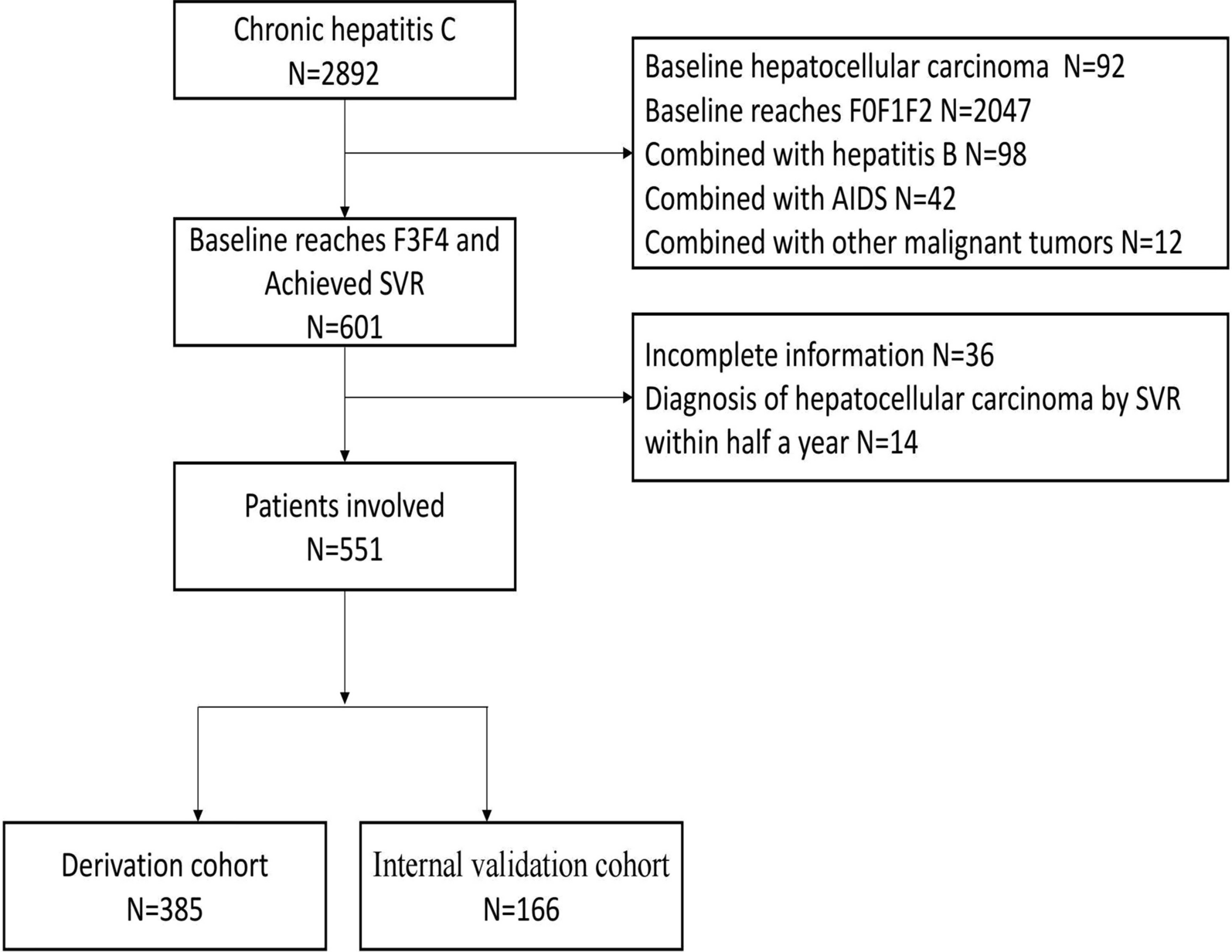
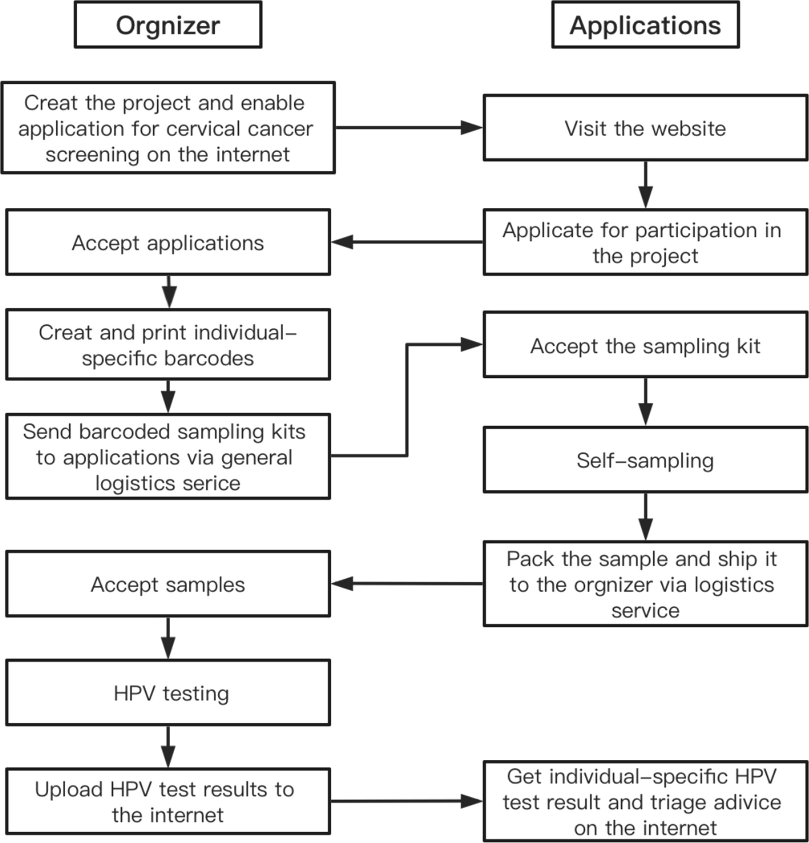


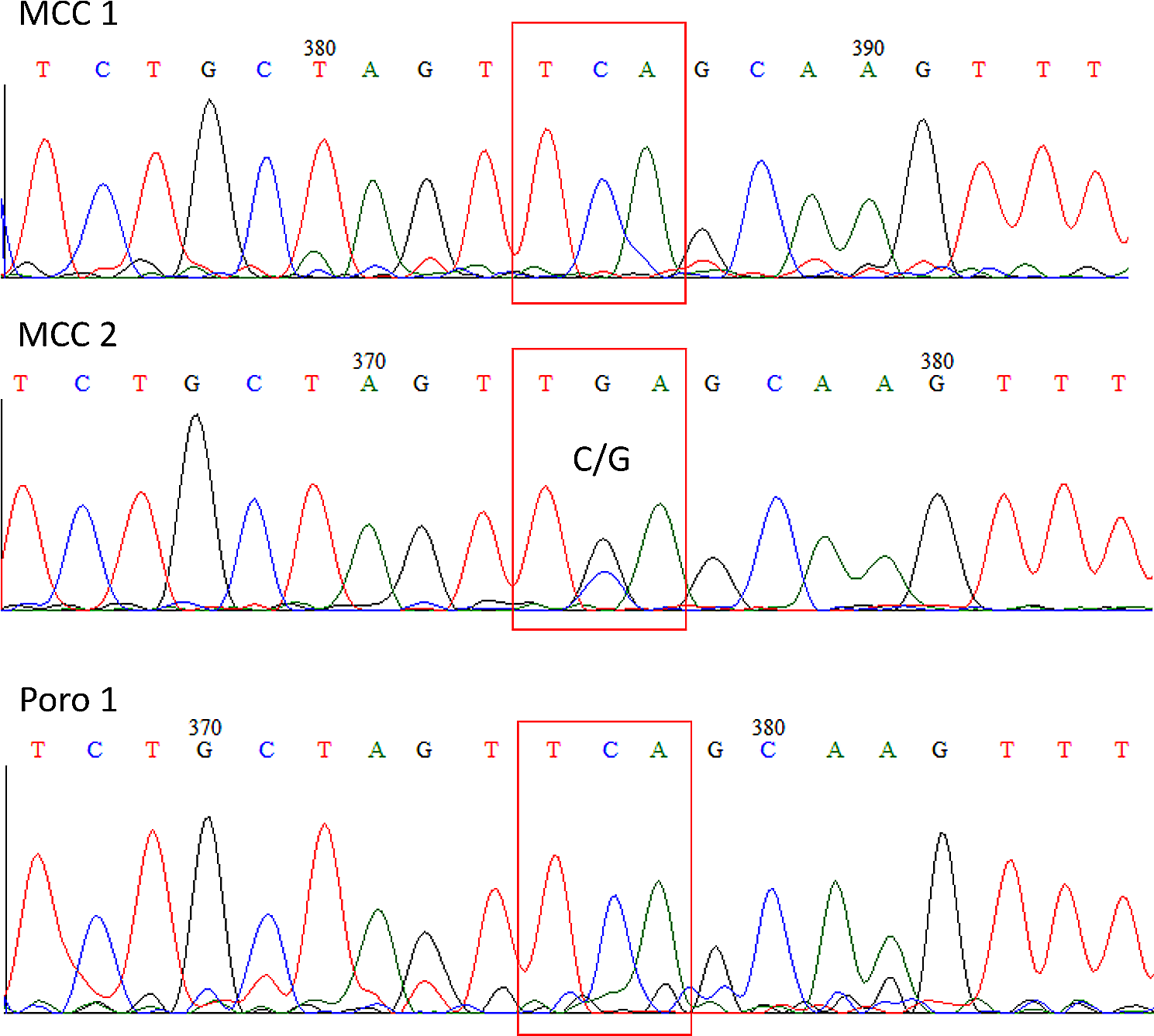


The pathologies described in this report regard women who had been to the outpatient clinic of a specialist in obstetrics and gynaecology. Following colposcopic examination, patients showed clinical signs of genital herpesvirus type 2 (HHV-2) and Papillomavirus (HPV)-related lesions. The patients’ clinical data are summarised in Table 1, and the colposcopic images of the pathologies treated are shown in Fig. 1. Patient ID and age are reported in Table 1, in columns A and B. All patients had vulvar or cervical lesions caused by genital HHV-2 (column C), and underwent cervical cancer screening, including cytology by ThinPrep specimens, HPV testing and typing, and cervical biopsy in one case (ID 32). HPV-DNA test was performed only in case of cytology abnormalities. The HPV-related pathologies detected are listed in column D. Atypical squamous cells of undetermined significance (ASC-US) were observed in ID6 Pap-test, while low-grade squamous intraepithelial lesions (L-SIL) were observed in ID 17, 29 and 37 Pap-tests. Patient ID29’s HPV-DNA test was negative, while the other mentioned patients had HPV-DNA test positive for the high-risk (HR) HPV16. HR-HPV66 and low-risk (LR)-HPV42 genotypes were also detected in cervical specimens from patients ID6 and ID17, respectively. Patient ID32 showed a high-grade intraepithelial lesion which was found to be CIN2 on histological examination after biopsy (column D). Patients ID12 and ID25 showed no cell abnormalities or HPV infection in Pap smear of cervical cells. HPV-related genital condylomatosis was observed in all patients except ID37 (column E), and candidiasis was observed in patients ID6, ID17, ID25, ID29 and ID32 (column F). The colposcopy images of clinical signs (blisters) of HHV-2 in vulva (ID12, 17, 25, 32 and 37) or cervix (ID6, 25 and 29), the perineal and anal condylomatosis (ID32), the vulvar microcondylomatosis (ID6, 12, 17, 25 and 29), and the HPV-related cervical lesions (ID6, 17, 29, 32 and 37) are shown in Fig. 1.
Table 1 Clinical data of patientsFig. 1
Colposcopy images of genital pathologies of women treated with antivirals. The columns report: A ID patient identification number; B images of clinical lesions in the external genitalia; C images of the cervix and Pap-test results, according to the Bethesda system are also reported for each patient; the magnification (5x, 10x and 20x) of images is indicated. Herpetic lesions are evident in ID6, 25 and 29 cervix and in ID12, 17, 25, 32 and 37 vulva. Colposcopic inspection was performed with the Binocular colposcope OP-C2, OPTOMIC with 5X, 10X and 20X magnifications. Digital images were archived using the Sinet healthnet suite 6.0 program by CSI NET srl
TreatmentsPatients were treated with oral and topical standard doses of Acyclovir (ACV), or its derivate Valacyclovir (VCV), which has a higher oral bioavailability [1]. ACV and VCV are the gold standard drugs for the treatment of HHV-1 and HHV-2 infections [2].
For each patient, systemic treatment with ACV or VCV, alone or in combination with topical treatment, was recommended for different periods in relation to the severity of the case and the patient’s health conditions, as deduced from the Summary of Product Characteristics (SPC) in force in Italy. A biweekly gynaecological visit, including a colposcopy examination when necessary, was scheduled as a follow-up of HPV-derived pathologies. Instead, patient ID32 with CIN2 diagnosis received a weekly visit in the first period of therapy. In Table 1, column G, the oral antiviral dosage is indicated for each patient. The topical administration of 5% ACV cream was suggested to the patients with HHV-2 vulvar lesions with the exception of the ID 12 patient who was affected by vulvodynia. In cases of candidiasis, the patients were treated topically with 1-week cycles of 1% Econazole nitrate (Pevaryl). The patients received instructions regarding daily personal hygiene (Additional file 1).
Observations at follow-upThe specialist offered to patients with multiple infections a gynaecological visit including a colposcopic examination, if necessary, every 1–2 weeks. Through these follow-ups, the specialist was able to balance the drug treatment period and observe disease improvements, including the remission of HPV-related cervical lesions and vulvar condylomatosis. During the antiviral treatment, the follow up gynaecological visits revealed not only the healing of the herpetic lesions, but also the remission of the cervical and vulvar HPV-related lesions.
In HHV-infected cells, for antivirals to function, they must be phosphorylated by viral thymidine kinase (HHV-TK) to inhibit DNA synthesis and viral replication by HHV-DNA polymerase, for which ACV has a high affinity [3]. However, it has been shown that cellular thymidine kinase can replace viral kinases [4], and ACV also showed inhibitory activity against cellular DNA Polymerases [5, 6]. Importantly, an in vitro study showed that the ACV treatment of HPV18-transformed HeLa cell line induced growth arrest, cell proliferation inhibition, and reduced cell survival with the formation of micronuclei [7]. Furthermore, the efficacy of ACV has also been demonstrated by intralesional administration in cutaneous warts [8, 9], as well as by postoperative therapy in Recurrent Respiratory Papillomatosis [10], both HPV-related pathologies. Two clinical trials on the efficacy of ACV against plantar warts are currently ongoing (NCT05429151; NCT05324904).
留言 (0)