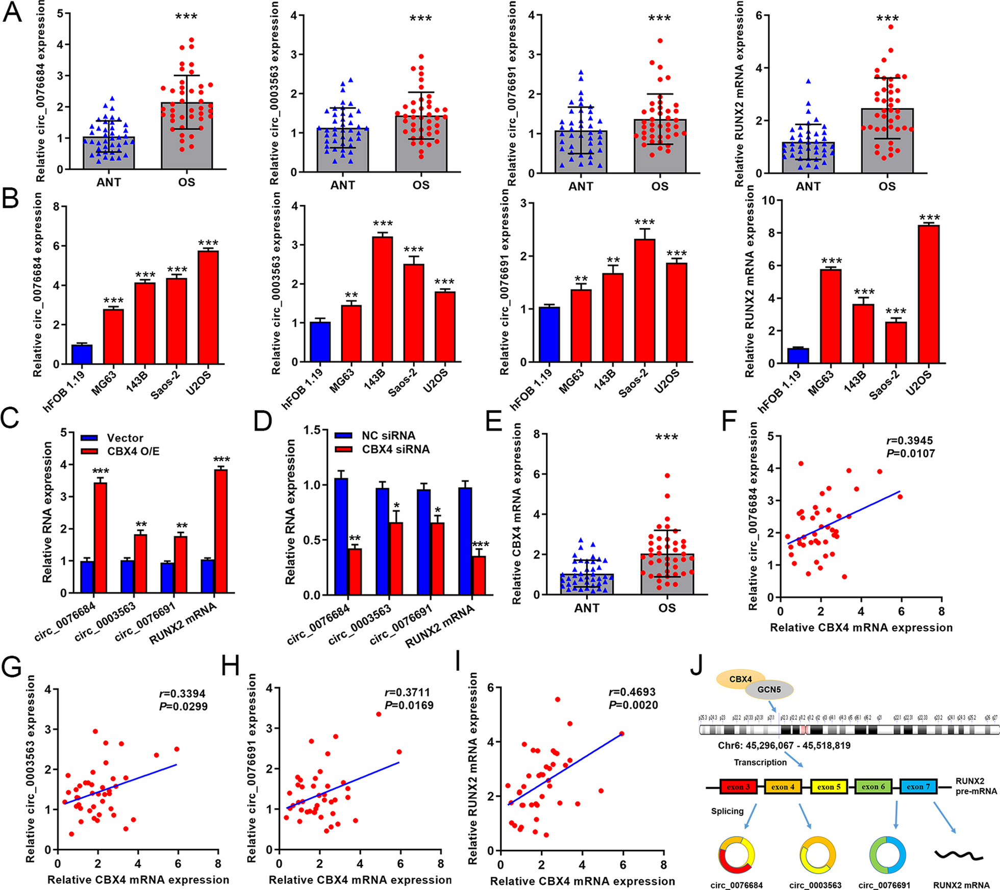
Lumbar degenerative disease is a common disorder that causes various neurological symptoms. With the advances in minimally invasive spinal surgery techniques, minimally invasive surgery has become the preferred treatment for lumbar degenerative disease. Since MIS-TLIF was first proposed by Foley in 2002 [10], it has gained in popularity owing to its great advantages. A comparative study showed that the MIS-TLIF group experienced less injury and recovered sooner after surgery for lumbar degenerative disease than open TLIF [15]. In addition, MIS-TLIF retains the advantage of direct and adequate decompression of the canal, which allows for more adequate nerve decompression through the intervertebral foramen and has a wider range of indications for more complex degenerative lumbar diseases [16]. As a result, MIS-TLIF has become an effective alternative to open surgery in recent years [17, 18].
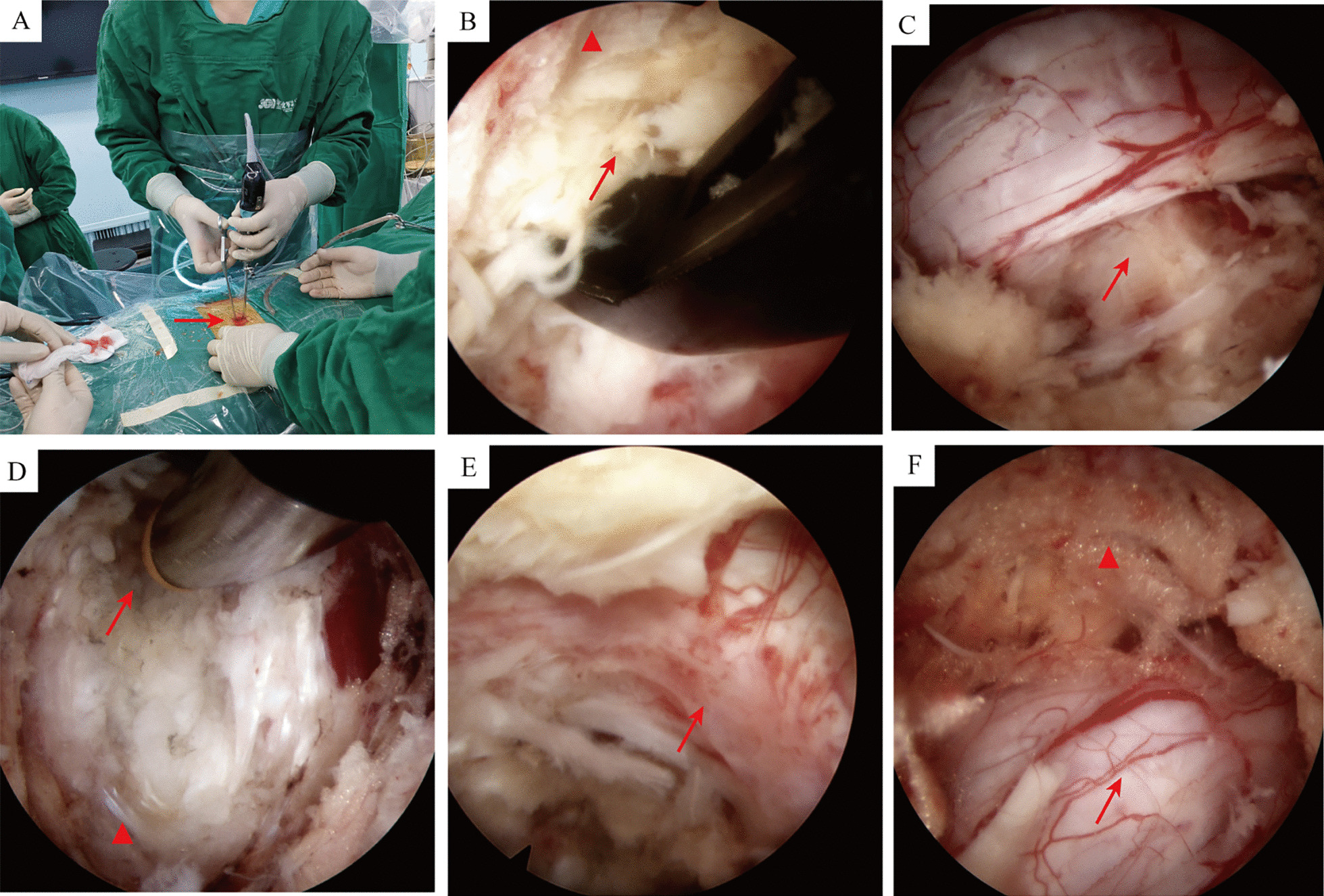
With the widespread use of endoscopy in spinal therapy, Osman et al. [19] first reported a technique of endoscopic transforaminal decompression, interbody fusion, and percutaneous pedicle screw implantation for lumbar degenerative disc disease, which is known as Endo-LIF. In the study by Osman et al. [19], 29.6% of patients achieved strong fusion and 36.2% had a stable internal fixation system; the reason for the relatively low fusion rate may be related to the absence of an implanted cage and autologous bone. Wang et al. [20] reported 10 cases of endoscopic transforaminal approach interbody fusion, with no intraoperative or postoperative complications and a 100% fusion rate. The authors concluded that Endo-LIF may be an alternative to conventional fusion therapy [20]. Endo-LIF uses a channel through Kambin’s triangle and thus does not require the osseous channel from the posterior or posterolateral approach that is needed in currently emerging endoscopic techniques, like unilateral biportal endoscopy. The absence of the need to remove the facet joint in Endo-LIF reduces the difficulty of the procedure and the probability of iatrogenic nerve injury and instability. Similarly to MIS-TLIF, Endo-LIF has the advantage of being minimally invasive, in addition to other advantages such as a quicker recovery, shorter duration of hospitalization, and lower costs [21]; however, no study has compared the specific clinical outcomes of MIS-TLIF and Endo-LIF.
In our study, we retrospectively compared a group of patients treated with Endo-LIF versus a group treated with MIS-TLIF. Compared with the MIS-TLIF group, the Endo-LIF group had significantly less intraoperative blood loss and a shorter hospital stay, but a longer operative time and fluoroscopy time. Although the VAS scores, ODI, MacNab criteria, and postoperative fusion rate at 1 year postoperatively were similar in both groups, the Endo-LIF group had less trauma to the surrounding tissues and had lower VAS scores for lower back pain than the MIS-TLIF group at all postoperative timepoints. Jung et al. [22] performed a meta-analysis to compare the results of full-endoscopic lumbar interbody fusion and MIS-TLIF for lumbar degenerative disease in a total cohort of 423 patients. The authors concluded that the immediate results of full-endoscopic lumbar interbody fusion were favourable in terms of blood loss and VAS for back pain compared with MIS-TLIF, although there were no differences between the two techniques in complications, short- or medium-term clinical outcomes, and fusion rates. Our findings are in agreeance with the findings of Jung et al. [21]
The complication rate for totally endoscopic lumbar interbody fusion has been reported to be 13.2% (range 0–38.6%) [23]. In our study, two patients had signs of exit nerve root injury after Endo-LIF, and their symptoms were relieved after 1 month of conservative therapy and functional exercise. To reduce this complication, we used a trephine instead of a drill to perform root-forming arthroplasty. In our clinical experience, none of the patients who have undergone this modified procedure have had extrusion of the exit nerve root. In addition, one patient in the Endo-LIF group had residual nucleus pulposus, which may have been due to incomplete microscopic nerve root and endplate treatment; secondary decompression is recommended for this issue. Further endoscopic exploration of the spinal canal and nerve root cleaning should be performed after the cage is inserted to prevent bone and nucleus pulposus from entering the spinal canal. We also noted that one patient in each group had non-fusion of the cage at the 1-year follow-up. We attributed this complication to the following three possible causes. (1) Cage subsidence owing to collapse of the endplate. In the process of propping up the vertebral space and bone grafting, endplate injury is a common complication, especially in osteoporotic patients. Therefore, it is important to match the cage perfectly with the channel. It is advisable to place the corresponding type of cage under fluoroscopic surveillance and protection with a nerve retractor. In addition, attention should be paid to the angle of bone graft entry to prevent accidental injury to the endplate. (2) Incomplete removal of the endplate, which is very likely to lead to non-fusion. In our experience, it is best to use a hook and curette instead of a reamer, as this will clean the endplate efficiently and thoroughly and make full use of the advantages of Endo-LIF to ensure satisfactory endplate cleaning by direct visualization. (3) Inappropriate autologous bone grafting. The choice of bone graft largely influences the postoperative fusion. While a large amount of autologous bone is obtained during MIS-TLIF, only a little of the facet joint is cut during Endo-LIF. Therefore, we empirically used allogeneic bone combined with decalcified dental matrix (BMP-2) in Endo-LIF to effectively shorten the fusion time and improve the success rate of fusion. The other common complications were cerebrospinal fluid leakage and dural tears, which did not occur at a high rate in the present study.
There are still some limitations of Endo-LIF. Firstly, for some severe lumbar degenerative diseases, such as severe spinal stenosis, severe lumbar spondylolisthesis, and foraminal stenosis, MIS-TLIF may be more appropriate because Endo-LIF may not be able to achieve adequate microscopic decompression. Secondly, the operative time was significantly longer in the Endo-LIF group than the MIS-TLIF group. Although endoscopic techniques have improved considerably, the surgical instruments used in Endo-LIF may require a longer operative time. Furthermore, Endo-LIF may require more fluoroscopic examinations to locate the incision and check the placement of the cage, which exposes the Endo-LIF group to a higher dose of radiation than the MIS-TLIF group. Thirdly, the amount of removal of facet joint was less in the Endo-LIF group than the MIS-TLIF group. BMP-2 was added to allogeneic bone grafts for better fusion in the Endo-LIF group, which might lead to statistical bias. Fourthly, Endo-LIF is technically challenging and requires a long learning period. The learning time of the technique and the surgeon’s skills also affect the postoperative recovery and complications [24]. Finally, the number of patients in the two groups was relatively small, and all patients in the study were only followed for less than 25 months. So that, a larger sample with a longer follow-up time are needed to make definitive clinical conclusions.


留言 (0)