During infection, pathogens must synthesize heme or acquire it from the host to survive and thus spread the disease. Heme is typically sequestered in high-affinity heme-proteins of the host; therefore, the heme biosynthesis is a more efficient strategy for bacterial pathogens [[1], [2], [3]]. Coproheme decarboxylase enzymes (ChdCs) catalyze the final step within the coproporphyrin-dependent heme b biosynthetic pathway, which is mainly, but not exclusively, used by monoderm bacteria [[4], [5], [6], [7]].
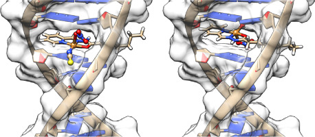
ChdCs accumulate heme b by the oxidative decarboxylation of propionate groups at positions 2 and 4 (p2 and p4) of coproheme to vinyls (v2 and v4) via two exogenous hydrogen peroxide (H2O2) molecules. The reaction involves first the cleavage of p2, i.e. the formation of a monovinyl monopropionyl deuteroheme (MMD, also known as harderoheme) reaction intermediate, which undergoes a 90° rotation. This reorientation allows p4 to get close to the catalytic residue and thus to be decarboxylated to v4, and eventually, heme b is formed [[8], [9], [10], [11], [12], [13]]. A schematic representation of the catalytic cycle of one propionate decarboxylation reaction in actinobacterial CdChdC is shown in Fig. 1.
From experimental and computational mechanistic studies on the representatives from Firmicutes (e.g. Listeria monocytogenes (Lm) [14], Staphylococcus aureus (Sa) [15] and Geobacillus stearothermophilus (Gs) [16]) and on actinobacterial Corynebacterium diphtheriae (Cd) [13,17] it was concluded that the transiently-formed MMD reaction intermediate rotates within the active site and not via a release and rebinding mechanism. For the ChdC from Corynebacterium diphtheriae (CdChdC), a combined spectroscopic and biochemical approach has been proved to be particularly effective in the characterization of the enzyme complexed with the coproheme, MMD and heme b in both the ferric and ferrous forms, during the catalytic reaction, and in the identification of the role of fundamental residues in the active site [13,17,18].
In ChdCs from Actinobacteria, unlike in Firmicutes, a distal histidine, located on a flexible loop connecting two ferredoxin-like domains, was identified as relevant for the enzymatic activity in addition to a catalytic Tyr (Y135 in CdChdC), which is completely conserved in all bacterial ChdCs [10,11]. This distal residue (H118 in CdChdC) acts as a distal base for deprotonation and subsequent heterolytic cleavage of hydrogen peroxide [10], leading to a significantly higher reactivity of CdChdC toward H2O2 [11,19]. Moreover, in the presence of H118 residue, a stoichiometric excess of hydrogen peroxide leads to the formation of iron chlorin-type heme d species [[20], [21], [22]]. Heme d formation was correlated with a potential protection mechanism from oxidative damage [23].
Actinobacterial ChdCs are also characterized by an arginine (R139 in CdChdC) instead of a catalytically important lysine (K151 in LmChdC and K149 in SaChdC), which is otherwise conserved in all the other clades [19]. R139 is involved in the extensive hydrogen bond (H-bond) network between the protein moiety and the propionate groups of the porphyrin, which stabilizes the substrate or the product within the active site both in the crystal [10] and in solution [18]. This arginine, bridging p2 and p4, has also been shown to be the most important residue for reactivity toward H2O2 in CdChdC [18].
Furthermore, the proximal side of ChdCs has been found to be less relevant than the distal side, since the proximal histidine does not have any H-bond interaction with the cavity residues [24,25]. Therefore, the proximal coordination is weak with respect to other structurally related enzymes (e.g. chlorite dismutases, Clds), as also shown by Fe-Im bond strength. In fact, in the ferrous heme b of ChdCs the wavenumber of the Fe-Im stretching mode was found between 210 (in CdChdC [17]) and 214–216 cm−1 (in LmChdC [26] and SaChdC [27], respectively), while in the heme b-Clds, it ranges from 222 cm−1 [28], 226 cm−1 [24], to 229 cm−1 [29].
Carbon monoxide (CO) has been extensively used both as a model exogenous ligand and as a probe sensitive to the nature and the architecture of the heme binding pocket in proteins. In fact, a positively charged electrostatic field or an H-bond between CO and a distal residue favors back-donation from the Fe dπ to the CO π* orbitals, strengthening the Fe − C bond and correspondingly weakening the C − O bond, thereby increasing the ν(FeC) stretching wavenumbers and decreasing the ν(CO) stretching wavenumbers. Conversely, a negative charged electrostatic field has the opposite effect. Variations in the donor strength of the trans-ligand affect the vibrational modes of carbon monoxide as well [30,31].
In this work, we investigated the role of key residues of the protein cavity of actinobacterial CdChdC in the stabilization of the incoming exogenous ligands by studying the CO adducts of the wild-type (WT) protein and selected variants in complex with coproheme, MMD and heme b using resonance Raman (RR) spectroscopy and Molecular Dynamics Simulation (MDS). In particular, we focused on the contributions of the distal H118 and of the amino acids of the protein moiety involved in hydrogen bonds with the propionate groups of the porphyrin during the catalytic reaction, as evolving from the coproheme substrate to the MMD reaction intermediate and finally to the heme b product (and heme d side-products). A comparative analysis of exogeneous ligand binding with data on representatives from ChdCs of the firmicute clade in terms of conserved residues was also carried out.


留言 (0)