
記住我


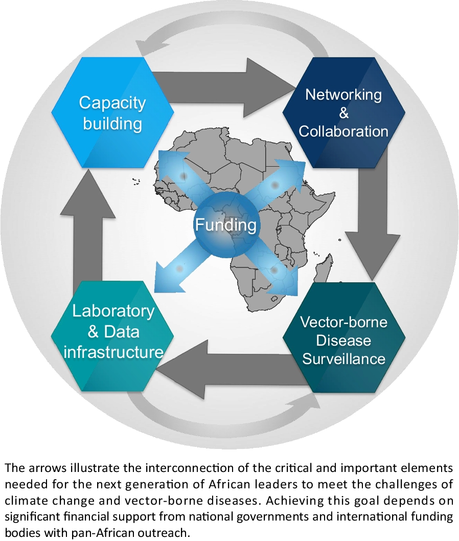
The study took place in six villages endemic for S. haematobium in the Kayes Region, western Mali [19]. This region is divided into three sanitary districts: Kayes, Bafoulabé and Diéma. The distances between the districts ranged from 20 to 45 km. The study villages were chosen according to their proximity to the water points (ponds in Diéma, Senegal River and its tributaries in Kayes and Bafoulabé). Agriculture and livestock are the population’s two main economic activities [20]. Two districts (Bafoulabé and Kayes) belong to the North-Soudanian climatic zone with two major seasons: the wet season from May–June to October with its beginning and end marked by torrential rains and thunderstorms, and the dry season from November to April–May. The mean annual rainfall is up to 1000 mm, which occurs mainly during the period from July to September. Diéma, located further north, has a Sahelian climate and two distinct seasons: a wet season from July to September–October and a dry season that spans the rest of the year. The annual rainfall is about 600 to 800 mm [20]. Two villages were selected in each district: Diakalèl and Koussané in Kayes; Babaroto and Saorané in Bafoulabé and Fangouné Bamanan and Débo Massassi in the Diéma district (Fig. 1).
Fig. 1
Localization of the study sites in the Kayes region in Mali (West Africa) (Source: MRTC – GIS; Author, Dolo M)
Type and period of the studyThe study was an observational cross-sectional study that took place from 7 to 21 November 2021. The studied population consisted of children from 6 to 14 years old, who attending in one of the six selected primary schools and who agreed to participate in the study. The minimum sample size was calculated based on the previous prevalence of the disease obtained in each school using the Schwartz formula, considering a 10% refusal rate and sampling errors. (National Schistosomiasis and Geohelminth Control Program, report 2015).
Data collection procedures (socio-demographic, socio-economic, paraclinical and clinical data collection procedures)A total of 1087 children were sampled. Children were randomly selected from the list of children in each class until the required number of samples were obtained. Sociodemographic data, including village, gender, age, were obtained using a structured questionnaire. Socioeconomic information was collected through questionnaires administered by a sociologist. The questions were close ended to facilitate the children's responses. For the parents' level of education (yes or no), type of toilet (modern or traditional), frequency of use of streams (No: Never frequents the river; Yes: I frequent the river several times a week), type of water used (drilling, river, rain, drinking), and reason for frequenting streams (swimming, fetching water, taking a bath). The teachers helped us if the child had difficulties answering. Clinical signs (abdominal pain, pollakiuria, dysuria) were collected through questioning and physical examination by a general practitioner. Microscopic hematuria was determined using Hemastix test strips (Ref: AZA—TESTUR10, Siemens Medical Solutions Diagnostics). Data were recorded on survey forms and each child was given a distinct identifier.
Parasitological examinationAll urine samples were collected between 10:00 am and 2:00 pm (the favorable period for the elimination of eggs in the urine) in the field, by trained laboratory technicians. From each subject, urine was collected in a properly labeled sample container. A filtration technique was used to analyze the samples. A total of 10 ml urine was taken from each sample flacon after mixing it. The mixed sample was filtered through a Whatman filter (CAT No. 1001-025, 25 mm) which was stained with a 3% ninhydrin solution. After drying, the filters were immersed into water and then examined under a microscope for characteristics of terminal spurred schistosome eggs [21]. The prevalence rate of schistosomiasis in the schools was defined as the number of positive individuals per school per total number of individuals examined per school and multiplied by 100. S. haematobium intensity was classified into three categories: (i) no eggs; (ii) slight (1–49 eggs per 10 ml of urine); and (iii) heavy (≥ 50 eggs per 10 ml of urine).
Ultrasound examinationUltrasound evaluations were carried out by a specialist with a portable ultrasound device, (Digital Ultrasonic Diagnostic Imaging System—Mindray, 15″ LCD display with tilt angle for better visualization, Lakeshore Burtchville, MI 480591-800-364-4942). All examinations were conducted by the same specialist at all sites. Several features were identified on ultrasound. The shape of the bladder was recorded, the detected lesions on the bladder wall were defined, and the degree of dilatation of the ureters and renal pelvis were measured, in accordance with current WHO guidelines [16]. The exact characterization of pathological changes was calculated using the global score as an index of the severity of morbidity and lesions. Children were asked to drink plenty of water before the ultrasound examination, which could only be conducted on a full bladder. Any marked abnormalities in the bladder, ureter and especially the renal pelvis, systematically prompted the resumption of ultrasound after urination to exclude the possibility of dilatation due to bladder and ureteral repletion. Urinary tract abnormalities were assessed based on the criteria defined by WHO. For successive analyses, children with at least one point on the WHO score were classified as having “urinary tract anomalies.” Bladder abnormalities were detected based on the shape and measurement of the bladder walls. A rectangular shape signified a normal bladder shape and corresponded to a score of (0), whereas bladder deformity corresponded to a rounded shape with a score of (1). Irregularity of the inner bladder wall with a thickening ≤ 5 mm was recorded as normal (score = 0), whereas a thickening > 5 mm was recorded as the presence of a lesion (focal, score = 1; multiple, score = 2). Posterior bladder wall thickness ≤ 5 mm was recorded as normal (score = 0) but posterior bladder wall > 5 mm was recorded as lesion (focal, score = 1; multiple, score = 2). A mass with local thickening ≤ 10 mm was normal (score = 0) in contrast to a mass with thickening > 10 mm was recorded as a mass (single, score = 1; multiple, score = 2). The polyp was defined based on wall growth with the presence being single or multiple. Dilation of each ureter and kidney was recorded separately. An absence of dilation corresponded to a score of (0), presence corresponded to a score of (3 = dilated; 4 = strongly dilated). Total scores were calculated based on the standard lesions observed to classify the likelihood of having schistosomiasis (≤ 1 = schistosomiasis unlikely; 2 = schistosomiasis likely; > 3 = schistosomiasis very likely) [16]. The sample size of children screened by ultrasound was 240, or 40 children per school.
Statistical analysisSocio-demographic, clinical and parasitological data were recorded on survey forms with identifiers for each child. The data was recorded in Excel. Calculations of prevalence, and intensity of infection were made using SPSS (IBM, version 23.0, Armonk,USA). Binomial analysis was performed on R software (Lucent Technologies, Jasmine Mountain, USA) to assess the relationship between clinical signs and urogenital schistosomiasis infection. Participants were divided into two age groups (6–10 and 11–14 years) for each sex. We performed statistical analyses to assess the relationship between parasitic infections and demographic, sociocultural, socioeconomic, clinical signs, and sonographic factors using a multivariate logistic regression. The differences in proportions were tested using the chi-square test or the exact Fisher test depending on the data. P values below 0.05 were considered significant.
留言 (0)