In this retrospective study, the FS index, area, and combined variables were investigated to establish whether scientifically sound sex estimation was possible among three populations. With the advent of new cutting-edge technologies in radiology, obtaining accurate and reliable measurements has become routine [2]. Lateral cephalometric is commonly used in dentistry, specifically by orthodontists, to diagnose, plan treatment, trace, and during orthognathic or implant surgery[7, 28]. The establishment of sex estimation by measuring 2D radiographs is deemed preferable to identification by photographic superimposition since radiographic evaluation is less time-consuming and technically exacting [28, 29].
The rationale for selecting these age groups was that the growth of this anatomical structure is slow [30], and the FSs complete their development and reach their maximum size at the age of 15 or 20 years [19, 23, 28, 30] and then remain stable until death [23, 31]. However, some of the radiographs could not be measured either because no FS was visible or because the radiograph did not meet the inclusion criteria. The radiographic diagnosis of the FS was performed according to previously reported literature that has identified the bilateral absence of FS [2]. The results of the present study revealed that the frequency of bilateral missing FS was identical in males among Bosnian and Nepalese (2.8%). It seems that the percentage of FS absences in females and males is higher in Chinese than in Bosnian and Nepalese subjects. Luo et al. [2] found that the frequency of bilateral FS absence was 9% for females and 5% for males. A greater frequency of bilateral absence was observed among females than males [30], which is identical to the observation of the present study. However, bilateral absence of FS was not observed in Manisa in Turkey among 100 cases when using paranasal computed tomography (CT) scans for identifying unknown bodies [11] and in 69 patients in Marseille in France for sex determination using 3D reconstructions [23]. According to the literature, bilateral absence of the FS is reportedly the least frequent, and the FS is present in 90% of adults[30].
It is important to emphasize that the FS index[2, 10] and FS area [2] have shown relatively higher accuracy for sex estimation in 2D radiographs [2, 10]. The rationale for using the FS index [10] and area was based on previously established criteria [2]. The methods of sex estimation using the FS index and FS area could be carried out due to their relative simplicity and short processing time, allowing for precise and reliable results [32, 33].
This study observed a high degree of variability between the individuals in each population group. Significant differences were observed in the study groups' FS index and FS area measurements between females and males. This result is in line with previously reported studies depicting a statistically significant sexual dimorphism of the FS index [10] and area [2]. The current outcome supports the dimorphic features of FS that have implications for individual identification [10, 22]. Various investigators have generally targeted the FS index [2, 10, 20,21,22,23] and both variables [2] to achieve sex estimation in their own population.
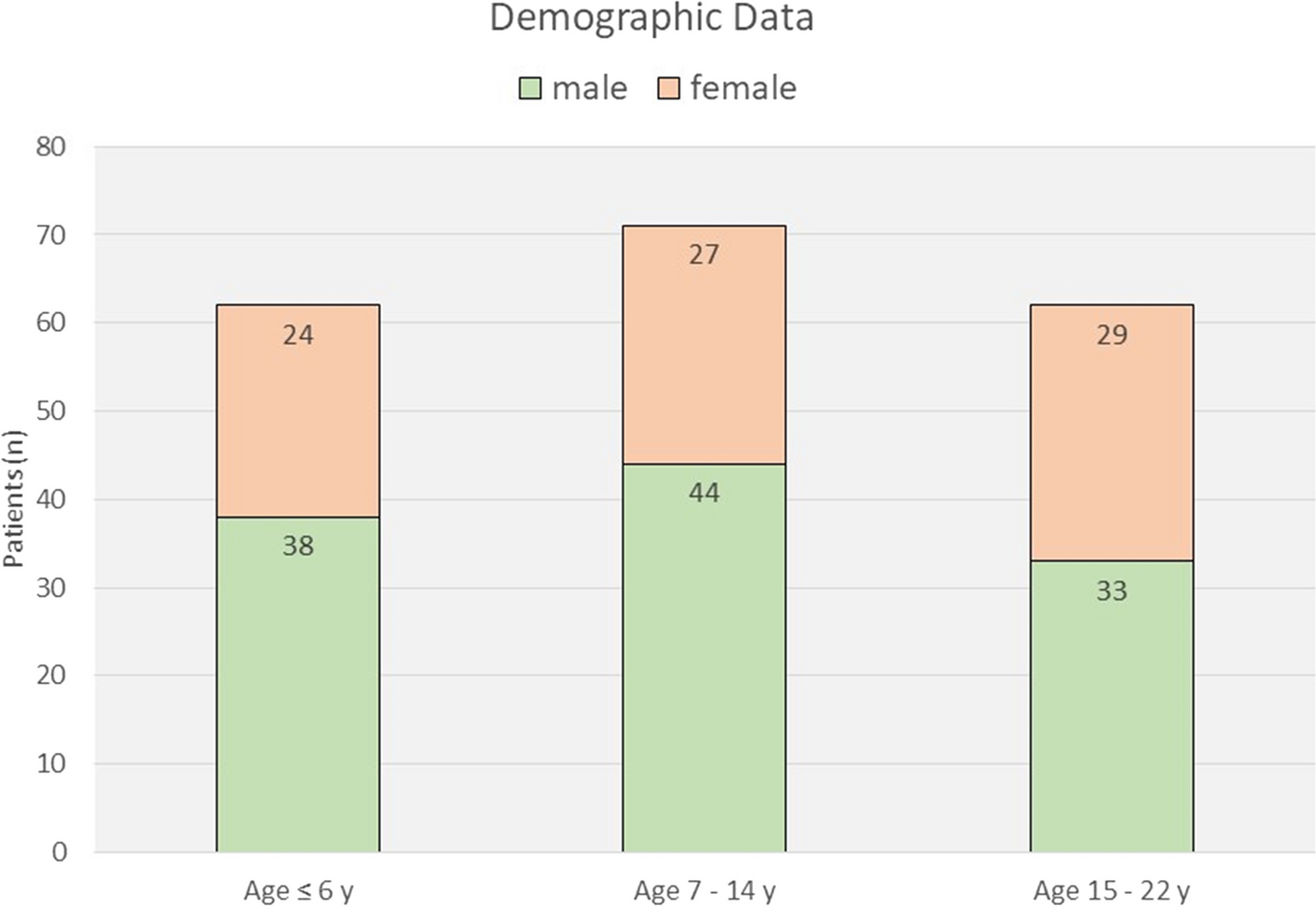
In this study, identical approaches were adopted in various population groups. The results indicated that the application of the FS index revealed the lowest sex estimation percentage compared to the proposed use of FS area and both variables with a difference of 3.7%, 2.3%, and 13.3% to FS area and with a difference of 5.5%, 8.2%, and 15.6% to both variables in Bosnian, Chinese, and Nepalese respectively. A study in India applied the FS index for sex estimation using 300 digital posteroanterior radiographs and achieved 64.6% sex discrimination with the logistic regression analysis [21]. A 2D radiograph was used among 216 Indian subjects, and the estimation function equation was used for the correlation between sex and FS index and achieved 67.6% correct sex estimation [10]. In this study, the outcomes of the FS index were lower than in previous studies when this approach was adopted, presenting 56.9%, 67.0%, and 62.8% in sex differentiation among Bosnian, Chinese, and Nepalese, respectively.
The results present the first attempt to estimate sex differentiation from the FS area. It was observed that the use of the FS area exhibits a lower percentage of correct sex estimation percentage than the application of both FS index and area, with a difference of 1.8%, 5.9%, and 2.3% in Bosnian, Chinese, and Nepalese, respectively. There is no previous supportive study to show the application of the FS area presents a higher percentage classification rate than the FS index. A study in France measured the total volume of the left and right FS with the application of 3D reconstruction of FS to determine sexual dimorphism among 69 cases and achieved 72.5% [23]. Although the results of the FS area indicated lower differences compared to the FS index, applying the FS index and area together is recommended based on 2D radiographic assessment [2]. Previous literature has improved the prediction model by combining FS index and area, which commit an overall sex classification accuracy of 76.6% among 475 cases in China [2]. It was found in a current study that, after being subjected to using FS index and area, the correct sex classification rate was 62.4%, 75.2%, and 78.4% in Bosnian, Chinese, and Nepalese, respectively. The use of the FS index and area presented the highest correct estimation percentage (Table 7), which implied that these variables were distinctly and quantifiably different at a high level and created a proper indicator, and promoted the discrimination rate compared to those in the other studies used merely the FS index [2]. The current findings highlighted a potential influence of the FS index and area on sex estimation, which rejected the hypothesis.
The current study compared outcomes with the current global datasets regarding sex estimation from the FS (Table 7) [2, 10, 20, 21, 23]. The correct estimation percentage in Caucasians (62.4%) in this study was lower than those studies conducted in China [2] (76.7%), France [23] (72.5%), and India (67.59% [10], 64.6%[21]). A previous study was conducted to determine the FS's dimorphic potential using a logistic regression model among 216 posterior-anterior Nigerian radiographs. It was concluded that the left-side FS width presented the highest accuracy of 60% in sex determination [22]. However, in the present study, China and Nepal demonstrated greater correct sex estimation with 75.2% and 78.4%, respectively. This discrepancy can be attributed to factors such as ethnicity, the use of various landmarks, the degree of radiographic enlargement, and techniques [24]. The remaining 37.6% Bosnian, 24.8% Chinese, and 21.6% Nepalese were not classified correctly. This may be attributed to high inter-individual variability in the morphology of the FS [10].
Assessing the FS index and area in other populations might be helpful, and familiarity with the FS values is relevant for successful surgery and anthropology [22]. Accurate selection of a reliable identification approach and appropriate measurement tools will assist in achieving reliable results. Applying the FS index and area technique can be considered an alternative identification modality to other procedures in sex estimation [2]. This highly versatile yet practical procedure allows forensic medicine and clinicians to rely on a simple, technique-insensitive, cost-effective material processing method.
Further research is required to explore and identify the difference in the FS of diseased persons between populations and factors that influence change in the FS that affects the decision of sex identification. The generalizability of these results is subject to certain limitations. For instance, 2D radiographs were selected in this study to measure FS and evaluate sex estimation, and this is mainly due to the high costs of CT examinations [28]. The limited number of lateral cephalometric studied in each population group is also a potential limiting factor.
The current study emphasized the implication of the FS as a positive tool in sex estimation through 2D radiographic assessment among various population groups. The bivariate models have borderline accuracy in the Nepalese population (just below 80% limit) while in the Chinese and Bosnian populations, the models are not recommended for use if there are alternative measurements in the cranium.


留言 (0)