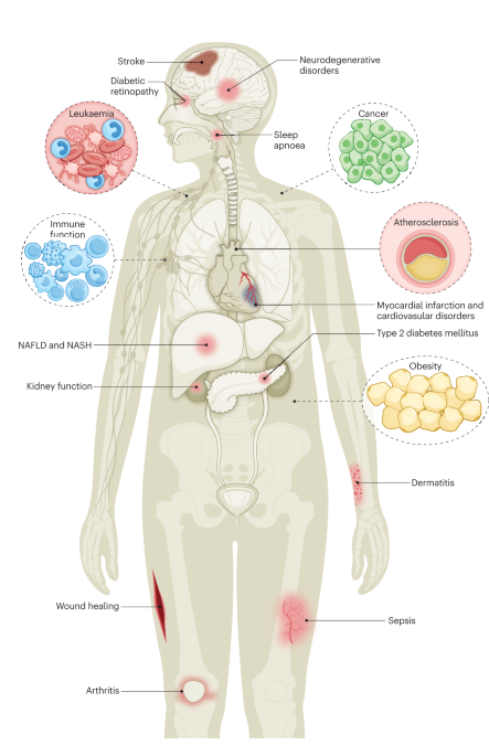
Epidemiology and risk factorsR1
Insulin resistance and features of the metabolic dysfunction are associated with specific alterations of iron metabolism regulation, which are epidemiologically linked with organ damage and clinical outcomes (U).
Not all patients with metabolic dysfunction or fatty liver present with increased serum levels of ferritin, suggesting that those with MHF constitute a subset with distinct risk factors, pathophysiology and clinical outcomes, possibly deserving specific management. Box 1 outlines the main genetic and acquired conditions associated with hyperferritinaemia and accumulation of iron in the body in individuals with metabolic dysfunction, fatty liver and insulin resistance.
Serum levels of ferritin were first described to be linked to insulin resistance in 1998 (ref. 27). The severity of insulin resistance3,4,5,6,7,8,9,10,11,28 and the severity of fatty liver disease, but not circulating biomarkers of inflammation, have been reported as determinants of MHF6,7,18,19,20,21,29,30. By contrast, genetic variants associated with iron metabolism, rather than those that affect lipid handling in the liver, were associated with increased serum levels of ferritin and MHF in patients with fatty liver31. This observation is in line with findings that indicate that serum levels of ferritin reflect hepatic and body iron stores more closely than the severity of liver lipid accumulation in individuals with metabolic dysfunction6,7,18,19,20,21. Furthermore, male sex, older age and moderate alcohol intake contribute to iron accumulation in fatty liver disease. This evidence is based on liver histology and MRI evaluations of cohorts of patients with fatty liver or multiple metabolic alterations6,7,18,19,20,21. Therefore, in patients with insulin resistance but without acute inflammatory conditions, severe alcohol abuse or poorly controlled diabetes mellitus, serum levels of ferritin are mainly determined by dysregulation of iron metabolism and next by the severity of insulin resistance.
In a large prospective cohort of middle-age healthy men conducted in South Korea, elevated serum levels of ferritin were independently associated with development of the metabolic syndrome during the 5-year follow-up period32. Furthermore, in a large prospective study on European cases of incident type 2 diabetes mellitus, increased serum levels of ferritin were associated with an increased risk of type 2 diabetes mellitus, even among individuals with no overt inflammation, liver disease, high alcohol consumption or obesity33.
Data from the past 25 years in European populations suggest that serum concentrations of ferritin are associated with insulin resistance and metabolic dysfunction, even in individuals without fatty liver or with liver levels of iron that are within the reference range3,4,5,6,7,8,9,10,11,27. However, in individuals with high serum concentrations of ferritin, the presence of fatty liver might indicate an increased risk of insulin resistance and the metabolic syndrome, whereas the presence of hepatic iron overload might indicate an increased risk of hyperglycaemia34. These data are in line with genetic evidence from the past decade that body stores of iron have a causal role in determining liver disease and the development of type 2 diabetes mellitus in the general population35,36,37.
By means of Mendelian randomization as a robust epidemiological approach to analyse the causal estimation of fatty liver disease associated with metabolic dysfunction (also known as metabolic dysfunction-associated fatty liver disease (MAFLD)) as an outcome using genetic variants, a study published in 2022 that included European individuals showed that the genetically predicted increase in liver levels of iron was associated with an increased risk of fatty liver disease. Although they need confirmation, these data support a causal association between deposition levels of iron in the liver and metabolic dysfunction38.
Box 1 Contributing factors
These factors contribute to or are associated with increased levels of ferritin or the development of metabolic hyperferritinaemia (MHF) and subsequent progression to iron accumulation (dysmetabolic iron overload syndrome; DIOS) in individuals with metabolic dysfunction or fatty liver disease.
Genetic
Male sex4,7,30
Heterozygous presence of the HFE p.C282Y pathogenic variant or homozygous presence of the p.H63D variant or, in particular, compound heterozygosity for p.C282Y/p.H63D variants7,30,101
PCSK7 variants102
Absence of TMPRSS6 p.A736V variant37,103
Heterozygous presence of SERPINA1 PiZ and PiS pathogenic variants104
Heterozygous pathogenic variants of β-globin gene (HBB), that is, β-thalassaemia trait18
Rare NMBR variants105
Heterozygous pathogenic variants of the gene that encodes ceruloplasmin31
Acquired
Severity of insulin resistance3,4,5,6,7,8,9,10,11,106
Severity of fatty liver disease6,7,18,19,20,21
Ageing4,7,30
Altered regulation of iron metabolism (increased absorption and cellular retention) associated with lipid accumulation14,40,41,42,107
Iron accumulation in the liver7,9,20,21
Moderate alcohol intake; 30 g per day in men and 20 g per day in women7
Hepatic copper deficiency and reduced ceruloplasmin activity51,52
PathogenesisR2
The pathophysiology of this alteration of iron metabolism regulation seems to be triggered by lipotoxicity in the presence of permissive environmental and genetic determinants, but additional studies are required to clarify the contribution of subclinical inflammation and the underlying mechanisms and implications (U).
Mechanism of iron accumulation
The pathogenesis of MHF is multifaceted, relating to the effect of common genetic variants on iron homeostasis, as well as hepatic, intestinal and adipose tissue factors. Systemic iron homeostasis is maintained through the hepcidin–ferroportin axis. Hepcidin is a liver peptide hormone, and the expression of hepcidin is upregulated by iron and inflammation. Hepcidin controls iron influx into the bloodstream from duodenal enterocytes and macrophages by binding, occluding and inducing the degradation of the iron exporter ferroportin. As a consequence of the increased hepcidin secretion, iron accumulates in cells expressing ferroportin, mainly macrophages and, to a lesser extent, hepatocytes. Genetic iron overload disorders (such as haemochromatosis) are most frequently caused by reduced hepcidin synthesis or impaired iron export39. In patients with MHF, hepcidin release in response to iron stores and the ability of hepcidin to downregulate intestinal iron absorption are generally preserved40. However, a subtle alteration in iron fluxes has been reported, whereby excess fatty acids have been linked to a reduced ability of hepcidin to limit intestinal iron absorption, while simultaneously increasing hepatic iron uptake and tissue deposition40,41,42,43.
Interestingly, patients with type 2 diabetes mellitus without clinical signs of inflammation show MHF with reduced hepcidin levels and increased systemic levels of iron44. Furthermore, the development of type 2 diabetes mellitus might be associated with impaired hepcidin release induced by hyperinsulinaemia45. In patients with metabolic dysfunction and hyperferritinaemia, body stores of iron have been associated with high dietary iron intake46 and a distinct microbiome profile47. In patients with MHF or obesity, development of mild iron accumulation in hepatocytes can lead to hepcidin upregulation and restrains further iron absorption48,49,50. Preserved regulation of hepcidin would favour the preferential retention of iron in liver macrophages (Kupffer cells), where ferroportin is highly expressed31,51,52. Within this context, the presence of inherited variants associated with impaired hepcidin would favour the development of more severe iron accumulation and DIOS (Table 1).
Table 1 The spectrum of iron metabolism in individuals with metabolic dysfunctionReduced expression of the ferroxidase ceruloplasmin, which cooperates with ferroportin for iron export in several cell lineages, including hepatocytes, might also favour iron accumulation in the liver. Furthermore, low-frequency inherited genetic variants of the CP gene (which encodes ceruloplasmin) that determine a mild functional impairment of ceruloplasmin were associated with MHF and more severe liver disease in patients with fatty liver disease31.
Role of excess iron in tissue damage and insulin resistance
Although the role of iron overload in determining liver disease (liver fibrosis progression and hepatocellular carcinoma) and pancreatic β-cell failure is established in haemochromatosis39, the potential effect of dysregulation of iron metabolism on the pathogenesis of dyslipidaemia and insulin resistance is less clear. A detailed discussion of the complex relationship between iron accumulation, activation of the BMP–SMAD signalling pathway, modulation of lipid metabolism and development of liver disease is presented in Supplementary Box 2. This process might involve the facilitation of ferroptosis as well as of other forms of cell death in hepatocytes and other liver cells53. Accumulation of iron in macrophages in the liver has been associated with more severe liver damage in patients with fatty liver disease20,54, compared with patients without iron accumulation in these cells. By catalysing the formation of reactive oxygen species (ROS), excess iron favours subclinical inflammation, which contributes to insulin resistance by directly downregulating insulin receptor expression and signalling and by worsening of alterations of glucose and lipid metabolism10,44,55,56, fibrogenesis and carcinogenesis14. Unlike in patients with haemochromatosis, whose macrophages are typically iron depleted because of hepcidin deficiency, iron accumulation in macrophages and hepatic stellate cells has been associated with a pro-inflammatory and pro-fibrotic response57,58. Moreover, excess fatty acids and lipotoxicity predispose to inflammation and type 2 diabetes mellitus by inducing macrophage iron accumulation via induction of FTH1, which encodes the ferritin H subunit (which has iron oxidase activity)59.
In addition to insulin resistance and dyslipidaemia, ferritin levels and levels of iron stores in the liver have also been associated with the build-up of iron in adipose tissue, which leads to insulin resistance, impaired adiponectin secretion and altered endocrine function in mouse models and in patients55,56,60, as well as impaired regulation of amino acid and phospholipid metabolism in patients with fatty liver61,62. Indeed, ferroportin is also expressed in adipocytes, where it can be targeted by hepcidin to exert its endocrine function55. Insulin resistance and the expansion of adipose tissue modulate the expression of iron-related genes, such as transferrin receptor 1 (TFRC) and FTH1 in adipose tissue in patients with metabolic alterations63,


留言 (0)