
記住我


Overall, low polymer concentration, freezing rate, or high gelation temperature should be preferred in scaffold designs with large macropores and elastic pore walls with injectability and convectional mass transport and cell infiltration applications. The cryogels with higher mechanical strength can be produced by increasing the amounts of crosslinker at low polymer concentration and relatively high gelling temperature to obtain thick pore wall thickness without compromising the pore size. Furthermore, more ingenious cryogelation strategies can be utilized to effectively tune the properties and functionality of silk-based cryogels.
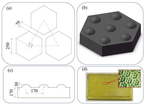
2.3. Silk-Based Composite CryogelsAs stated above, silk-based cryogels have many remarkable features making them attractive for a wide range of biomedical applications. However, as material requirements are increasing, the bar for designing materials for cutting-edge biomedical applications is often high, making it difficult to achieve using a single material source [51,52]. Therefore, natural polymers, including silk, are often combined with synthetic polymers to improve their overall mechanical and structural performance [53,54]. Additionally, SF alone suffers from a lack of inherent antimicrobial and antioxidant properties that could be improved with metal/metal oxide nanoparticles or other antimicrobial/antioxidant agents [55,56]. Therefore, many researchers have turned to having SF or sericin crosslinked incorporating other natural/synthetic polymers, inorganic compounds, and other functional groups to create cryogels with multi-functionality. Neo et al. [57] examined the efficacy of composite cryogel with a silk-based composite consisting of polyvinyl alcohol (PVA) and SF. In the study, PVA-SF composite cryogels were produced to eliminate the cell adhesion challenges of PVA cryogels for nucleus pulpous tissue engineering. With the addition of SF, the mechanical strength, as well as the swelling and cell adhesion capacity of the cryogels, were improved. In a study to increase cell adhesion and proliferation in silk fibroin cryogels, biodegradable and biocompatible collagen and its derivative were prepared. Backer et al. [37] found that adding CS to pure silk fibroin not only doubles the porosity of the cryogel but also quadruples the pore surface area. In another study, SF composite cryogel combined with SF, CS, agarose, and hydroxyapatite had enhanced cell adhesion, superior mechanical strength, and bone tissue mineralization properties. Moreover, the authors observed a significant increase in cell viability and proliferation of the composite cryogel when compared to the pure SF cryogel [58]. Yu et al. [59] added tannic acid/ferric ions to create photo-responsive antibacterial CS/SF (CS/SF) cryogels. They compared the SF and CS/SF cryogels and correlated the decrease in pore size with the hydrogen bonds formed in the structure. Another CS/SF cryogel-based study was presented by Han et al. [60] by adding polydopamine (PDA) nanoparticles as a photo-responsive antimicrobial agent to CS/SF cryogels, and they obtained enhanced cell and tissue affinity. On the other hand, Zhao et al. [61] improved the mechanical properties of cryogels used in 3-D tissue regeneration studies by adding cellulose acetate nanofibers (NFs) to CS/SF cryogel scaffolds. Compared to pure scaffolds, the composites have a rough surface, enlarged pore size, and a six-fold increase in compression modulus. Unlike the composite studies described above, Yetiskin et al. [48] have combined the concepts of a DN strategy and composite material by polymerizing acrylic acid and octadecyl acrylate in the pores of SF cryogel. They produced a cryogel with met-SF in the presence of BDDE at −18 °C. The fabricated cryogel was then immersed in a solution of acrylic acid and octadecyl acrylate as monomers, N,N-dimethylacrylamide (DMAAm) as a spacer, N,N’-methylenebisacrylamide (MBAAm) as a chemical crosslinker and ammonium persulfate (APS)-TEMED as a free-radical initiator system. They named this new material class organohydrogel. This new class of materials, containing both hydrophilic and lyophilic phases, is of great interest due to its tunable and programmable mechanical and rheological properties. These materials, which can be easily shaped above the melting point (Tm = 49–54 °C) of the crystalline regions they contain, also have shape memory properties. 3. Biomedical Application of Silk-Based CryogelsAs a protein-based natural biopolymer approved by the Food and Drug Administration (FDA), silk-based materials have been extensively utilized to fabricate many commercial medical products such as sutures, wound dressing bandages, gels, and masks [55,62,63]. However, emerging biomedical advances, including tissue engineering, controlled drug delivery, and cell-based therapy, require materials with unique structural and physiochemical properties that can hardly be achieved by conventional fabrication techniques [4,64,65]. The cryogelation could surpass these limitations by offering many desirable traits for the silk-based materials, such as a highly interconnected microporous structure, a tunable biodegradation rate, superior mechanical strength, and injectability that greatly extend their application in those advanced biomedical fields (Table 1) [4,12]. In this section, we summarize proof-of-concept studies that have been performed on the clinical usage of silk-based cryogels in tissue engineering, wound healing, drug delivery, and other biomedical application. 3.1. Tissue EngineeringTissue engineering is currently one of the most rapidly developing technologies that aim to develop engineered tissues consisting of cells, growth factors, and scaffolds to restore the structure and/or function of defected tissues [64]. The scaffold is vitally important in tissue engineering, which should be biocompatible, biodegradable, and its 3-D structure and biomechanical properties should be similar to the native extracellular matrix (ECM) [64,79]. Meanwhile, cell adhesion, growth and migration capacity, oxygen, nutrition transport efficiency, and prevascularization speed are other critical factors in scaffold design [80,81]. Inevitably, silk-based cryogels have been favored for engineering wide ranges of tissue constructs, including intervertebral disc (IVD), cartilage, liver, muscle, and bone [14,22,57]. Neo et al. [57] studied the cell attachment capacity, mechanical properties, and surface characteristics of physically cross-linked SF/PVA cryogels towards the regeneration of nucleus pulposus (NP) tissue. They showed that the addition of only 20 wt% of silk significantly improves surface wettability, water adsorption, rehydration capacity, and, eventually, the cell adhesion and proliferation ability of the composite cryogels. Furthermore, the cell-laden SF/PVA cryogels also exhibited compressive modulus and hoop stress values comparable to the native human NP tissue. In another study, Lee et al. [72] investigated SF/PVA cryogels with different mass ratios for cartilage tissue engineering applications through both in vitro and in vivo studies. The cryogels with an equal ratio of SF and PVA displayed the most suitable structural and surface characteristics for the proliferation, migration, and differentiation of chondrocytes to regenerate auricular cartilage. They also fabricated ear-shaped cryogel grafts and transplanted them into rats after seeding rat isolated chondrocytes. The implanted grafts offered enough mechanical strength to retain their shape and phenotypical stability over the culture period and facilitated the formation of a mature cartilage with a lacunar structure without causing any immune response or graft rejection. Li et al. [73] incorporated silver and strontium co-doped hydroxyapatite (AgSrHA) nanoparticles into SF/CS network to formulate a super resilient cryogel that can be used for bone tissue engineering. The presence of silver/ strontium dopants not only improved the cytocompatibility and bioactivity of the cryogel scaffold against bone marrow stromal cells (BMSCs) but also provided a durable antibacterial activity and enhanced osteoinductivity. Furthermore, the bone regeneration efficacy of the developed cryogel was also demonstrated in vivo experiments using a rat skull model. Wang et al. [70] designed a multifunctional scaffold by loading annulus fibrosus cell-derived exosomes into an SF cryogel to seal annulus fibrosus (AF) defects and promote their regeneration. The anti-inflammatory and antioxidant activity, biocompatibility, and fibrocartilage differentiation ability of the designed scaffold were studied by in vitro experiments. The animal experiment further proved that the engineered cryogel scaffold could effectively reverse the degeneration of AF defects in the rat and facilitate their regeneration by regulating the immune microenvironment of the IVD and reducing oxidative stress. Zhang et al. [14] fabricated a series of SSFs injectable cryogels that could be used for engineering wide ranges of tissues. They demonstrated that the mechanical properties of the cryogels can be tuned by adjusting SSFs concentration or crosslinking condition, which can effectively regulate the differentiation behaviors of BMSCs. Moreover, they suggested that the fabricated cryogels can be further functionalized with large variety of bioactive ingredients without compromising their structural and mechanical properties.One of the critical challenges in applying cryogels for tissue engineering is that the shapes of tissue defects are mostly irregular, and by the conventional molding method, it is very difficult to fabricate cryogels that can fill exactly in the defect site [82,83]. To solve this issue, Wang et al. [74] recently presented a 3-D cryoprinting technique to fabricate injectable cryogels composed of laponite nanoparticles (LAP) and SF in predesigned sizes and shapes (Figure 2). In vitro studies and animal model experiments proved that the printed cryogels can promote attachment, propagation, migration, and osteogenic differentiation of the BMSCs. What is more, the printed cryogels can be implanted into the host tissue defects through subcutaneous injection without causing any significant inflammation response, showing their great application potential in bone tissue engineering. 3.2. Wound HealingThe effective treatment of non-healing wounds, such as chronic wounds, large or deep wounds, and burn injuries, has been one of the primary concerns in the clinic over the past several decades [84,85]. Such type of wounds require novel bioactive materials that prevent wound infection, ischemia, and oxidative stress and facilitate their re-epidermization by triggering cell proliferation and migration [86,87]. Being a well-known wound healing material since ancient time, silk-based advanced biomaterials in various forms, including cryogels, have shown their incredible potential in the treatment of different types of wounds. For example, Lan et al. [75] studied the burn healing efficacy of an SF-based cryogel embedded with antibiotic gentamycin sulfate (GS)-loaded gelatin microspheres (GM). The SF/GS/GM composite cryogel showed strong antimicrobial activity against several surgical site infection (SSI)-associated pathogens, such as Pseudomonas aeruginosa (P. aeruginosa), Staphylococcus aureus (S. aureus) and Escherichia coli (E. coli). The in vivo rat model study showed that the cryogel dressing could significantly reduce P. aeruginosa-associated burn infection and accelerate re-epithelization over the full-thickness burn area. Similarly, Han et al. [60] developed muscle-inspired, antimicrobial, antioxidant, super-elastic, and smart wound dressing by impregnating photo-responsive PDA nanoparticles within an SF/CS cryogel. They proved that the designed cryogel could quickly kill SSI-associated pathogens upon near-inferred (NIR) light exposure and alleviate the oxidate stress at the wound site by suppressing the reactive oxygen species level. In vivo experiment further demonstrated that the combination of cryogel dressing with NIR-assisted photothermal therapy could remarkably accelerate the regeneration of full-thickness epidermal defects. In addition to non-healing wounds, traumatic hemorrhage is another serious global concern, which requires effective hemostatic dressing materials that can rapidly stop bleeding to enhance survival rate [88,89,90]. Recently, Zhu et al. [76] designed an antibacterial and hemostatic cryogel by polymerizing methacryloyl-modified silk sericin, and in situ embedding silver ions into the polymer network at subzero temperature. The cryogel exhibited high blood adsorption capacity, outstanding antimicrobial performance, and much faster hemostatic ability than commercial gelatin in rat tail amputation, liver injury, and femoral artery injury models. They suggested that the rapid hemostatic activity of the cryogel could be due to the high platelets’ adhesion ability of silk sericin. 3.3. Drug and Cell DeliveryThere have been increasing demands for pharmaceutical products to prevent, treat and cure various diseases by restoring, recovering or modifying organ function [91]. Although pharmaceutical research mostly focused on discovery of new drugs for particular application, much evidence showed that their therapeutic efficiency and side effects strongly depend on the mode of their delivery [92,93]. In this regard, effective drug delivery systems (DDS) often require a biocompatible and biodegradable drug carrier that allows the proper encapsulation of the drugs to prevent them from burst release, control their spatiotemporal delivery, and retain their effective concentration [94,95]. Silk-based cryogels possess the capability to load both hydrophilic and hydrophobic drugs, drug stabilizing ability, and stimuli-responsive properties, making them an excellent DDS [14,62,95]. For example, Zhang et al. [14] showed that hydrophobic curcumin (Cur), which has antioxidant, antibacterial, and anti-inflammatory effects, can be uniformly distributed within the SSF cryogel network. The exposure of Cur-laden SSF to protease XIV caused the degradation of SSF and the sustainable release of Cur to maintain its high antioxidant capacity. In another study, self-assembled SF cryogels were applied for enzyme-responsive delivery of BMSCs-derived exosomes [69]. The integrity and bioactivity of exosomes did not deteriorate during the encapsulation, and their cryogel degradation-dominated release profile was demonstrated both in vitro and in vivo. They also suggested that controlled release of the exosomes could promote cell migration, formation of new blood vessels, and ingrowth of myofibroblasts to improve tissue rehabilitation. In addition to being used for DDS, silk-based cryogels have also been utilized to deliver therapeutic cells to diseased organs or tissues for cell-based therapy [77,78]. Tyeb et al. [77] combined stem cell-based therapy with tissue engineering to efficiently repair diabetic wounds. They first fabricated multifunctional scaffolds by coating highly cell-adhesive laminin on antioxidant gelatin-silk sericin cryogels (GSL). Then, adipose-derived stem cells (ADSC) were cultured on the GSL scaffold that can enhance cell proliferation by paracrine factors and differentiate them into fibroblasts, keratinocytes, and vascular endothelial cells to improve the healing efficiency of chronic wounds significantly. In vivo diabetic rat ulcer model study demonstrated that the ADSCs-laden GSL scaffolds exhibit significantly higher cell regeneration, collagen remodeling, and angiogenesis efficacy compared to other control groups. 4. Conclusions and Future DirectionIn this review, for the first time, we summarize cutting-edge research using silk-based cryogels showing their potential to transform various bioengineering applications. We discuss how purifying silk into its two main constituents can yield biomaterial precursors that can be converted into multifunctional scaffolds. We highlight the advantages of cryogelation as a fabrication method avoiding other more traditional approaches that are plagued with processability challenges that also include generating mechanically inferior scaffolds. Cryogel scaffolds made using various crosslinking mechanisms during cryogelation can be optimized for pore size and network interconnectivity. We show the advantages of chemical versus physical crosslinking and how nanomaterial nanocomposite can be included to further optimize the properties of cryogels and improve their mechanical and other properties. Taken together, this clearly shows the advantage of cryogels over their hydrogel counterparts that exhibit lower mechanical properties, including minimal compressibility, toughness, and the ability to uptake or release water within seconds, quickly respond to external stimuli, and as composites in load-bearing applications. Furthermore, by changing the synthesis parameters, including the temperature of cryogelation and solute concentration, it is possible to truly generate macroporous scaffolds able to undergo syringe injectability, tunable degradation and highly interconnected pore architecture. These properties, therefore, could propel the use of cryogels in a myriad of biomedical applications, including tissue engineering, drug delivery, and wound healing. We finally discussed specific applications where cryogels have unique advantages. For example, silk cryogels have been used in IVD, cartilage, liver, muscle, and bone tissue engineering applications [14,22,57,74]. Cryogels made out of silk-based biomaterials consistently outperformed other scaffolds by offering unique properties where their macroporous structure supports cell growth and migration including nutrient and waste transport, and can improve the speed of scaffolds vascularization [70]. Similarly, in wound healing applications, the combination of silk-based composite cryogels can offer high blood adsorption capacity due to its highly porous features [59,76]. Furthermore, such silk cryogels exhibit outstanding antimicrobial performance with faster hemostatic ability than current commercial products [75,76]. Finally, the unique macroporous cryogel network structure has major advantages for cell and drug delivery [58,69]. For example, silk-based cryogels can be engineered to minimize burst release, provide spatiotemporal control, as well as stimuli-responsive drug delivery of both hydrophilic and hydrophobic drugs [14,62,94,95]. The cryogel macropores likewise allow for the loading of whole therapeutic cells, such as stem cells, into the scaffolds post fabrication that could be used in repairing diseased tissues or organs, including in the treatment of diabetic wounds. Although much progress has been made using silk-based cryogels, some challenges still remain. Currently, silk-based cryogels exhibit unique and improved physical properties, but further progress is needed for some complex bioengineering applications. For example, in many biomedical applications, sterilization of the scaffold is required for the clinical application of such scaffolds. While autoclave sterilization has been demonstrated for other cryogel-based scaffolds [29,96], research on silk-based cryogels autoclave processability is lacking. Therefore, currently, some biomedical applications might be out of reach for silk-based cryogels. Similarly, injectable silk-based cryogels need to be improved in terms of their potential stimuli responsiveness using triggers such as oxygen [97], pH [98], and temperature [48] that could propel applications in immunotherapy and cancer treatments. In addition, the use of oxygen-releasing silk cryogels could aid in wound healing applications when combined with improvements in cell adhesion and antimicrobial activity by the use of various silk nanocomposites [16]. On the other hand, when using silk sericin in drug delivery applications, mechanisms that are based on degradation using non-enzymatic approaches of silk could significantly improve their utility [99]. Finally, we were able to review only a handful of references that use silk-based cryogels in 3-D printing applications [74]. Therefore, we believe there is a significant gap in the application of silk-based cryogels when compared to other biomaterial precursors. This includes the application of other natural and synthetic polymers for use in 3-D cryogenic bioprinting for both disease modeling and regenerative medicine [100,101]. To achieve this, making silk-based cryogel bioinks will require more fine-tuning of their mechanical properties that could support high print fidelity and accurate structures reconstruction while retaining their biocompatibility, biodegradability, and biomimicry. In conclusion, silk-based cryogels are unique biomaterials that have the potential to revolutionize many biomedical fields, and we hope for major developments in their application and utility.
留言 (0)