


記住我
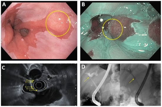


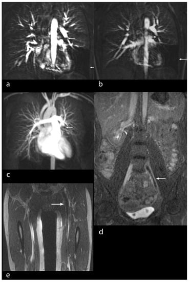

Figure 1. CT displaying an AL with extravasation of contrast agent and free air in the mediastinum indicated by the blue arrow.
Figure 2. Endoscopy showing a circumferential anastomotic leakage after oesophagectomy indicated by the blue arrow. Yellow arrow is pointing to the true lumen. The radiologist and the endoscopist were blinded to their respective findings.
Table 1. Patient and tumour characteristics.
Table 1. Patient and tumour characteristics.
Variables Male28 (84.8%)Female5 (15.2%)Age58.5 (32–83)BMI a25.5 (16–47)ASA b I1 (3%)ASA II18 (54.5%)ASA III14 (42.5%)Tumour location Middle third1 (3%)Distal third32 (97%)Tumour histology Adenocarcinoma30 (91%)Squamous cell carcinoma3 (9%)Open procedure28 (84.8%)Hybrid procedure5 (15.2%)R0-Resection33 (100%)Chemotherapy16 (48.5%)Radiochemotherapy5 (15.2%)No neoadjuvant therapy12(36.3%)Table 2. Efficiency and Feasibility of UE and CT.
Table 2. Efficiency and Feasibility of UE and CT.
NoUECTLeaks333323 010Sensitivity 100 %69.7%Technical feasibility333333
留言 (0)