MAIT cells play a pivotal role in immunity. Indeed, MAIT cells are implicated in cancers as well as in an array of diseases such as bacterial, viral, and parasitic infections, and allergic, autoimmune, and metabolic diseases [
7]. As is the case for other innate-like T cells, MAIT cells are characterized for the semi-invariant T cell receptor (TCR) alpha variable (TRAV)—1-2-TCR alpha joint (TRAJ)-33, -12, and -20 for human and TRAV1-TRAJ33 for mouse—and for the biased use of TCR beta repertoires [
8,
9]. An early study identified CD161 (killer cell lectin-like receptor), CD26 (dipeptidyl peptidase IV), and CD243 (ATP-dependent multidrug efflux pump) as the markers for MAIT cells [
1]. MAIT cells can express activation markers (CD44, CD25, CD38, CD39, CD69 and human leukocyte antigen (HLA)-DR) upon activation. Furthermore, MAIT cells harbor the costimulatory/coinhibitory molecules, CD27, CD28, programmed cell death receptor (PD)-1, and cytotoxic T-lymphocyte-associated antigen (CTLA-4) [
10,
11,
12,
13], the chemokine receptors, CCR5, CCR6, CCR9, and CXCR6, and the cytokine receptors, interleukin (IL)-1 receptor (R), IL-7R, IL-12R, IL-15R, IL-18R, and IL-23R [
7]. MAIT cells are, however, unique in terms of transcription factor expression. In fact, MAIT cells could harbor promyelocytic leukemia zinc finger (PLZF), retinoic acid receptor-related orphan receptor (ROR)γt, T-bet, and Eomesdermin (Eomes). PLZF is required for functional maturation and for exerting the innate-like features of MAIT cells, such as immediate production of inflammatory cytokines and cytotoxic molecules. Whereas RORγt is required for the development of T helper (Th) 17 cells and of group 3 innate lymphoid cells (ILC3s) [
14], T-bet and Eomes are required for Th1 development and the cytotoxic activity of NK cells and CD8 T cells, respectively [
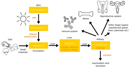
15].Development of MAIT cells is contingent upon non-polymorphic major histocompatibility complex I-related molecule (MR1) and microbiota, and it consists of three stages as defined by the expression of CD24 and CD44 (stage 1–3), in which stage 3 consists of extrathymic expansion and maturation [
17,
18]. MR1 presents the non-peptide antigens: vitamin B2 metabolites, such as 5-(2-oxoethylideneamino)-6-D-ribitylaminouracil (5-OE-RU) and 5-(2-oxopropylideneamino)-6-D-ribitylaminouracil (5-OP-RU), and vitamin B6 metabolites, such as 6-formyl pterin (6-FP). Whereas 5-OE-RU and 5-OP-RU function as agonists, 6-FP antagonizes the function of MAIT cells [
19,
20]. Development of MAIT cells in the murine thymus is dependent on 5-OP-RU synthesized by bacteria harboring the vitamin B2 biosynthesis pathway [
21]. Accordingly, germ-free mice lack MAIT cells [
17,
22].There exist two different pathways for MAIT cell activation, one dependent on TCR and the other one independent of TCR. The former is triggered through the presentation of antigens, such as 5-OE-RU and 5-OP-RU, loaded onto MR1 in most, if not all, cells and is not limited to professional antigen presenting cells (APCs) such as dendritic cells, macrophages, and B cells. Such TCR engagement results in activation of MAIT cells, as manifested by upregulation of CD25, CD69, CD38, and HLA-DR, followed by TNF-α and/or IL-17 production [
1,
19]. MAIT cells are also activated by the cytokines IL-12, IL-15, and IL-18, which are TCR-independent in nature [
23]. Intriguingly, TCR-dependent activation results in more production of inflammatory cytokines than TCR-independent activation [
24].

留言 (0)