
記住我


There are technical points in the hip embolization that must be highlighted. First, in patients where pain and inflammation are more prominent in gluteal muscles, ascending embolization of superior and/or inferior gluteal arteries may be necessary. In this cohort, two patients needed this approach, since embolization of regular branches was not enough to relief their pain. The embolization of gluteal branches was not pre-planned and was performed in the same session, and the decision-making was based by patient symptoms, which were not improving during the embolization. For this reason, we believe the full collaboration of patient is extremely necessary and is that why the procedures were performed under local anesthesia without sedation. In most patients, however, there are several collaterals in this area, and catheterization of the ascending branch of the LFCA is adequate to relief all areas of pain (Fig. 3).
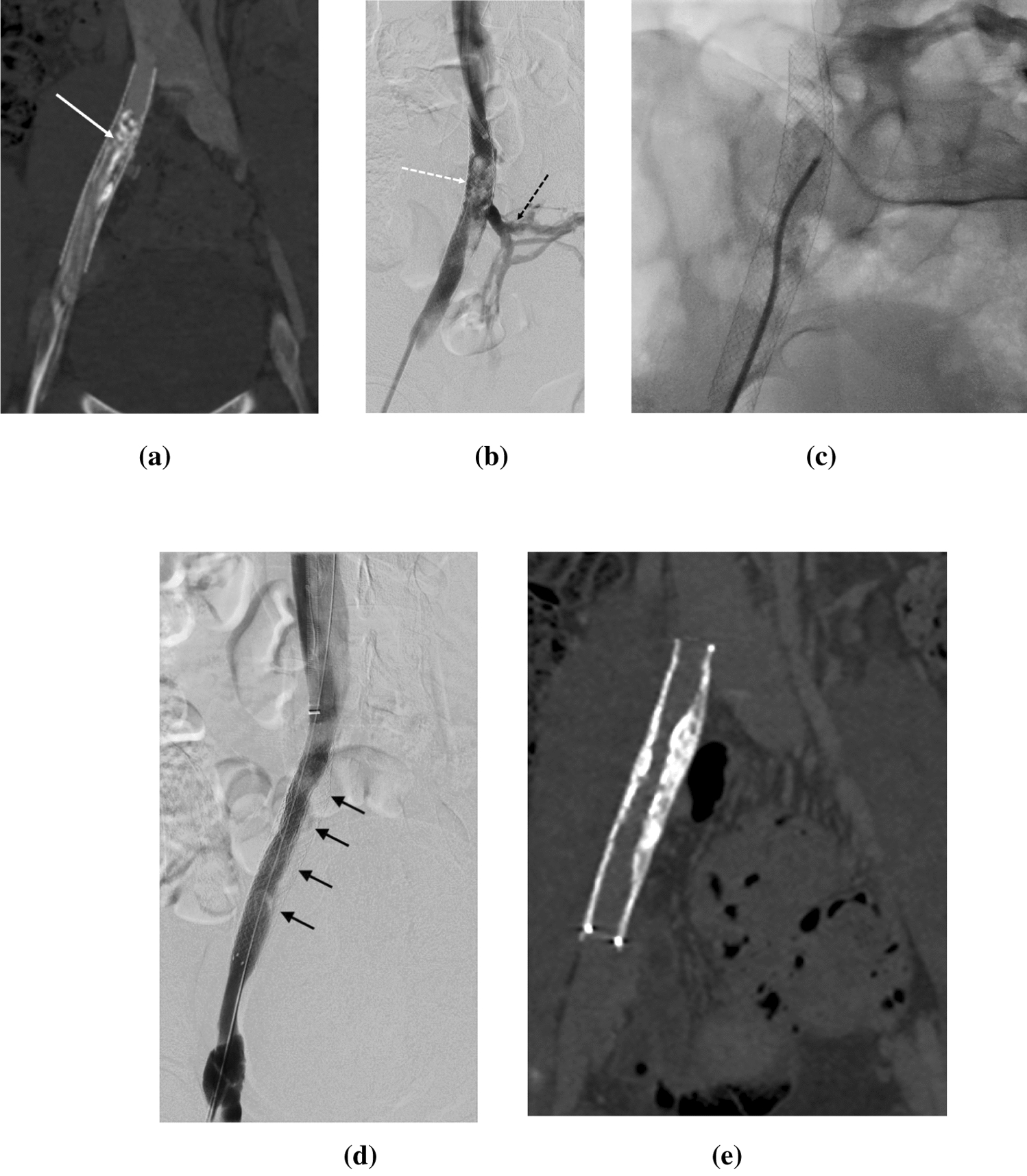
Fig. 3
Illustrative image of anterior (A) and posterior (B) vascularization of the hip. 1—deep femoral artery, 2—medial circumflex femoral artery, 3—lateral circunflex femoral artery, 4—superior branch of the lateral circunflex femoral artery, 5—transverse branch of the lateral circunflex femoral artery, 6—inferior branch of the lateral circunflex femoral artery, 7—superior gluteal artery, 8—inferior gluteal artery. *—Gluteus minimus muscle, o—piriformis muscle
Second, in contrast with genicular artery embolization, the tumor-like blush is not as common as expected, and corkscrew-like arteries were found. Third, LCFAE favors the use of imipenen/cilastatin, since this embolic agent has the theoretical advantage of preventing ischemia, as demonstrated by Woodhams et al. [11] In this territory, there is a particular concern of the orthopedic team, since there is an unknown risk of aseptic hip necrosis (AHN). In this short-term cohort, we do not have any clinical sign of osteonecrosis or AHN, and no patient was submitted to hip replacement due to worsening of the symptoms following embolization. Just one MRI was performed before one year [7], with no sign of osteonecrosis. These findings so far are similar of GAE, where no osteonecrosis was found [12]. There were two posterior tight numbness, probably due to inadvertent embolization of sciatic nerve branches. Both had spontaneous improvement, with no additional complications. After this first two events, the authors did not have any similar complication.
This study has limitations. In addition to the small sample size and short-term follow-up, MRI was not performed in most patients after 6 months, compromising information about osteonecrosis. In addition, despite that hip OA and GTPS are vastly associated, the analysis of both treatments in this study may be a confounder. Multiple embolic agents used and the lack of information of medications used before and after the first patients can also be cofounders of the results.
留言 (0)