
記住我

Polyphosphates and DNA appeared on the podium of hemostasis and thrombosis in the past 2 decades, and hence joined the well-known therapeutic heparins, as biorelevant polyanions in this field. Although both size and sulfation pattern have been appreciated as key determinants of heparin's anticoagulant properties, our knowledge on the mostly prothrombotic effects of polyphosphates and DNA is still emerging.
After a brief summary of how these polyanions of different chemical structure have entered the field of hemostasis and thrombosis, this review will summarize recent literature data on the size-dependence, and electric charge-dependence of their modulatory actions on fibrin structure and lysis.
 Box 1:
 no caption available
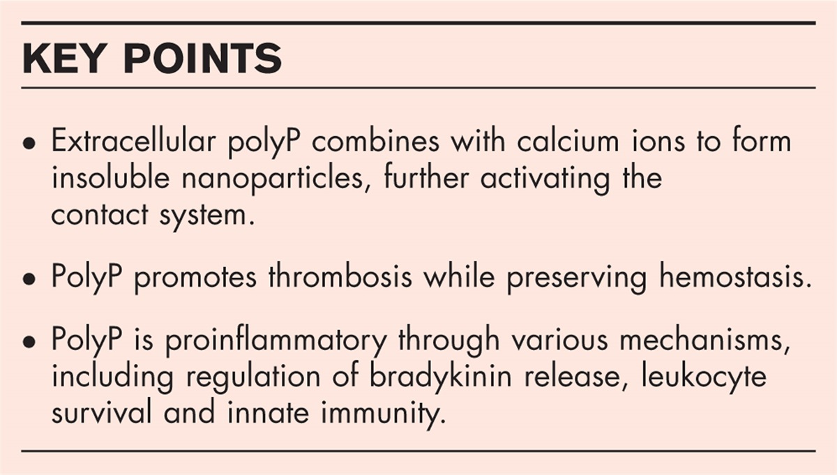
POLYPHOSPHATES: THE BIG, THE SMALL AND THE PARTICULATEPolyphosphates (PolyP) are linear polymers of phosphate residues linked with high-energy phosphoanhydride bonds, which carry one negative charge/phosphate unit at physiological pH. Microorganisms store very long polyphosphates containing hundreds to thousands of phosphate units most probably as a storage pool of energy during starvation [1]. Among mammalian cells, platelet dense granules have been found to contain large amounts of polyphosphates together with Ca2+-ions, which are secreted upon platelet activation and granule release [2]. As opposed to microorganisms, the size of platelet polyphosphates was estimated to be in the range of 60–100 phosphate monomers, and multiple prothrombotic interactions between polyphosphates and the hemostatic system have been described over the years, many of them being dependent on polymer length [3]. Contact pathway activation in a FXII-dependent manner is also profoundly dependent on polyphosphate length: while polymers up to 30 mers did not shorten the clotting time of recalcified plasma, 60–100 mers typically found in platelets showed about five-fold higher activity than kaolin, whereas the bacterial PolyP – longer than 1000 mers were about 3000-fold more potent [4]. Since FXII-dependent contact activation of the coagulation cascade plays apparently no significant role in the process of physiological hemostasis, but its downregulation dampens thrombosis in animal models, specific FXII-inhibitor, FXI-inhibitor and polyphosphate-inhibitor may offer a new promising therapeutic regimen of antithrombotics without the dangerous side effect of bleeding [5–7]. After the publication of numerous data on the prothrombotic, antifibrinolytic and inflammatory effects of linear ‘soluble’ polyphosphates, platelet-released polyphosphates were visualized to remain bound to platelet membrane surface as mostly Ca2+-polyphosphate nanoparticles much bigger in size than the secreted linear soluble polymers [8]. It was argued that the true form of platelet polyphosphates is this nanoparticular form, the idea being based mostly on their high potency in FXII-dependent coagulation activation, the very low water-solubility of Ca2+-polyphosphate mixtures and the relatively low amount of polyphosphate detected in the linear soluble form after platelet degranulation.
DNA: THE HUGE SLIMEThe polyanionic nature of nucleic acids is due to the phosphate-sugar backbone, which contains one negative charge/nucleotide unit at physiological pH. Based on a crude comparison of the chemical structures of linear polyphosphates and DNA one could speculate, that about 3 phosphate units in polyphosphates might be as long as one nucleotide, that is, the linear negative charge density of polyphosphates is three-fold higher than that of nucleic acids (Fig. 1). Cell-free DNA (cfDNA) of various sizes can be released in multiple cellular processes. DNA displaying a ladder pattern of DNA at 150 interval (1 nucleosomal unit) is released by apoptotic cells, whereas DNA fragments released during necrosis and the formation of neutrophilic extracellular traps (NETs) can be much larger, in excess of 10 000 basepairs. Extracellular DNA in NETs forms a sticky network that traps pathogens, but it also activates the contact pathway of coagulation and promotes thrombosis in multiple ways [9,10▪]. Elevated cfDNA levels in plasma have been detected in a number of diseases, including sepsis and patients with higher cfDNA levels are more likely to face severe complications and death [11].
 FIGURE 1:
 Relative size and charge density of biorelevant polyanions affecting the structure and function of fibrin. The drawn structures are scaled to indicate the equivalent relative size of the molecules.
HEPARINS AND RELATIVES COME IN DIFFERENT SIZE AND CHARGEHeparins belong to the class of glucosaminoglycans (GAGs), which are linear polymers of repeating disaccharide units and carry multiple negative charges. The family of GAGs include heparan sulfate, chondroitin sulfate, keratan sulfate and hyaluronic acid, all of them contain the negatively charged uronic acid in every other position, and all but hyaluronic acid are also sulfated, which adds to their polyanionic nature (Fig. 1). Endogenous heparin is synthesized mostly in mast cells, and resembles heparan sulfate among GAGs, but heparin is shorter and more sulfated than heparan sulfate. The exact regulation of mammalian GAG synthesis is not yet fully understood, but all GAGs are well known for their heterogenity with respect to both length and sulfation pattern. Therapeutically applied heparins are largely purified from porcine or bovine intestinal mucosa, or bovine lungs, tissues naturally rich in mast cells. The naturally occurring unfractionated heparin (UFH) has been used in the clinical practice as an anticoagulant for long time, since it facilitates the inactivation of thrombin by antithrombin [12]. The ubiquitously expressed heparan sulfate proteoglycans exert a similar anticoagulant mechanism on the luminal surface of the endothelium, which contributes to the physiological nonthrombogenic nature of the vessel wall. Heparin binds antithrombin with a well characterized pentasaccharide sequence, and the heparin-bound antithrombin shows a higher reactivity to thrombin and FXa. UFH is long enough to bind thrombin as well, which results in an overall 4000-fold increase in the thrombin inactivation rate in the ternary complex. To reduce heterogenity, potential contaminants from the animal sources, and to improve the pharmacokinetic properties, shorter heparins were derived from UFH (low-molecular-weight heparins, LMWHs). LMWHs differ from UFH in their anticoagulant properties: the shorter length hampers their thrombin-binding ability, which results in an increasing anti-FXa/antithrombin ratio of the inhibitory potency of heparin-bound antithrombin. In addition to UFH and LMWHs, second-generation LMWH (or ultralow molecular weight heparins) and the synthetic pentasaccharide sequence (fondaparinux) have been also introduced in the clinical practice as anticoagulants [13]. In addition to being an anticoagulant, about 70% of the molecules in UFH preparations do not bind antithrombin and this fraction is termed ‘inactive’ or ‘low-affinity material’. As it turns out, such ‘nonanticoagulant’ heparin molecules exert a wide variety of other biological activities: they can modulate growth factor availability, cancer metastasis, inflammatory reactions to mention a few and some of these effects depend on the length of the heparin molecule [14▪].
SIZE-DEPENDENT AND CHARGE-DEPENDENT EFFECTS OF POLYANIONS ON THE FORMATION, STRUCTURE, AND LYSIS OF CLOTSPolyphosphates, DNA and various GAG groups have all been studied over the years, as to their modulatory effects on clot formation and structure with consequent changes in the fibrin susceptibility to lysis. The presence of the kringle domains in addition to the serine-protease domain and the optimal 3D-structure of plasmin enables its high processivity while solubilizing the fibrin polymer, hence altered network structure greatly influences lytic susceptibility [15,16]. Some previous studies looked specifically at the role of polyanion size and/or charge, while in other studies one can mine the relevant information and draw a conclusion retrospectively.
The size-dependent nature of the interactions between polyphosphates and the coagulation system was early recognized [4], but comparative data in the field of fibrin structure and fibrinolysis have been emerging only recently. PolyP of 75 units enhances clot turbidity, and increases fiber thickness, as well as the mechanical strength of purified fibrin clots, which are also more resistant against incorporated plasmin-mediated or tPA-mediated fibrinolysis [17]. As a mechanistic background of the observed antifibrinolytic effects, impaired binding of tPA and plasminogen to fibrin, as well as a mildly inhibited plasminogen activation were demonstrated [18]. Comparative studies using PolyPs shorter than 100 mers (typical for platelets) and 700 mers (typical for microorganisms and cancer cells) revealed some fundamental size-dependent differences [19▪]. Short PolyPs accelerate fibrin formation, whereas PolyP700 delays clotting and results in a significantly higher final clot turbidity. All PolyPs enhance the mechanical stability of fibrin clots, but with different mechanisms: shorter PolyPs decrease fibrin deformability, whereas PolyP700 increase the maximal bearable deformation of the structure. Despite the size-dependent variabilities in the structures of PolyP/fibrin composite clots, PolyPs of all sizes delay plasmin-mediated clot lysis. This antifibrinolytic effect is not observed with plasmin derivatives lacking kringle domains, which are known to mediate plasmin binding to lysine residues, suggesting that these polyanions modify the kringle-dependent access of fibrinolytic proteases to fibrin fibers [19▪]. The antifibrinolytic effect of polyphosphates is observed with the nanoparticular (100–200-nm diameter) form of polyphosphates as well [20▪].
Incorporation of DNA into fibrinogen clotted with thrombin yields a mechanically less stable clot structure, which consists of longitudinally disarranged fibrin monomers [21]. In the plasma environment, clots containing DNA are characterized by thicker fibers and a tighter network, which could restrict the lytic function of surface-applied plasmin or tPA [11,22]. Despite the DNA-mediated structural changes, additional antifibrinolytic actions of DNA seem to be more important: these include the formation of a nonproductive ternary complex between plasmin, DNA and fibrin, and the binding of fibrin degradation products, which slows down the disassembly of the fibrin/DNA network. In terms of size and charge, we could anticipate a relatively lower longitudinal charge-density along a DNA-chain than along a polyphosphate-chain, while for disruption of fibrinolysis a minimum length of 300 bp is required [11].
Among the GAGs, we have studied chondroitin sulfate, dermatan sulfate, the chondroitin sulfate/dermatan-sulfate chain of decorin and various size-variants of hyaluronic acid (25, 500, 1500 kDa), which are nonsulfated polyanions [23,24]. Although the exact size of the chondroitin sulfate, dermatan-sulfate in the applied preparations were not directly measured, they were even smaller than the 25-kDa hyaluronic acid-variants based on their migration in gel electrophoresis. The two large-sized hyaluronic acid variants delay fibrin formation, and increase fibrin fiber diameter and clot porosity. Such a pronounced structural change is not seen with the smallest (25 kDa) hyaluronic acid-variant, chondroitin sulfate or dermatan-sulfate. However, they all result in a decreased shear-resistance of composite fibrin clots, and they all inhibit tPA-mediated plasminogen activation, both in solution and on composite fibrin surfaces. The plasmin-mediated fibrinolysis is impaired by large-sized hyaluronic acid and not affected by 25-kDa hyaluronic acid, whereas chondroitin sulfate/dermatan-sulfate rather enhance it.
Yet another recently studied class of polyanions is the therapeutically applied heparin variants. The largest one, UFH behaves similarly to the large hyaluronic acid-variants with respect to delay in clotting kinetics and formation of thicker fibrin fibers enclosing larger pores, but as a difference, the UFH/fibrin composite structure is mechanically more stable than pure fibrin. The UFH-mediated structural changes are largely dependent on the sulfation pattern, as indicated by the distinct effects of partially and fully desulfated UFH-variants. Considering molecular weights, even the ‘large’ UFH with its average molecular weight of 15 kDa is smaller than the 25-kDa hyaluronic acid-variant, whereas LMWH and the pentasaccharide are even smaller, about 5 and 1.7 kDa, respectively. LMWH and the pentasaccharide both accelerate fibrin formation and result in a tighter network with smaller pores. In terms of biomechanics, the LMWH/fibrin clots are still more stable than pure fibrin, but the pentasaccharide/fibrin clots are essentially identical to pure fibrin [19▪]. In contrast to results gained with isolated proteins in the absence of antithrombin, all therapeutic anticoagulants modify the plasma clot structures from treated patients by decreasing thrombin generation in an antithrombin-dependent way, which results in thicker fibers enclosing larger pores and a consequent higher lysability with tPA [25,26].
Based on these data, one could speculate that very large sized polyanions like hyaluronic acid or DNA, disturb the arrangement of fibrin monomers and protofibrils within the fibers, which weakens the clot and geometrically destroys the fibers as substrates for plasmin, ultimately delaying fibrinolysis. PolyP700 delays clotting and thickens fibers similarly to 500-kDa hyaluronic acid, which is of about the same size, but PolyP700 is clearly more densely decorated with negative charges, which explains its clot-strengthening effect. UFH, albeit a much shorter but highly sulfated polymer, can cause charge-dependent structural changes and strengthen the network by electrostatic interactions. The smallest polyanions, PolyP100, PolyP45, LMWH and the pentasaccharide share many common modulatory effects. They accelerate fibrin formation and favor a tighter, mechanically more stable network. These effects are not mirrored with the similarly sized, but less charged GAGs, such as chondroitin sulfate, dermatan-sulfate or with the nonsulfated hyaluronic acid variant. Pentasaccharide, albeit heavily charged, may be too small to sustain the electrostatic interactions necessary for a mechanically stabilized fibrin network (Table 1). In addition to the characteristic structural changes in polyanion-fibrin clots, their mostly increased resistance to fibrinolysis can stem from masking of kringle-dependent binding sites necessary for the efficient action of plasmin, as seen in clots containing DNA, large hyaluronic acid and PolyPs [11,18,24].
Table 1 - Size-dependence and charge-dependence of polyanion effects on fibrin clot formation, structure and fibrinolysis Polyanions Clotting Fiber thickness and porosity (Ks) Critical stress to disintegrate clot Fibrinolysis HA 1500 kDaHA 500 kDaDNA DelayedDelayedNo change ↑Fibers, ↑Ks↑Fibers, ↑Ks↑Fibers, ↓Ks LowerLowerLower DelayedDelayedDelayed PolyP700 Delayed ↑Fibers, ↓Ks Higher Delayed CSDSHA 25 kDa DelayedNo changeNo change →Fibers, → Ks→Fibers, → Ks↑Fibers, → Ks LowerLowerNo change FasterFasterNo change UFH 15 kDa Delayed ↑Fibers, ↑Ks Higher Delayed Partially desulfated UFHFully desulfated UFH AcceleratedNo change ↑Fibers, ↓Ks↑Fibers, →Ks LowerNo change No changeNo change LMWH 5 kDaPolyP <100 mer AcceleratedAccelerated ↑Fibers, ↓Ks↑Fibers, →Ks HigherHigher DelayedDelayed PS 1.7 kDa Accelerated ↑Fibers, ↓Ks No change Delayed The table compares the polyanion effects on clotting and lysis times, fibrin fiber thickness, network porosity and critical shear stress necessary to disintegrate clots in rheometry (Ks indicates the Darcy constant reflecting pore size, whereas ↑, ↓, → arrows indicate increase, decrease or no change, respectively in comparison to the additive-free fibrin). Data obtained in comparable in-vitro experimental setups with polyphosphates of indicated polymer size (PolyP700, PolyP [17,19▪,21,23,24,35]. The additives are listed according to their relative size (polyanion length or molecular mass) in decreasing order.Sepsis is characterized by the activation of inflammatory and procoagulant pathways, endothelial cell injury, multiorgan dysfunction and death [27]. Extracellular histones released from dying macrophages have been identified as major mediators of death in sepsis [28]. Both histone-neutralizing antibodies and the histone-cleaving activated protein C improve survival rates of septic mice, baboons and humans [28–30]. The potential bleeding complications seen with use of activated protein C, warranted a quest for other means to neutralize histone-effects. In this context, heparins, as polyanions were expected to neutralize positively charged histones in a charge-dependent manner. Heparins play an important role in the prevention of thromboembolic complications in critically ill patients and their administration in severe sepsis or sepsis associated with disseminated intravascular coagulation, has been found to improve survival [31,32]. Beside its anticoagulant activities, heparin displays anti-inflammatory actions including antihistone effects which are beneficial in diseases such as asthma or COVID-19, but concerns for bleeding complications have limited its value and once more, raised the unmet need for the development of nonanticoagulant heparin derivatives [33,34▪▪]. An important aspect to consider is that the anticoagulant and histone-neutralizing properties of heparin might be separable. Various histone effects are charge-dependently neutralized by UFH, its nonanticoagulant, nonantithrombin-binding fraction and a partially desulfated, low-anticoagulant UFH variant, whereas fully desulfated UFH demonstrates neither anticoagulant, nor antihistone activity [35,36,37▪]. Histone-polyanion interactions appear to be more complicated than a simple charge-neutralization, and structural elements required for histone-neutralization may depend even on the specific histone-effect under consideration. Thus, UFH, LMWH, pentasaccharide and a nonantithrombin-binding LMWH variant counteract the histone-mediated inhibition of activated protein C generation, and all these heparins with the exception of the pentasaccharide form reverse tissue factor upregulation in cultured endothelial cell and histone-mediated cytotoxicity [38▪▪]. However, the recently reported effects of histones on fibrin structure and function (mechanical strengthening, fibrinolytic resistance) are often not counteracted, but rather enhanced by DNA, LMWH, pentasaccharide and short PolyPs [21,35,39▪].
CONCLUSIONTargeting polyanions (cfDNA from NETs or other origin, polyphosphate and GAGs) in arterial, venous, infection-associated and cancer-associated thrombosis will remain a promising therapeutic approach with the hope of developing antithrombotics that do not interfere with physiological hemostasis [40▪▪,41,42▪▪,43▪]. Exploration of appropriate tools to eliminate the harmful effects of cfDNA and polyphosphates, or construction of a polyanion of optimal size and charge to confer potent histone-neutralizing properties would be of great clinical benefit.
AcknowledgementsNone.
Financial support and sponsorshipThe current work was supported by the Hungarian National Research, Development and Innovation Office (NKFIH) #137563 and the Thematic Institutional Excellence funding scheme of the Ministry of Innovation and Technology in Hungary for the Molecular Biology thematic programme of Semmelweis University (TKP2021-EGA-24).
Conflicts of interestThere are no conflicts of interest.
REFERENCES AND RECOMMENDED READINGPapers of particular interest, published within the annual period of review, have been highlighted as:
▪ of special interest
▪▪ of outstanding interest
REFERENCES 1. Morrissey JH, Choi SH, Smith SA. Polyphosphate: an ancient molecule that links platelets, coagulation, and inflammation. Blood 2012; 119:5972–5979. 2. Ruiz FA, Lea CR, Oldfield E, et al. Human platelet dense granules contain polyphosphate and are similar to acidocalcisomes of bacteria and unicellular eukaryotes. J Biol Chem 2004; 279:44250–44257. 3. Morrissey JH, Smith SA. Polyphosphate as modulator of hemostasis, thrombosis, and inflammation. J Thromb Haemost 2015; 13: (Suppl 1): S92–S97. 4. Smith SA, Choi SH, Davis-Harrison R, et al. Polyphosphate exerts differential effects on blood clotting, depending on polymer size. Blood 2010; 116:4353–4359. 5. Labberton L, Kenne E, Long AT, et al. Neutralizing blood-borne polyphosphate in vivo provides safe thromboprotection. Nat Commun 2016; 7:12616. 6. Bickmann JK, Baglin T, Meijers JCM, et al. Novel targets for anticoagulant lacking bleeding risk. Curr Opin Hematol 2017; 24:419–426. 7. Srivastava P, Gailani D. The rebirth of the contact pathway: a new therapeutic target. Curr Opin Hematol 2020; 27:311–319. 8. Verhoef JJF, Barendrecht AD, Nickel KF, et al. Polyphosphate nanoparticles on the platelet surface trigger contact system activation. Blood 2017; 129:1707–1717. 9. Gould TJ, Lysov Z, Liaw PC. Extracellular DNA and histones: double-edged swords in immunothrombosis. J Thromb Haemost 2015; 13: (Suppl 1): S82–S91. 10▪. Rangaswamy C, Englert H, Deppermann C, et al. Polyanions in coagulation and thrombosis: focus on polyphosphate and neutrophils extracellular traps. Thromb Haemost 2021; 121:1021–1030. 11. Gould TJ, Vu TT, Stafford AR, et al. Cell-free DNA modulates clot structure and impairs fibrinolysis in sepsis. Arterioscler Thromb Vasc Biol 2015; 35:2544–2553. 12. Hirsch J, Warkentin TE, Shaughnessy SG, et al. Heparin and low-molecular-weight heparin: mechanism of action, pharmacokinetics, dosing, monitoring, efficacy, and safety. Chest 2001; 119: (1, Suppl): 64S–94S. 13. Walenga JM, Lyman GH. Evolution of heparin anticoagulants to ultralow-molecular-weight heparins: a review of pharmacologic and clinical differences and applications in patients with cancer. Crit Rev Oncol Hematol 2013; 88:1–18. 14▪. Beurskens DMH, Huckriede JP, Sckrijver R, et al. The anticoagulant and nonanticoagulant properties of heparin. Thromb Haemost 2020; 120:1371–1383. 15. Weisel JW, Litvinov RI. The biochemical and physical process of fibrinolysis and effects of clot structure and stability on the lysis rate. Cardiovasc Hematol Agents Med Chem 2008; 6:161–180. 16. Kolev K, Tenekedjiev K, Komorowicz E, Machovich R. Functional evaluation of the structural features of proteases and their substrate in fibrin surface degradation. J Biol Chem 1997; 272:13666–13675. 17. Smith SA, Morrissey JH. Poyphosphate enhances fibrin clot structure. Blood 2008; 112:2810–2816. 18. Mutch NJ, Engel R, de Willige SU, et al. Polyphosphate modifies the fibrin network and down-regulates fibrinolysis by attenuating binding of tPA and plasminogen to fibrin. Blood 2010; 115:3980–3988. 19▪. Komorowicz E, Balázs N, Tanka-Salamon A, et al. Biorelevant polyanions stabilize fibrin against mechanical and proteolytic decomposition: Effects of polymer size and electric charge. J Mech Behav Biomed Mater 2020; 102:103459. 20▪. Lovas M, Tanka-Salamon A, Beinrohr L, et al. Polyphosphate nanoparticles enhance the fibrin stabilization by histones more efficiently than linear polyphosphates. PLoS One 2022; 17:e0266782. 21. Longstaff C, Varjú I, Sótonyi P, et al. Mechanical stability and fibrinolytic resistance of clots containing fibrin, DNA, and histones. J Biol Chem 2013; 288:6946–6956. 22. Varjú I, Longstaff C, Szabó L, et al. DNA, histones and neutrophil extracellular traps exert antifibrinolytic effects in a plasma environment. Thromb Haemost 2015; 113:1289–1298. 23. Rottenberger Z, Komorowicz E, Szabó L, et al. Lytic and mechanical stability of clots composed of fibrin and blood vessel wall components. J Thromb Haemost 2013; 11:529–538. 24. Komorowicz E, Balázs N, Varga Z, et al. Hyaluronic acid decreases the mechanical stability, but increases the lytic resistance of fibrin matrices. Matrix Biol 2017; 63:55–68. 25. Varin R, Mirshahi S, Mirshahi P, et al. Clot structure modification by fondaparinux and consequence on fibrinolysis: a new mechanism of antithrombotic activity. Thromb Haemost 2007; 97:27–31. 26. Bridge KI, Philippou H, Ariens RAS. Clot properties and cardiovascular disease. Thromb Haemost 2014; 112:901–908. 27. Lorente JA, García-Frade LJ, Landín L, et al. Time course of hemostatic abnormalities in sepsis and its relation to outcome. Chest 1993; 103:1536–1542. 28. Xu J, Zhang X, Pelayo R, et al. Extracellular histones are major mediators of death in sepsis. Nat Med 2009; 15:1318–1321. 29. Taylor FB Jr, Chang A, Esmon CT, et al. Protein C prevents the coagulopathic and lethal effects of Escherichia coli infusion in the baboon. J Clin Invest 1987; 79:918–925. 30. Bernard GR, Vincent JL, Laterre PF, et al. Recombinant human protein C Worldwide Evaluation in Severe Sepsis (PROWESS) study group. Efficacy and safety of recombinant human protein C for severe sepsis. N Engl J Med 2001; 344:699–709. 31. Cornet AD, Smit EGM, Beishuizen A, Groeneveld ABJ. The role of heparin and allied compounds in the treatment of sepsis. Thromb Haemost 2007; 98:579–586. 32. Zarychanski R, Abou-Setta AM, Kanji S, et al. The efficacy and safety of heparin in patients with sepsis: a systematic review and metaanalysis. Crit Care Med 2015; 43:511–518. 33. Young E. The anti-inflammatory effects of heparin and related compounds. Thromb Res 2008; 122:743–752. 34▪▪. Buijsers B, Yanginlar C, Maciej-Hulm ML, et al. Beneficial nonanticoagulant mechanisms underlying heparin treatment of COVID-19 patients. EBoMedicine 2020; 59:102969. 35. Longstaff C, Hogwood J, Gray E, et al. Neutralisation of the anticoagulant effects of heparin by histones in blood plasma and purified systems. Thromb Haemost 2016; 115:591–599. 36. Wildhagen KCAA, de Frutos PG, Reutelingsperger CP, et al. Nonanticoagulant heparin prevent histone-mediated cytotoxicity in vitro and improves survival in sepsis. Blood 2014; 123:1098–1101. 37▪. Hogwood J, Pitchford S, Mulloy B, et al. Heparin and nonanticoagulant heparin attenuate histone-induced inflammatory responses in whole blood. PLoS One 2020; 15:e0233644. 38▪▪. Sharma N, Haggstrom L, Sohrabipour S, et al. Investigations of the effectiveness of heparin variants as inhibitors of histones. J Thromb Haemost 2022; 20:1485–1495. 39▪. Komorowicz E, Balázs N, Tanka-Salamon A, et al. Size- and charge-dependent modulation of the lytic susceptibility and mechanical stability of fibrin-histone clots by heparin and polyphosphate variants. J Thromb Haemost 2021; 19:1307–1318. 40▪▪. Wolach O, Martinod K. Casting a NET on cancer: the multiple roles for eutrophil extracellular traps in cancer. Curr Opin Hematol 2022; 29:53–62. 41. Thalin C, Hisda Y, Lundström S, et al. Neutrophil extracellular traps. Villains and targets in arterial, venous, and cancer-associated thrombosis. Arterioscler Thromb Vasc Biol 2019; 39:1724–1738. 42▪▪. Ligi D, Maniscalco R, Plebani M, et al. Do circulating histones represent the missing link among COVID-19 infection and multiorgan injuries, microvascular coagulopathy and systemic hyperinflammation? J Clin Med 2022; 11:1800. 43▪. Moiana M, Aranda F, de Larranaga G. A focus on the roles of histones in health and diseases. Clin Biochem 2021; 94:12–19.
留言 (0)