
記住我

Acute lymphoblastic leukemia is the most common pediatric cancer and, in the majority of cases, originates within the B cell lineage (Hunger & Mullighan, 2015). Genome-wide studies demonstrated that 60% of all B-cell precursor acute lymphoblastic leukemia (B-ALL) cases carry genetic alterations in genes coding for regulators of B cell development, with the most commonly affected transcription factor genes being PAX5, EBF1, and IKZF1 (Ikaros) (Kuiper et al, 2007; Mullighan et al, 2007, 2008). Mutations leading to aberrant activation of tyrosine kinase and cytokine receptor signaling were also identified in about 15% of all B-ALL cases, which are collectively referred to as Philadelphia chromosome-like (Ph-like) B-ALLs and have a poor prognosis (Roberts et al, 2012, 2014; Ofran & Izraeli, 2017). One Ph-like B-ALL subtype is characterized by the PAX5-JAK2 rearrangement (Nebral et al, 2009; Roberts et al, 2012), which we have studied here with regard to its molecular and oncogenic function in a mouse model.
The transcription factor Pax5 is an essential regulator of B cell commitment and development (Nutt et al, 1999; Horcher et al, 2001; Medvedovic et al, 2001). It acts as a transcriptional repressor to suppress B-lineage-inappropriate genes (Delogu et al, 2006) and functions as an activator to induce gene expression required for B cell development and function (Schebesta et al, 2007; Revilla-i-Domingo et al, 2012). Notably, mature B cells upon conditional loss of Pax5 cannot only be converted into functional T cells via dedifferentiation to uncommitted bone marrow progenitor cells, but also give rise to an aggressive progenitor cell leukemia (Cobaleda et al, 2007). Hence, Pax5 maintains B cell identity throughout B-lymphopoiesis and functions as a tumor suppressor in the B-lymphoid lineage.
PAX5 was identified as a haploinsufficient tumor suppressor gene in human B-ALL, as heterozygous PAX5 deletions, rearrangements, and loss-of-function mutations are present in one third of all cases (Kuiper et al, 2007; Mullighan et al, 2007). PAX5 translocations occur at a frequency of 2.5% in human B-ALLs and are currently known to involve 28 different partner genes, generating novel PAX5 fusion proteins (Gu et al, 2019). The different partner genes code for proteins of diverse functions such as transcription factors (exemplified by ETV6), signal transducers (JAK2), chromatin regulators (BRD1), structural proteins (ELN), and proteins of unknown function (NOL4L) (Cazzaniga et al, 2001; Nebral et al, 2009; Coyaud et al, 2010). The PAX5 fusion proteins contain the N-terminal DNA-binding paired domain, but lack the potent C-terminal transactivation domain of PAX5 (Nebral et al, 2009; Coyaud et al, 2010) and were therefore thought to act as dominant-negative proteins to prevent genomic DNA binding of the full-length PAX5 protein expressed from the second allele (Kawamata et al, 2012; Fortschegger et al, 2014). However, we could demonstrate in a mouse model that the PAX5-ETV6 fusion protein does not interfere with the expression of regulated Pax5 target genes and hence does not act as a dominant-negative protein (Smeenk et al, 2017). Instead, PAX5-ETV6 functions as a potent oncoprotein to promote B-ALL development in combination with loss of the tumor suppressors Cdkn2a and Cdkn2b (Smeenk et al, 2017). Similarly, heterozygous loss of Pax5 is not sufficient for tumor formation, as it additionally requires constitutive activation (ca) of STAT5 for leukemia development in transgenic caStat5b Pax5+/− mice (Heltemes-Harris et al, 2011). Hence, heterozygous PAX5 alterations promote B-ALL development in cooperation with a second oncogenic “hit.”
The Janus kinase 2 (JAK2) belongs to a family of nonreceptor tyrosine kinases that mediate signal transduction downstream of many cytokine and growth hormone receptors, regulating hematopoiesis, immunity, growth, and development (Chen et al, 2012; Villarino et al, 2017). Upon signaling, JAK2 phosphorylates STAT transcription factors in the cytoplasm, which promotes their dimerization and translocation to the nucleus, where they control the expression of genes involved in cell survival, differentiation, and metabolism (Malin et al, 2010; Villarino et al, 2017; de Araujo et al, 2019). In addition to this canonical JAK-STAT signaling function, JAK2 was also shown to be present in the nucleus and to directly phosphorylate histone H3 on tyrosine 41 (H3Y41ph), which, in turn, prevents interaction of the heterochromatin protein 1α (HP1α) with H3, thus leading to the activation of oncogenes such as Lmo2 and Myc (Dawson et al, 2009; Rui et al, 2010). Hence, these studies uncovered a second role of JAK2 as an “epigenetic writer” that stimulates expression of leukemogenic genes. JAK2 has directly been implicated in the development of Ph-like B-ALL by activating mutations, which are predominantly located in its autoinhibitory pseudokinase domain (JH2) (Chen et al, 2012). JAK2 also participates in translocations with at least 22 different partner genes, which all contain the catalytically active JAK2 kinase domain (JH1) as a common denominator (Nebral et al, 2009; Chen et al, 2012; Roberts et al, 2012; Akkari et al, 2020).
The PAX5-JAK2 rearrangement codes for a fusion protein consisting of the DNA-binding paired domain of PAX5 fused to only the kinase domain (JH1) of JAK2 (Nebral et al, 2009) (Appendix Fig S1A). As shown by detailed characterization in transfected cell lines, PAX5-JAK2 is a monomeric nuclear protein that can bind Pax5 recognition sequences and possesses constitutive kinase activity. Consequently, PAX5-JAK2 activates STAT5 by phosphorylation, which likely induces a STAT5-dependent gene program (Schinnerl et al, 2015). Moreover, JAK2 inhibitors efficiently block the constitutive kinase activity of PAX5-JAK2 in transfected cells, suggesting that these inhibitors may be beneficial for the treatment of PAX5-JAK2+ B-ALL (Roberts et al, 2014; Schinnerl et al, 2015). As the PAX5-JAK2 rearrangement inactivates one functional PAX5 allele, resulting in haploinsufficiency, and simultaneously leads to STAT5 activation, it may function as a dual-hit mutation to promote aggressive B-ALL. However, the function of PAX5-JAK2 in B cell development and leukemogenesis has not yet been investigated in an in vivo model system.
Here, we have generated a mouse model that expresses the PAX5-JAK2 protein under the control of the Pax5 locus. Pax5Jak2/+ mice exhibited normal B cell development up to 3 weeks of age, but thereafter rapidly developed an aggressive B-ALL tumor in the absence of another cooperating exogenous gene mutation. The DNA-binding function and kinase activity of Pax5-Jak2 both contributed to leukemia development, as evidenced by mutation of the paired domain of Pax5 or the catalytic center of Jak2. Unexpectedly, the wild-type Pax5 allele was lost in all Pax5Jak2/+ B-ALLs by acquired uniparental disomy, which facilitated efficient binding of Pax5-Jak2 to its genomic recognition sequences, thus pointing to an important oncogenic function of the fusion protein in the nucleus. While we could not find evidence for an epigenetic role of Pax5-Jak2 in the nucleus, STAT5 was highly phosphorylated in the earliest pre-leukemic B220low B cells of 4-week-old mice. Consistent with this finding, activated STAT5 target genes were upregulated in Pax5Jak2/+ B-ALLs. Together, these data indicate that the constitutively active Pax5-Jak2 kinase maintains active STAT5 at high levels in the nucleus, thus leading to continuous expression of STAT5 target genes in Pax5Jak2/+ B-ALL cells.
Results Pax5-Jak2 expression from the Pax5 locus leads to development of an aggressive B-ALLTo study the role of PAX5-JAK2 (Appendix Fig S1A) in B-ALL development, we used ES cell targeting to generate a mouse model by inserting human cDNA sequences, starting with exon 4 and encoding the remaining PAX5-JAK2 protein, into the mouse Pax5 locus to recapitulate the corresponding human rearrangement as closely as possible (Fig 1A and Appendix Fig S1B and C). Additionally, we inserted an IRES-luciferase indicator gene downstream of the Jak2 sequence and turned the endogenous Pax5 exon 4 into a loxP-stop-loxP (LSL) cassette by the insertion of a stop codon together with six polyadenylation sequences to generate the Pax5LSL-Jak2-Luc allele, which expresses the DNA-binding paired domain of Pax5 instead of the full-length Pax5-Jak2 protein (Appendix Fig S1B). The Pax5LSL-Jak2 allele was subsequently created by Dre recombinase-mediated deletion of the IRES-luciferase gene (Appendix Fig S1B). To enable the expression of the Pax5-Jak2 fusion, we eliminated the LSL cassette by ubiquitous Cre expression from the Meox2 locus (Tallquist & Soriano, 2000) in Meox2-Cre Pax5LSL-Jak2/+ or Meox2-Cre Pax5LSL-Jak2-Luc/+ mice, which will be thereafter referred to as Pax5Jak2/+ or Pax5Jak2-Luc/+ mice, respectively. The LSL cassette was efficiently deleted in cells of Pax5Jak2/+ and Pax5Jak2-Luc/+ mice, as shown by PCR analysis (Appendix Fig S1D). Immunoblot analysis of nuclear extracts with an anti-Pax5 antibody recognizing the N-terminal paired domain demonstrated that the Pax5-Jak2 and wild-type Pax5 proteins were similarly expressed in pro-B cells of 3-week-old Pax5Jak2/+ mice (Fig 1B and Appendix Fig S1E and F). These results therefore identified the Pax5Jak2/+ mouse as a valid model for studying the developmental and leukemogenic role of Pax5-Jak2 in B cells.

Figure 1. B-ALL development in Pax5Jak2/+ mice
Schematic diagram of the Pax5Jak2 allele. Human cDNA sequences, starting in PAX5 exon 4 and encoding the remaining PAX5-JAK2 protein, were inserted in frame into exon 4 of the mouse Pax5 locus to generate the Pax5Jak2 allele (Appendix Fig S1B). The C-terminal tag sequence (grey) contains an epitope for the V5 antibody, two cleavage sites for the TEV protease, and a biotin acceptor sequence (Biotin). A black oval denotes the B cell-specific enhancer (En) in intron 5 (Decker et al, 2009). pA, polyadenylation site. Notably, the human and mouse Pax5 protein sequences encoded from exon 1 to exon 5 contain only one amino acid substitution (human Ser13 vs. mouse Ile13 in exon 1) located upstream of the paired domain (Adams et al, 1992). Expression of the Pax5 protein in Pax5Jak2/+ pro-B cells. Nuclear extracts of short-term cultured pro-B cells from 3-week-old Pax5Jak2/+ and Pax5+/+ mice were analyzed by immunoblot analysis with an anti-Pax5 antibody recognizing the N-terminal paired domain. The size of marker proteins is indicated in kilodaltons (kDa) to the left. Flow-cytometric analysis of bone marrow cells from the two hindlegs of 3-week-old Pax5+/+ and Pax5Jak2/+ mice. The percentage of the cells in each gate or quadrant is indicated. Absolute cell numbers of the indicated cell types were determined by flow-cytometric analysis of the bone marrow from 3-week-old Pax5+/+ and Pax5Jak2/+ mice. Average cell numbers are shown with SEM and were statistically analyzed by multiple t-tests (unpaired two-tailed with Holm-Šídák's correction); ns, not significant (P > 0.05). See Methods section for flow-cytometric definition of the different cell types. One of 3 independent experiments is shown. Flow-cytometric analysis of bone marrow cells and splenocytes from Pax5Jak2/+ mice at the age of 4 or 6 weeks. The newly emerging B220low B cells are highlighted by black boxes. Kaplan-Meier survival analysis of Pax5Jak2/+ (black) and control Pax5+/+ (grey) mice. n, number of mice analyzed. A P value of < 0.0001 was determined for the survival curves by statistical analysis with the log-rank (Mantel-Cox) test. Size comparison of the spleen and lymph nodes from a control Pax5+/+ mouse and a Pax5Jak2/+ tumor mouse. Eosin-hematoxylin-stained sections of the lung and liver of a Pax5Jak2/+ tumor mouse. Infiltrating and blasting tumor cells are indicated by an arrow. Flow-cytometric analysis of lymph node cells from a control Pax5+/+ mouse and a 10-week-old Pax5Jak2/+ tumor mouse. Flow-cytometric analysis of B220lowCD19+ B cells from the bone marrow of a 4-week-old Pax5Jak2/+ mouse.Source data are available online for this figure.
Flow-cytometric analysis of B cell development in 3-week-old mice revealed that total B, pro-B, large and small pre-B, as well as immature B cells were present at similar numbers in the bone marrow of Pax5Jak2/+ and control Pax5+/+ mice (Fig 1C and D). These data therefore indicate that heterozygous expression of Pax5-Jak2 had no apparent effect on B cell development in young mice.
The first sign of aberrant B cell development appeared in the bone marrow of Pax5Jak2/+ mice at around 4 weeks of age with the emergence of B220low B cells in some of these mice (Fig 1E). At 6 weeks of age, most B lymphocytes in the bone marrow were B220low and proved to be tumorigenic, as their transplantation into wild-type C57BL/6 mice resulted in tumor development within 72 days (Appendix Fig S1G). With some delay, these tumorigenic B220low B cells also appeared in the spleen (Fig 1E), in agreement with their generation in the bone marrow. The expansion of these tumorigenic B220low B cells led to the rapid death of Pax5Jak2/+ mice with a median survival of 74 days, as shown by Kaplan–Meier survival analysis (Fig 1F). Moribund Pax5Jak2/+ mice exhibited enlarged lymph nodes and splenomegaly (Fig 1G) as well as infiltration of leukemic cells in other organs such as the lung and liver (Fig 1H). Flow-cytometric analyses revealed that the leukemic B cells from lymph nodes were blasting, as shown by their large size, and expressed surface markers characteristic of early B lymphopoiesis, such as CD93, IL-7Rα (CD127), and Flt3 (CD135) (Fig 1I). The cell surface phenotype of the Pax5Jak2/+ tumors was B220lowCD93+IL7Rlow/+Flt3+Kitlow/+IgMlow/−CD2−IgD−CD21−CD23−, while the expression of CD19 was variable, with some tumors being positive, negative, or mixed (CD19+ to CD19−) (Fig 1I and Appendix Fig S1H). Moreover, the Pax5Jak2/+ tumor cells from lymph nodes gave rise to overt leukemia within 30 days after transplantation in wild-type C57BL/6 mice, thus highlighting their aggressive nature (Appendix Fig S1G). Notably, the B220low B cells in the bone marrow of 4-week-old Pax5Jak2/+ mice already expressed CD93, IL-7Rα, and Flt3 (Fig 1J) and thus had a similar cell surface phenotype as the B-ALL tumors in the lymph nodes of Pax5Jak2/+ mice (Fig 1I). In summary, these data demonstrate that Pax5-Jak2 expression from the Pax5 locus initially did not interfere with normal B cell development in young mice, but then rapidly led to the development of an aggressive B-ALL tumor.
The Pax5Jak2 allele does not provide any canonical Pax5 functionTo gain insight into the role of Pax5-Jak2 in early B cell development, we performed RNA-sequencing (RNA-seq) with ex vivo sorted pro-B cells (CD19+B220+Kit+CD2−IgM−) from the bone marrow of Pax5Jak2/+ and control Pax5+/+ mice at the age of 3 weeks, before leukemic B220low cells were detected. Differentially expressed genes were identified by an expression difference of > 2-fold, an adjusted P value of < 0.05 and an expression value of > 5 TPM in at least one of the two cell types (Dataset EV1). Very few gene expression changes were observed between Pax5Jak2/+ and Pax5+/+ pro-B cells. Only 35 and 28 genes were up- or down-regulated, respectively, in Pax5Jak2/+ pro-B cells compared with control Pax5+/+ pro-B cells (Fig 2A), consistent with the absence of a B cell developmental phenotype in Pax5Jak2/+ mice (Fig 1C and D). For comparative analyses, we identified 331 activated and 327 repressed Pax5 target genes with an expression difference of > 3-fold by performing RNA-seq analysis with ex vivo sorted Pax5−/− and Pax5+/+ pro-B cells as well as Bio-ChIP-seq analysis with ex vivo sorted Pax5Bio/BioRag2−/− pro-B cells (Appendix Fig S2A and Dataset EV2; see Appendix Supplementary Methods). Comparison of the differentially expressed genes identified in Pax5Jak2/+ pro-B cells with these regulated Pax5 target genes revealed that 26 (74%) of the 35 upregulated genes corresponded to repressed Pax5 target genes, while 25 (89%) of the 28 downregulated genes qualified as activated Pax5 target genes (Fig 2B and Dataset EV1). Interestingly, a previous RNA-seq comparison of Pax5+/− and Pax5+/+ pro-B cells identified a similarly low number of Pax5-regulated genes (Smeenk et al, 2017). Together, these data suggest that the Pax5Jak2 allele behaves like a Pax5 null allele with regard to Pax5 function.

Figure 2. Gene regulation in Pax5Jak2/+ pro-B cells and Pax5Jak2/+ B-ALL cells
A. Scatter plot of gene expression differences between Pax5Jak2/+ and Pax5+/+ pro-B cells, which were isolated from the bone marrow of 3-week-old mice prior to RNA-sequencing. The expression data of individual genes (indicated by dots) are plotted as mean normalized rlog (regularized logarithm) values. Genes with an expression difference of > 2-fold, an adjusted P value of < 0.05 and a TPM value of > 5 (in at least one cell type) are colored in blue and red, corresponding to upregulation or downregulation in Pax5Jak2/+ pro-B cells (Dataset EV1). B. Bar graph indicating how many of the up- or down-regulated genes in Pax5Jak2/+ pro-B cells correspond to repressed (orange) or activated (green) Pax5 target genes identified in pro-B cells (Appendix Fig S2A). C. Scatter plot of gene expression differences between Pax5Jak2/+ and Pax5+/−Cdkn2ab+/− B-ALL cells of tumors that were isolated from lymph nodes prior to RNA-sequencing. Genes with an expression difference of > 3-fold, an adjusted P value of < 0.05 and a TPM value of > 5 (in at least one tumor cell type) are colored in blue and red, corresponding to upregulation or downregulation in Pax5Jak2/+ B-ALL cells (Dataset EV3). D–G. GSEA analysis of 327 repressed (D) or 330 activated (F) Pax5 target genes identified in pro-B cells (Appendix Fig S2A), as compared with the ranked log2-fold gene expression changes in Pax5Jak2/+ (PJ) B-ALLs versus control (Ctrl) Pax5+/−Cdkn2ab+/− B-ALLs (left). NES, normalized enrichment score. The expression of five genes that are upregulated in Pax5Jak2/+ B-ALL cells (upper row) and Pax5−/− (KO) pro-B cells (lower row) is shown in (E). Likewise, the expression of five genes that are upregulated in control B-ALL cells (upper row) and Pax5+/+ (WT) pro-B cells (lower row) is shown in (G). TPM, transcripts per million. Mean TPM values with SEM are shown for the following RNA-seq experiments: 3 (WT pro-B), 2 (KO pro-B), 2 (Ctrl B-ALL), and 4 (PJ B-ALL). H. Expression of the Ptprc (CD45) gene from exon 3 to exon 8, as determined by RNA-seq of Pax5Jak2/+ and Pax5+/−Cdkn2ab+/− B-ALL cells. The alternatively spliced exons 4 (A), 5 (B), and 6 (C) are indicated in orange in the respective exon-intron structure of the Ptprc gene. Ptprc transcripts of B220+Pax5+/−Cdkn2ab+/− B-ALL cells contain all three exons, thus giving rise to expression of the CD45 isoform RABC (known as B220). In contrast, reads at exon 4 are barely detectable and reads at exon 6 are strongly reduced in the mRNA of Pax5Jak2/+ B-ALL cells, which likely gives rise to the CD45 isoforms RBC and RB (see Appendix Fig S2I).To corroborate this finding, we generated Pax5Jak2/− and control Pax5Prd/− mice, which were unable to generate CD19+ B cells in the bone marrow (Appendix Fig S2B), suggesting that the Jak2 kinase domain does not provide transcriptional activity to the Pax5-Jak2 fusion protein. Importantly, B cell development in both mouse strains was arrested at an uncommitted B220+CD19− progenitor cell stage, expressing Pax5-Jak2 or the Pax5 paired domain (Prd) from the Pax5 locus (Appendix Fig S2C and D). We conclude therefore that Pax5-Jak2 does not function as a transcriptional regulator, as the Jak2 kinase domain cannot substitute for the loss of the central and C-terminal Pax5 sequences encoding a potent transactivation domain (Dörfler & Busslinger, 1996).
Pax5-dependent gene expression signature of Pax5Jak2/+ B-ALL cellsTo investigate the oncogenic function of Pax5-Jak2 in the Pax5Jak2/+ B-ALL model, we next performed RNA-seq analysis with tumors isolated from the lymph nodes of moribund Pax5Jak2/+ mice. As reference tumors, we analyzed B-ALLs that developed in the lymph nodes of Pax5+/−Cdkn2ab+/− mice, a tumor model that lacks constitutively activated JAK-STAT signaling (Smeenk et al, 2017). The Pax5Jak2/+ and control Pax5+/−Cdkn2ab+/− B-ALL tumors were assigned by principal component analysis (PCA) between pro-B and large pre-B cells in early B cell development (Appendix Fig S2E) and yet differed from each other in expression (Appendix Fig S2F). Moreover, the Pax5Jak2/+ B-ALL cells were of oligoclonal origin, as they predominantly expressed a few VH genes of the immunoglobulin heavy-chain locus (Appendix Fig S2G). By defining differentially regulated genes by an expression difference of > 3-fold and the aforementioned criteria, we identified 254 upregulated and 144 downregulated genes in Pax5Jak2/+ B-ALLs relative to the control B-ALLs (Fig 2C and Dataset EV3). Unexpectedly, gene set enrichment analyses (GSEA) revealed that 76 repressed Pax5 target genes, identified in pro-B cells (Appendix Fig S2A and Dataset EV2), were significantly enriched as upregulated genes in Pax5Jak2/+ B-ALL tumors (Fig 2D), which is also exemplified by the expression of 5 representative genes in the respective pro-B and B-ALL cells (Fig 2E). Likewise, 39 activated Pax5 target genes were significantly enriched as downregulated genes in Pax5Jak2/+ B-ALL tumors (Fig 2F and G). Hence, 29% of the differentially expressed genes in Pax5Jak2/+ B-ALL cells correspond to regulated Pax5 target genes. These observations strongly suggest that the function of wild-type Pax5 may also be compromised in Pax5Jak2/+ B-ALL tumors. While the Ptprc (CD45) gene was similarly expressed in the Pax5Jak2/+ and control B-ALL tumors (Appendix Fig S2H), the increased expression of the repressed Pax5 target gene Hnrnpll (Fig 2E) may explain the decreased B220 expression on Pax5Jak2/+ B-ALL cells, as the RNA-binding protein hnRNPLL regulates the alternative splicing of exons 4–6 of the Ptprc (CD45) mRNA (Oberdoerffer et al, 2008) (Fig 2H and Appendix Fig S2I). Low expression of hnRNPLL, as observed upon Pax5-mediated repression in control B-ALL and B cells, leads to the inclusion of all three exons in the Ptprc mRNA giving rise to the CD45 isoform RABC (known as B220), while increased expression of hnRNPLL, as detected in Pax5Jak2/+ B-ALL cells, results in skipping of individual exons (Fig 2H), giving rise to other CD45 isoforms (Oberdoerffer et al, 2008), including RBC and RB (Appendix Fig S2I). Hence, these expression data provided a molecular explanation why B220 downregulation can be used as a surrogate marker for Pax5Jak2/+ B-ALL tumors, and furthermore indicated that the function of wild-type Pax5 may be impaired in Pax5Jak2/+ B-ALL tumors.
Loss of wild-type Pax5 in B-ALL by uniparental disomy of the Pax5Jak2 alleleTo study why the function of Pax5 may be impaired in Pax5Jak2/+ B-ALL cells, we next compared the RNA-seq expression pattern at the Pax5 locus in Pax5+/+ pro-B cells and Pax5Jak2/+ B-ALL tumors. Whereas all 10 Pax5 exons were expressed in Pax5+/+ pro-B cells, abundant expression was detected in Pax5Jak2/+ B-ALL cells only from exon 1 to exon 5, which code for the N-terminal paired domain present in the Pax5-Jak2 fusion protein (Fig 3A). Consistent with the absence of wild-type Pax5 mRNA, Pax5Jak2/+ B-ALL cells failed to express full-length Pax5 protein, in contrast to the Pax5-Jak2 protein, as shown by immunoblot analysis with a Pax5 paired domain-specific antibody (Fig 3B). Absence of the wild-type Pax5 protein was confirmed by intracellular Pax5 staining of Pax5Jak2/+ B-ALL cells with a C-terminal Pax5-specific antibody that is unable to detect the Pax5-Jak2 protein (Fig 3C).

Figure 3. The loss of wild-type Pax5 accelerates B-ALL development in Pax5Jak2/+ mice
A. RNA-seq expression profile at the Pax5 locus in Pax5Jak2/+ B-ALL cells (black) and Pax5+/+ pro-B cells (grey). The exon-intron structure and the two alternative promoters at exons 1A and 1B of Pax5 are shown below. B. Immunoblot analysis of nuclear extracts isolated from Pax5+/+ (WT) pro-B cells and Pax5Jak2/+ lymph node tumor cells. The wild-type Pax5 and Pax5-Jak2 proteins were detected with an antibody raised again the N-terminal (N-term) paired domain of Pax5. The size of marker proteins is indicated in kilodaltons (kDa) to the left. C. Flow-cytometric analysis of Pax5 protein expression by intracellular Pax5 staining of lymph node B220+CD19+ B cells from a Pax5+/+ mouse (grey filled) and B220lowCD19+ B-ALL cells from a 10-week-old Pax5Jak2/+ tumor mouse (black). The full-length Pax5 protein was detected with an antibody that recognizes C-terminal Pax5 sequences that are absent in the Pax5-Jak2 protein. D. Flow-cytometric analysis of bone marrow cells from 4-week-old Pax5ihCd2/+ and Pax5ihCd2/Jak2 mice. The expression of human (h) CD2 from the Pax5ihCd2 allele is shown for CD19+B220+ (grey) and CD19+B220low (black) B cells. E. Loss of the wild-type Pax5 allele in CD19+B220low B cells from the bone marrow of 6-week-old Pax5Jak2/+ mice. Genomic DNA isolated from flow cytometry-sorted CD19+B220+ (+) and CD19+B220low (low) B cells was analyzed by PCR with primers amplifying exon 4 of the wild-type Pax5 allele and unique sequences of the Pax5Jak2 allele (Table EV1), as shown in the schematic diagram of the two Pax5 alleles. The positions of the PCR fragments corresponding to the two Pax5 alleles are indicated to the right of the agarose gel. F. Copy number analysis of the Pax5Jak2 allele by quantitative PCR analysis with primers amplifying unique sequences across the Pax5 exon 5-Jak2 exon 19 junction of the Pax5Jak2 allele. The PCR analysis was performed with genomic DNA isolated from lymph node (LN) Pax5Jak2/+ B-ALL cells (n = 3 mice), from flow cytometry-sorted CD19+B220+ and CD19+B220low B cells from the bone marrow (BM) of 6-week-old Pax5Jak2/+ mice (n = 2 each) and from CD19+B220+ B cells from Pax5+/+ bone marrow (n = 2). The PCR data were normalized to the product amplified from a control region located 35 Mb upstream of Pax5, and the ratio obtained for B220+ B cells from Pax5Jak2/+ bone marrow was set to 1 copy number of the Pax5Jak2 allele. G. Kaplan–Meier survival analysis of Cd79a-Cre Ikzf1neo/+Pax5LSL-Jak2/+ (black) and Cd79a-Cre Pax5LSL-Jak2/+ (grey) mice. Statistical analysis of the survival curves was performed with the log-rank (Mantel-Cox) test; ****P < 0.0001. n, number of mice analyzed. H. Flow-cytometric analysis of B220 and CD19 expression in B-ALL tumor cells from the lymph node of a Cd79a-Cre Ikzf1neo/+Pax5LSL-Jak2/+ mouse (black; left). Pax5 expression in these B-ALL tumor cells (black line) and control Pax5+/+ lymph node B cells (grey filled) was determined by intracellular Pax5 staining (right).Source data are available online for this figure.
To investigate the developmental onset of the Pax5 expression loss, we took advantage of the Pax5ihCd2 allele, which carries an IRES-hCd2 gene in the 3’ untranslated region of Pax5 and thus reports Pax5 expression by giving rise to the expression of human (h) CD2 (Fuxa & Busslinger, 2007). To this end, we generated Pax5Jak2/ihCd2 mice and analyzed the bone marrow of these mice at the age of 4 weeks by flow cytometry, which revealed that hCD2 was expressed by B220+ cells but was already lost in all B220low cells (Fig 3D). These data further confirmed that the downregulation of B220 expression is an ideal surrogate marker for monitoring the loss of wild-type Pax5 expression in Pax5Jak2/+ B-ALL cells.
We next examined whether the wild-type Pax5 allele was lost in leukemic Pax5Jak2/+ B cells. For this, we sorted B220+ and B220low B cells from the bone marrow of 6-week-old Pax5Jak2/+ mice and analyzed genomic DNA of these cells by PCR with specific primers that amplified exon 4 of the wild-type Pax5 allele or unique sequences of the Pax5Jak2 allele, respectively (Fig 3E). While the Pax5Jak2 allele was identified in both B220+ and B220low B cells, the Pax5 exon 4 could only be amplified from the B220+ B cells in all Pax5Jak2/+ mice analyzed (Fig 3E). Moreover, copy number alteration in the Pax5-containing genomic region could not be observed by whole genome sequencing at a 10-fold coverage. As wild-type Pax5 sequences are present on both sides of the Jak2 cDNA insertion in the Pax5Jak2 allele (Fig 1A), it is conceivable that the Jak2 insertion may be copied by interchromosomal homologous recombination into the wild-type Pax5 allele by a process known as acquired uniparental disomy or copy-neutral loss of heterozygosity (Tuna et al, 2009). To investigate a possible copy-neutral gain of the Pax5Jak2 allele, we performed quantitative PCR analyses with primers that amplified the unique sequences at the Pax5 exon 5–Jak2 exon 19 junction of the Pax5Jak2 allele (Fig 3F). To allow for normalization of the PCR data, we amplified a control region in intron 2 of the Car8 gene, located 35 Mb upstream of Pax5, and then set the ratio obtained with B220+ B cells from Pax5Jak2/+ bone marrow to 1 copy number for the Pax5Jak2 allele (Fig 3F). This analysis revealed the gain of a second Pax5Jak2 allele in B220low B cells and B-ALL cells of Pax5Jak2/+ mice (Fig 3F). Conversely, the loss of the wild-type Pax5 allele in B220low B cells and B-ALL cells was confirmed by quantitative PCR analyses with primers amplifying a 150-bp sequence in Pax5 intron 3 that was absent on the Pax5Jak2 allele (Appendix Fig S3A). Together, these data revealed a strong selection pressure to lose the wild-type Pax5 allele and to gain a second Pax5Jak2 allele by acquired uniparental disomy in leukemic B220low B cells of the Pax5Jak2/+ mouse model.
Pax5 loss is not essential for B-ALL formation, but accelerates tumor progressionTo investigate whether the loss of Pax5 is a prerequisite for leukemia formation, we reasoned that ectopic transcription of Pax5 from a heterologous locus may maintain Pax5 expression in B cells of Pax5Jak2/+ mice. For this, we took advantage of the Ikzf1neo allele, which contains a loxP-flanked neomycin (neo) resistance gene upstream of a Pax5 mini-gene in the Ikaros (Ikzf1) locus (Souabni et al, 2002). Cre-mediated deletion of the neo cassette results in Pax5 expression from the Ikzf1Pax5 allele (Souabni et al, 2002). As ectopic Pax5 expression in the T-lymphoid lineage leads to the development of an aggressive T cell lymphoma (Souabni et al, 2007), we used the Cd79a-Cre line (Hobeika et al, 2006) to convert the Ikzf1neo to the Ikzf1Pax5 allele only at the onset of B cell development. Hence, we generated Cd79a-Cre Ikzf1neo/+Pax5LSL-Jak2/+ and control Cd79a-Cre Pax5LSL-Jak2/+ mice, which we monitored for the development of B cell leukemia. Kaplan–Meier survival analysis revealed that the control Cd79a-Cre Pax5LSL-Jak2/+ mice had a median survival of 79 days (Fig 3G) and thus died as rapidly as Pax5Jak2/+ mice (Fig 1F). Cd79a-Cre Ikzf1neo/+Pax5LSL-Jak2/+ mice also developed B-ALL, although with a longer latency and incomplete penetrance, as 27% of these mice were still alive after one year (Fig 3G). B-ALL tumors from the lymph nodes of Cd79a-Cre Ikzf1neo/+Pax5LSL-Jak2/+ mice had a similar cell surface phenotype as Pax5Jak2/+ B-ALL tumors except for higher expression of B220 and CD19 (Fig 3H), although they still lost the wild-type Pax5 allele (Appendix Fig S3B). Consistent with normal B220 expression, intracellular Pax5 staining revealed that the B-ALL tumors of Cd79a-Cre Ikzf1neo/+Pax5LSL-Jak2/+ mice expressed high Pax5 levels similar to the wild-type B cells (Fig 3H). In summary, we conclude that the loss of Pax5 expression was not strictly required for leukemia formation, although it clearly accelerated tumor development.
To determine whether the wild-type PAX5 allele is present or absent in human PAX5-JAK2+ B-ALL cells, we interrogated the RNA-seq data of 8 human PAX5-JAK2+ B-ALL tumors to identify sequence reads spanning the unique exon junctions of the wild-type PAX5 gene (exon 5-exon 6) and PAX5-JAK2 rearrangement (PAX5 exon 5-JAK2 exon 19). Sequence reads could be detected at both unique exon junctions in all human PAX5-JAK2+ B-ALLs, in contrast to the absence of sequence reads at the Pax5 exon 5-exon 6 junction in the murine Pax5Jak2/+ tumors (Appendix Fig S3C, left). Interestingly, the PAX5-JAK2 transcripts were increased in 6 of the 8 B-ALLs, resulting in an average percentage of 68.4% for PAX5-JAK2 mRNA compared with 31.6% for full-length PAX5 mRNA (Appendix Fig S3C, right), whereas a similar analysis of PAX5-ETV6+ B-ALLs revealed a 1:1 ratio of both PAX5 transcripts (Smeenk et al, 2017). Analysis of the RNA-seq expression pattern at the human PAX5 locus corroborated that all 10 PAX5 exons were expressed in PAX5-JAK2+ B-ALLs (Appendix Fig S3D), contrary to the situation observed in murine Pax5Jak2/+ B-ALLs (Fig 3A). These data therefore demonstrate that the wild-type PAX5 allele is not lost in human PAX5-JAK2+ B-ALLs. The discrepancy between the human PAX5-JAK2 rearrangement and the murine Pax5Jak2/+ model is likely caused by the insertion of the Jak2 cDNA sequence in the mouse Pax5 locus, which provides an ideal substrate for acquired uniparental disomy due to the presence of Pax5 sequence homologies on both sides of the Jak2 cDNA insertion.
The Jak2 kinase activity is required for the development and maintenance of Pax5Jak2/+ B-ALLWe next investigated whether the kinase activity of Pax5-Jak2 is essential for leukemia development. As mutation of the full-length JAK2 protein at lysine (K) 882 to glutamic acid (E) in the ATP-binding loop was previously shown to abolish its kinase activity (Feng et al, 1997), we introduced the equivalent K272E mutation in the Pax5Jak2 allele to generate a kinase-dead (KD) Pax5-Jak2 protein (Fig 4A and Appendix Fig S4A). The Pax5-Jak2-KD protein was expressed in pro-B cells of Pax5Jak2-KD/+ mice, albeit at a 4-fold lower level relative to the wild-type Pax5 protein, as shown by immunoblot analysis of nuclear pro-B cell extracts (Appendix Fig S4B). All B cell subsets were present at normal frequencies and did not downregulate B220 expression in the bone marrow and spleen of Pax5Jak2-KD/+ mice at the age of 2 months (Fig 4B and C). Notably, no mice succumbed to leukemia during the observation period of 12 months (Appendix Fig S4C). While the 4-fold lower expression of the Pax5-Jak2-KD protein in pro-B cells is expected to significantly delay the tumor onset, the complete absence of B-ALL in 1-year-old Pax5Jak2-KD/+ mice nevertheless indicates a critical role of the Pax5-Jak2 kinase activity in the initiation of leukemia development.

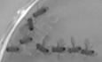
Figure 4. The development of Pax5Jak2/+ tumors depends on the Jak2 kinase activity
A. Schematic diagram of the kinase-dead (KD) Pax5-Jak2 fusion protein, which is encoded by the Pax5Jak2-KD allele and contains the K272E mutation known to abolish the Jak2 kinase function. B, C. Flow-cytometric analysis of bone marrow and spleen from Pax5Jak2-KD/+ (black; n = 11) and control Pax5+/+ (grey; n = 7) mice at the age of 6–8 weeks. The frequencies of the indicated B cell types are shown for each organ (B). The data are presented as mean percentages with SEM and were statistically analyzed by multiple t-tests (unpaired and two-tailed with Holm-Šídák's correction); ns (P > 0.5), *P < 0.05, **P < 0.01. See Methods for flow-cytometric definition of the different cell types. The expression of CD19 and B220 on bone marrow B cells of the indicated genotypes is shown in (C). D, E. Tumor progression in mice, transplanted with Pax5Jak2-Luc/+ tumor cells, in the presence or absence of the JAK1/2 inhibitor ruxolitinib. At day 14 after cell transfer, the transplanted mice were treated twice daily with ruxolitinib or vehicle, and the tumor mass was monitored by bioluminescence measurements. Images of three representative mice with or without ruxolitinib treatment are shown in (D). The bioluminescence measurements of transplanted mice treated with ruxolitinib (black) or vehicle (grey) at the indicated days after cell transfer are shown as mean radiance values with SEM (E). n, number of mice analyzed. Statistical data are shown as mean value with SEM and were analyzed by the mixed-effect model REML with Geisser–Greenhouse’s correction and Tukey’s multiple comparison test: *P < 0.05, ***P < 0.001. F. Kaplan–Meier survival an
留言 (0)