
記住我


Despite recent improvements in targeting metastatic melanoma, resistance to inhibition of the BRAFV600 oncogenic pathway occurs in most patients treated with MAPK-inhibiting drugs. Melanoma cells adopt various means to evade therapy, including transcriptional reprogramming leading to phenotypic dedifferentiation and acquisition of mesenchymal and pro-fibrotic features. This state of cellular resistance is highly invasive and displays an increased ability to produce and remodel the extracellular matrix (ECM), creating a drug-tolerant microenvironment. However, the molecular networks that define this pro-fibrotic cellular behavior and promote resistance are still unclear.
ResultsWe show that the anti-fibrotic drug nintedanib prevents the fibrotic reaction and improves MAPK-targeting therapy efficacy, retarding the onset of resistance in a mouse melanoma model. Expression screening and mechanistic studies identified the pro-fibrotic miR-143/-145 cluster as a driver of nintedanib-sensitive mesenchymal resistant phenotype. Using a combination of gain- and loss-of-function approaches, we dissected the molecular and cellular processes regulated by these FibromiRs and demonstrate that during drug adaptation, melanoma cells upregulate the miRNA cluster, which drives a phenotypic switch toward a dedifferentiated therapy-resistant state. The miR-143/-145 cluster also induces ECM production and promotes cell migration and invasion through the activation of focal adhesion dynamics and mechanotransduction pathways. Finally, Fascin actin-bundling protein 1 (FSCN1) was identified as a key functional target of miR-143-3p and miR-145-5p for the acquisition of the pro-fibrotic therapy-resistant phenotype.
ImpactOur study highlights non-genetic mechanisms of therapeutic resistance in melanoma and deciphers a regulatory cascade involving the miR-143/-145/FSCN1 pro-fibrotic axis in the acquisition of a therapy-resistant cellular state. It also provides a scientific rationale for designing clinical trials with nintedanib and potentially other anti-fibrotic agents to overcome resistance in patients with BRAF-mutated melanoma. Finally, our findings might have implications for other MAPK-driven cancers and fibrosis-related diseases.
IntroductionBecause of its high mutational burden, metastasis propensity, and resistance to treatment, cutaneous melanoma is one of the most aggressive human cancers and the deadliest form of skin cancer (Shain & Bastian, 2016). Melanoma is a non-epithelial tumor that originates from neural crest-derived and pigment-producing melanocytes in the skin. Genetic alterations in the BRAF, NRAS, or NF1 genes define melanoma subtypes and lead to the MAPK pathway hyperactivation (Flaherty et al, 2012; Cancer Genome Atlas, 2015). Current therapeutic options for BRAFV600E/K metastatic melanoma include MAPK-targeted therapies, which show remarkable efficacy during the first months of treatment (Chapman et al, 2011; Robert et al, 2019). However, the majority of patients treated with a combination of BRAF inhibitor (BRAFi) and MEK inhibitor (MEKi) inevitably relapse within months (Long et al, 2017). Genetic mechanisms of resistance cannot singly explain the acquisition of therapy resistance in melanoma, and non-genetic heterogeneity actively participates in drug tolerance (Rambow et al, 2019; Marine et al, 2020). Extensive studies have been carried out to dissect the non-mutational mechanisms of resistance (Rambow et al, 2018; Tsoi et al, 2018). Genetic and non-genetic mechanisms of resistance are frequently linked and not mutually exclusive (Marine et al, 2020). Non-genetic resistance is due to the intrinsic melanoma cell phenotypic plasticity, i.e., ability to undergo transcriptional and epigenetic reprogramming in response to environmental challenges or upon therapy (Arozarena & Wellbrock, 2019). These adaptive mechanisms exploit the developmental plasticity of melanoma cells and often result in an undifferentiated state characterized by upregulation of receptor tyrosine kinases (RTK) such as PDGFRβ or AXL, downregulation of melanocyte differentiation transcription factors MITF and SOX10 (Sun et al, 2014), and acquisition of mesenchymal and invasive features (Nazarian et al, 2010; Villanueva et al, 2010; Girotti et al, 2013; Muller et al, 2014; Fallahi-Sichani et al, 2017; Rambow et al, 2018; Tsoi et al, 2018; Rathore et al, 2019).
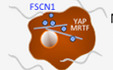
Tumors are shaped dynamically by reciprocal crosstalk between cancer cells and the extracellular matrix (ECM) through cellular–ECM interactions and stromal matrix remodeling. Recent findings indicated that elevated ECM production and remodeling contribute to adaptive and acquired resistance to BRAFi therapy by conferring a drug-protective niche to melanoma cells (Fedorenko et al, 2016; Titz et al, 2016; Girard et al, 2020; Marusak et al, 2020). Moreover, we recently reported that undifferentiated mesenchymal-like BRAFi-resistant cells exhibit myofibroblast/cancer-associated fibroblast (CAF)-like features leading to pro-fibrotic ECM reprogramming in vitro and in vivo (Diazzi et al, 2020; Girard et al, 2020). Cell-autonomous ECM deposition and remodeling abilities adopted by melanoma cells after MAPKi treatment result in cross-linked collagen matrix and tumor stiffening fostering a feedforward loop dependent on the mechanotransducers YAP and MRTFA and leading to therapy resistance (Girard et al, 2020). Thus, this pro-fibrotic-like response, typical of the early adaptation and acquired resistance to MAPK inhibition, provides a therapeutic escape route through the activation of alternative survival pathways mediated by cell-matrix communications. However, the signaling networks underlying the acquisition of this undifferentiated, mesenchymal-like melanoma cell state and drug-resistant behavior remain unclear.
We reasoned that therapeutic approaches aimed at preventing this targeted therapy-induced abnormal pro-fibrotic reaction could represent rationale combination strategies to normalize the fibrous stroma and overcome non-genetic resistance in BRAFV600E-mutated melanomas. We show here that the anti-fibrotic drug nintedanib (BIBF1120, Ofev®) improves the response of the BRAFi/MEKi-targeted therapy in a preclinical model of melanoma and in BRAF-mutated cell lines by preventing MAPKi-induced lineage dedifferentiation, ECM reprogramming, and mesenchymal traits. We also identified the master regulator associated with the acquisition of this pro-fibrotic and dedifferentiation program, pointing the miR-143/-145 cluster as a driver of the phenotype switching to a drug-resistant mesenchymal-like cell state.
Results Nintedanib/BIBF1120 prevents MAPKi-induced pro-fibrotic-like response, enhances targeted therapy efficiency, and delays tumor relapseIn order to limit ECM reprogramming and collagen remodeling associated with therapy resistance and relapse in melanoma, we tested the effect of the anti-fibrotic drug nintedanib (BIBF1120), a triple inhibitor of PDGFR, VEGFR, and FGFR used to treat idiopathic pulmonary fibrosis (IPF) in combination with BRAFi/MEKi in a syngeneic model of transplanted murine YUMM1.7 Braf-mutant melanoma (Meeth et al, 2016). YUMM1.7 cells were subcutaneously injected, and tumors were treated with vehicle, BIBF1120 alone, a combination of BRAFi plus MEKi, or the triple combination (Fig 1A). BIBF1120 did not display any anti-melanoma effect when administered alone, slightly slowing down tumor growth but not triggering tumor volume decrease. Administration of the BRAFi/MEKi initially reduced tumor growth, but after three weeks of treatment, tumor growth resumed and 100% of tumors relapsed. Importantly, combination of MAPK-targeted therapies and BIBF1120 significantly delayed relapse and led to complete remission in 33% of mice (2 out of 6; Figs 1B and C, and EV1A). Overall, the combined treatment significantly improved mouse survival (Fig 1C) without body weight loss or sign of toxicity throughout the study (Fig 1D). As previously described in melanoma xenograft models (Girard et al, 2020), an extensive deposition of collagens and increased expression of ECM remodeling and myofibroblast markers were observed in YUMM1.7 tumors treated with the combination of BRAFi and MEKi as revealed by picrosirius red staining of collagen fibers and qPCR analysis of typical molecular markers of tumor fibrosis. This response was significantly reduced by the co-administration of BIBF1120 (Figs 1E–G and EV1B). Thus, combination of targeted therapy with the anti-fibrotic drug nintedanib prevents the appearance of a pro-fibrotic matrix observed upon MAPK-targeted therapy exposure and significantly delays the onset of resistance in vivo.

Figure 1. Nintedanib/BIBF1120 prevents MAPKi-induced ECM remodeling, decreases resistance to targeted therapy, and delays tumor relapse
A–G. Mouse YUMM1.7 melanoma cells were subcutaneously inoculated into C57BL/6 mice, and when tumors reached 100 mm3, mice were treated with vehicle (Ctrl), nintedanib/BIBF1120 (BIBF), MAPKi (BRAFi, vemurafenib and MEKi, trametinib), or BRAFi/MEKi plus BIBF (n = 6). (B) Representative median graphics showing tumor growth following treatment (n = 6). Two-way ANOVA was used for statistical analysis. **P ≤ 0.01. (C) Kaplan–Meier survival curves of mice treated with the indicated therapies (n = 6). The log rank (Mantel–Cox) statistical test was used for MAPKi vs MAPKi/BIBF1120. ****P ≤ 0.0001. (D) Mouse body weight was measured at the indicated times. Data shown are mean ± SD (n = 6). (E, F) Tumor sections were stained with picrosirius red and imaged under polarized light. (E) Representative image of collagen fiber network in tumors from mice under the different treatments. Scale bar 200 μm. (F) Quantification of collagen fiber thickness (n = 6 for control, BIBF, and BRAFi/MEKi groups and n = 5 for BRAFi/MEKi + BIBF group). Two-way ANOVA statistical test was used for statistical analysis of mature collagen fiber thickness quantification. **P ≤ 0.01, ***P ≤ 0.001, and ****P ≤ 0.0001. Significance was calculated against the control group. Statistical significance of BIBF vs BIBF + BRAFi/MEKi was also calculated. (G) Heatmap showing the differential expression of ECM and myofibroblast/CAF genes in mice treated with MAPK-targeted therapies with or without BIBF compared to control mice (log2 ratio, n = 5). H–J. Human M238R cells and/or parental M238P cells were analyzed for different parameters. (H) Heatmap and Western Blot showing the expression of ECM, myofibroblast/CAF and phenotype switch markers in M238R compared to M238P cells. Heatmap represents the mean of expression of 3 independent experiments by RT-qPCR. (I) Heatmap showing the expression of ECM, myofibroblast/CAF and phenotype switch markers in M238R treated with BIBF (2 µM, 72 h) or vehicle alone by RT-qPCR (n = 3). (J) Crystal violet viability assay of M238R cells treated with BRAFi/MEKi (BRAFi, Vemurafenib and MEKi, Trametinib) (1 µM), BIBF (2 μM) or with BRAFi/MEKi (1 μM) plus BIBF (2 μM) for 72 h. Paired Student t-test was used for statistical analysis. ****P ≤ 0.0001. Significance was calculated against the control group. Statistical significance of BIBF vs BIBF + BRAFi/MEKi was also calculated. Data is represented as mean ± SD from a triplicate representative of 3 independent experiments.Source data are available online for this figure.
Click here to expand this figure.
Figure EV1. Administration of nintedanib/BIBF1120 resensitizes melanoma cells to MAPK-targeted therapies, delays tumor relapse, and normalizes MAPKi-induced ECM remodeling and miR-143/-145 expression
A, B. YUMM1.7 cells were subcutaneously inoculated into C57BL/6 mice, and when tumors reached 100 mm3, mice were treated with the indicated therapies. (A) Individual graphics showing tumor growth following treatment. (B) Normalized expression of myofibroblast/CAF and ECM-related genes assessed by RT-qPCR in individual tumors treated as indicated. Data are represented as median with range (n = 5). One-way ANOVA was used for statistical analysis. *P ≤ 0.05, **P ≤ 0.01, and ****P ≤ 0.0001. Significance was calculated against the control group. Statistical significance of BRAFi/MEKi vs BRAFi/MEKi + BIBF was also calculated. C–E. Human M238P cells were treated with BRAFi (vemurafenib) + MEKi (trametinib; 1 μM), BIBF1120 (2 µM) or BRAFI + MEKi (1 µM) plus BIBF (2 µM) for 72 h. (C) Heatmap showing the expression of ECM, myofibroblast/CAF markers, and phenotype switch markers by RT-qPCR (n = 3). (D) Crystal violet viability assay of M238P cells treated with MAPK-targeted therapies as above. Paired Student's t-test was used for statistical analysis. ****P ≤ 0.0001. Data are represented as mean ± SD from a triplicate representative of three independent experiments. (E) Western blot showing the expression of ECM, myofibroblast/CAF markers, and activation levels of signaling pathways (AKT and ERK1/2) in the different conditions. F–H. Human M238R cells were treated with BIBF1120 (2 µM) or with CP673451 (2 µM) for 72 h. (F) Western blot showing activation levels of signaling pathways (PDGFR and AKT) in the different conditions. (G) Heatmap showing the expression of ECM, myofibroblast/CAF markers, and phenotype switch markers by RT-qPCR in M238R cells treated with the indicated inhibitors (n = 3). (H) Crystal violet viability assay of M238R cells treated with the indicated inhibitors. Paired Student's t-test was used for statistical analysis. ****P ≤ 0.0001. Data are represented as mean ± SD from a triplicate representative of three independent experiments.We next examined the impact of nintedanib on ECM reprogramming and cell phenotype switching in the context of early adaptation and resistance to MAPK-targeted therapy in human BRAFV600E-mutated melanoma M238P cells. BIBF1120 strongly attenuated targeted drug-induced ECM/myofibroblast-related signatures, prevented the undifferentiated AXLhigh–MITFlow phenotype switch (Fig EV1C) and potentiated the effect of the BRAFi/MEKi cocktail on M238P cell viability (Fig EV1D). The efficacy of the described treatment to reduce upregulation of fibronectin (FN1) and LOXL2 expression was confirmed at protein levels by Western blot analysis (Fig EV1E). Of note, a strong activation of AKT induced by the BRAFi/MEKi cocktail was fully inhibited by BIBF1120, suggesting that the anti-fibrotic drug is able to counteract the rewiring of alternative survival pathway observed upon MAPK oncogenic pathway inhibition (Fig EV1E) (Nazarian et al, 2010).
We finally evaluated the effect of BIBF1120 on the undifferentiated mesenchymal-like resistant M238R cells obtained through chronic exposure of the M238P cells to the BRAFi vemurafenib (Nazarian et al, 2010) and that displayed cross-resistance to MEKi (Atefi et al, 2011). We recently reported that this RTK-driven resistant cell line exhibits low expression of the differentiation factor MITF and high AXL levels and displays a strong myofibroblast-like phenotype with expression of classical ECM and contractile markers such as smooth muscle actin-α (αSMA) and myosin light chain 2 (MLC2), as well as ECM remodeling activities compared with parental M238P cells (Fig 1H) (Girard et al, 2020). BIBF1120 was able to attenuate melanoma-undifferentiated state markers and expression of ECM and myofibroblast/CAF-related signature (Fig 1I), but also significantly decreased cell viability and resistance to BRAFi (Fig 1J). To address the specific contribution of PDGFRβ inhibition in nintedanib/BIBF1120 effects, we compared the effect of the selective PDGFR inhibitor CP673451 with BIBF1120 in M238R-resistant cells. The two inhibitors showed similar efficiency in causing a strong decrease in phospho-PDGFRβ and phospho-AKT levels (Fig EV1F). However, while selective inhibition of PDGFR attenuated the myofibroblast-like signature typical of resistant cells (Fig EV1G) and significantly decreased cell viability (Fig EV1H), CP673451 was found less efficient than BIBF1120 in inducing phenotypic switch toward a more differentiated cell state (Fig EV1G). Altogether, these findings indicate that an anti-fibrotic therapy is able to revert the undifferentiated mesenchymal resistant phenotype and potentiate targeted therapy in human melanoma cells.
Suppression of MAPKi-induced resistant pro-fibrotic phenotype by nintedanib is associated with loss of miR-143/145 cluster expressionNext, we investigated the molecular mechanisms associated with the emergence of MAPKi-induced mesenchymal and pro-fibrotic phenotype and its inhibition by nintedanib/BIBF1120. Because several microRNAs (miRNAs), named FibromiRs, have been shown to play key roles in the initiation and progression of fibrotic processes in various organs (Ishida & Selaru, 2013; Pottier et al, 2014; Hanna et al, 2019; Savary et al, 2019), we performed an expression screening to compare the level of these FibromiRs in BRAFV600E mutant melanoma cells sensitive to MAPK-targeted therapies (M229P, M238P, M249P) compared to their corresponding resistant counterparts (Nazarian et al, 2010). The screening identified miR-143-3p and miR-145-5p, localized within the miR-143/145 cluster on chromosome 5 as the best hits with a strong upregulation in AXLhigh MITFlow mesenchymal-like resistant M238R and M229R cells tested compared to parental cells (Figs 2A and EV2A). Similar results were obtained in the mesenchymal resistant UACC62R cells (Misek et al, 2020) (Fig EV2A). In contrast, acquisition of resistance through secondary NRAS mutation was not associated with increased expression of miR-143-3p and miR-145-5p in the non-mesenchymal AXLlow–MITFhigh M249R cells (Figs 2A and EV2A). Upregulation of miR-143/145 cluster expression was also observed in BRAFi/MEKi double-resistant (DR) melanoma clones described in Shen et al (2019). Interestingly, expression levels of the two miRNAs were more pronounced in acquired DR melanoma cells displaying a mesenchymal-like cell state (Fig EV2B). We next examined whether a treatment with BRAFi, MEKi, or a combination of both was able to modulate the expression of the cluster. The two drugs, alone or in combination, significantly increased miR-143-3p and miR-145-5p expression levels in all BRAFV600E mutant melanoma cells tested including patient-derived short-term melanoma cultures (Fig EV2C–G). This strong induction was abolished when the BRAFi/MEKi treatment was combined with BIBF1120, both in melanoma cell lines cultured in vitro (Fig 2B) and in the YUMM.1.7 syngeneic model (Fig 2C) presented in Fig 1. Overall, the expression of the miR-143/-145 cluster paralleled the phenotypic switch associated with a mesenchymal resistant phenotype.

Figure 2. Expression of miR-143/-145 is correlated with the undifferentiated mesenchymal-like MAPKi-resistant phenotype and is negatively regulated by nintedanib/BIBF1120
A. Heatmap showing the differential expression of a selection of miRNAs known as “FibromiRs” in human BRAFV600E mutant melanoma cells sensitive to MAPK-targeted therapies (M229, M238, M249) and the corresponding BRAFi-resistant cells. Expression of indicated FibromiRs was evaluated by RT-qPCR; log2 (resistant vs parental). Expression level of AXL and MITF phenotypic markers for each resistant cell line is indicated. B. Relative miRNA expression levels were quantified in M238P cells treated for 72 h with BIBF1120 (BIBF, 2 μM), MAPKi (BRAFi, vemurafenib and MEKi, trametinib; 1 µM), or with MAPKi (1 μM) plus BIBF (2 µM) by RT-qPCR and normalized to miR-16-5p. Data are represented as mean ± SEM from a triplicate representative of three independent experiments. One-way ANOVA was used for statistical analysis. **P ≤ 0.01 and ***P ≤ 0.001. Significance was calculated against the control group. Statistical significance of BRAFi/MEKi vs BRAFi/MEKi + BIBF was also calculated. C. Expression of miR-143-3p and miR-145-5p in control mice and mice treated with the indicated therapies (see legend of Fig 1 for details) was quantified by RT-qPCR. Data are represented as mean ± SEM from two independent experiments performed on six mice, with two sites of injections. One-way ANOVA was used for statistical analysis. *P ≤ 0.05 and ****P ≤ 0.0001. Significance was calculated against the control group. Statistical significance of BRAFi/MEKi vs BRAFi/MEKi + BIBF was also calculated. D. Relative miRNA expression levels were quantified in M238P cells stimulated for 48 h with TGF-β (10 ng/ml) or PDGF-BB (20 ng/ml) by RT-qPCR and normalized to miR-16-5p. Data are represented as mean ± SEM from a triplicate representative of three independent experiments. P-values were calculated using paired Student's t-test. *P ≤ 0.05 and ***P ≤ 0.001. E. Relative miRNA expression levels were quantified in M238R cells treated for 48 h with the triple kinase inhibitor nintedanib/BIBF1120 (BIBF, 2 μM), the TGF-β receptor kinase inhibitor SB431542 (SB, 10 µM), and the pan-AKT inhibitor GSK690693 (GSK, 10 µM) by RT-qPCR. Data are represented as mean ± SEM from a triplicate representative of three independent experiments. P-values were calculated using paired Student's t-test. *P ≤ 0.05, **P ≤ 0.01, and ***P ≤ 0.001. F, G. Phenotype switch/invasive/ECM markers (F) and relative miRNAs expression levels (G) were quantified in therapy-naïve (N) and therapy-resistant (R) PDX samples. The log2 fold change or the ratio of the fold change R vs N is shown for each couple of samples.Source data are available online for this figure.
Click here to expand this figure.
Figure EV2. High expression of miR-143/-145 is correlated with an undifferentiated/mesenchymal-like BRAFi-resistant phenotype in melanoma cells
A. Relative miRNA expression levels were quantified in parental (P) and paired resistant (R) cells (M238, UACC62, M229, M249) by RT-qPCR. Log2 (R/P) is shown for each cell line. Data are represented as mean ± SE from a triplicate representative of at least three independent experiments. Paired Student's t-test was used for statistical analysis. ****P ≤ 0.0001. B. Heatmap showing the expression of ECM, myofibroblast/CAF, and phenotype switch markers, and miRNAs by RT-qPCR in A375 cells resistant to BRAFi and MEKi (cobimetinib 1 µM and trametinib 0.1 µM). C–G. Relative miRNA expression levels were quantified in human melanoma cell lines (M238P, UACC62P, 1205Lu) or short-term patient-derived cell lines (MM034, MM099) treated or not for 72 h with MAPK-targeted therapies BRAFi (vemurafenib 3 µM), MEKi (trametinib 1 µM), or BRAFi + MEKi (1 µM) by RT-qPCR and normalized to miR-16-5p. H. Relative miRNA expression levels were quantified in M238R cells treated or not for 72 h with BIBF1120 (2 µM) or CP673451 (2 µM). (C–H) Data are represented as mean ± SE from a triplicate representative of at least three independent experiments. Paired Student's t-test was used for statistical analysis. *P ≤ 0.05, **P ≤ 0.01, ***P ≤ 0.001, and ****P ≤ 0.0001.Given the critical role of RTK upregulation such as PDGFRβ and of the pro-fibrotic TGF-β signaling pathway overactivation in mesenchymal resistance (Nazarian et al, 2010; Sun et al, 2014; Diazzi et al, 2020; Girard et al, 2020), we stimulated MAPKi-sensitive melanoma cells with PDGF-BB or with TGF-β and analyzed miR-143-3p and miR-145-5p expression. Both TGF-β and PDGF-BB triggered a strong upregulation of miR-143/-145 expression in M238P cells (Fig 2D). Conversely, treatment of mesenchymal BRAFi-resistant M238R cells with BIBF1120 but also with the TGF-β receptor inhibitor SB431542, the pan-AKT inhibitor GSK690693 or the PDGFR inhibitor CP673451 significantly decreased the expression of the two mature miRNAs (Figs 2E and EV2H), indicating that both PDGF and TGF-β pathways control the expression of the miR-143/-145 cluster in melanoma cells.
Finally, we investigated the expression of these miRNAs in several patient-derived xenograft (PDX) samples that acquired resistance to BRAFi/MEKi combotherapy and exhibited distinct phenotypic and molecular profiles (Fig 2F) (Marin-Bejar et al, 2021). Upregulation of miR-143/-145 cluster between therapy naïve and resistant cells was observed in two different PDX samples, MEL015 and MEL003, with a predominant invasive/undifferentiated transcriptome profile (Fig 2F and G) (Marin-Bejar et al, 2021). The MEL015-resistant model also presented elevated expression of ECM remodeling, myofibroblast, and pro-fibrotic markers such as COL1A1, LOXL2, CYR61, THBS1, and MYL9. In contrast, we did not observe an upregulation of the cluster in drug-resistant lesions from the two additional PDX models, MEL006 and MEL047, in which the mesenchymal-like signature is not overrepresented (Fig 2F and G). These data indicate that upregulation of the pro-fibrotic miR-143/-145 cluster is also observed in PDX MAPKi-resistant melanomas associated with an invasive/undifferentiated transcriptome profile.
miR-143/-145 cluster promotes melanoma cell dedifferentiation toward a pro-fibrotic mesenchymal-like state and resistance to MAPK therapeuticsTo confirm a potential link between the miR-143/-145 cluster and ECM reprogramming, we first used a gain-of-function approach consisting in the transient overexpression of miR-143-3p or miR-145-5p in various therapy-naïve melanoma cells (Appendix Fig S1A). The results showed increased expression of transcripts related to ECM structure and remodeling, as well as myofibroblast/CAF markers in cells overexpressing either miRNA compared to miR-neg control cells (Fig 3A). Conversely, we next tested whether miR-143-3p or miR-145-5p inhibition can reverse the phenotypic pro-fibrotic response induced by oncogenic BRAF inhibition in M238P melanoma cells. BRAFi treatment was combined with locked nucleic acid (LNA)-modified antisense oligonucleotides (ASOs) designed against miR-143 (LNA-143), miR-145 (LNA-145), or a control LNA ASO (LNA-Ctrl). RT-qPCR analysis showed that the BRAFi-induced ECM- and myofibroblast/CAF-related gene signature was significantly inhibited by LNA-143 and LNA-145 ASOs (Fig 3B). These results were confirmed at protein level by Western blot analysis of cell lysates and conditioned media of ECM proteins and cross-linking enzymes, as well as myofibroblast/CAF markers using same gain- or loss-of-function approaches (Fig 3C–D and Appendix Fig S1B).

Figure 3. miR-143/-145 cluster promotes ECM reprogramming, melanoma cell dedifferentiation, and drug resistance
Heatmap showing the differential expression of a selection of ECM-related genes, cytoskeleton, and myofibroblast markers in three distinct cell lines (M238P, UACC62P, and M229P) transfected with the indicated mimics (control (miR-neg), miR-143 or miR-145 mimics, 72 h, 30 nM), assessed by RT-qPCR (n = 3). M238P cells were treated 72 h with BRAFi (vemurafenib, 3 μM) in the presence or the absence of LNA-based anti-miR-143 (LNA-143) or anti-miR-145 (LNA-145; 50 nM) or a combination of the two. ECM marker RT-qPCR data are represented as mean ± SD from a triplicate representative of at least three independent experiments. One-way ANOVA was used for statistical analysis. *P ≤ 0.05, **P ≤ 0.01, and ***P ≤ 0.001. Western blot analysis of ECM remodeling markers on total cell lysates from M238P cells transfected as in (A). Western blot analysis of ECM remodeling markers on total cell lysates from cells treated with or without BRAFi and the indicated combination of inhibitors as in (B). Proliferation curves using time-lapse analysis of cells with the IncuCyte system. Graph shows quantification of cell confluence. Two-way ANOVA was used for statistical analysis. ****P ≤ 0.0001. Migration assay performed in Boyden chambers. Representative images showing migrating cells in the different conditions. The histogram represents the quantitative determination of mean ± SD from six independent fields representative of three independent experiments, using ImageJ software. Paired Student's t-test was used for statistical analysis. *P ≤ 0.05 and ***P ≤ 0.001. Western blot analysis of phenotype switch markers on lysates from M238P cells treated as in (A). Crystal violet viability assay of M238P cells treated 72 h with the different combinations of inhibitors in the presence or not of BRAFi (vemurafenib, 3 μM). Data are represented as mean ± SD from a triplicate representative of at least three independent experiments. One-way ANOVA was used for statistical analysis. ****P ≤ 0.0001.Source data are available online for this figure.
We next investigated whether the cluster contributed to the acquisition of the slow cycling, undifferentiated, and invasive cell state. Melanoma cells experienced reduced cell proliferation after ectopic expression of miR-143-3p or miR-145-5p as visualized by Western blot analysis of cell cycle markers (Appendix Fig S2A) and by analysis of cell confluence by live-cell imaging (Fig 3E and Appendix Fig S2B) with an accumulation of cells in the G0/G1 phase and a decreased percentage of cells in S phase (Appendix Fig S2C). Acquisition of the slow cycling cell state was not linked to cell death, as confirmed by annexin V/DAPI staining (Appendix Fig S2D). Inhibition of proliferation was also accompanied by enhancement of cell migratory abilities, as shown using Boyden chamber assays (Fig 3F and Appendix Fig S2E), as well as by the acquisition of a less differentiated phenotype, with decreased levels of MITF and SOX10, and increased levels of AXL, PDGFRβ, EGFR, NGFR, and SOX9 (Fig 3G and Appendix Fig S2F). Lentivirus-mediated stable overexpression of the two miRNAs in two distinct melanoma cell lines reproduced increased ECM protein production, inhibition of cell proliferation, and transition to an undifferentiated/invasive phenotype (Fig EV3A–E) observed upon transient transfection. Acquisition of this features was also linked to a decreased intrinsic sensitivity to BRAFi/MEKi treatment, as measured by crystal violet survival assays performed on melanoma cells stably overexpressing miR-143/-145 cluster compared to control cells (Fig EV3F). Conversely, targeting the two miRNAs by ASOs in combination with BRAFi improved the efficacy of the targeted drug (Fig 3H and Appendix Fig S3A and B), demonstrating that miR-143/-145 cluster upregulation in response to BRAFV600E pathway inhibition represents a pivotal adaptive resistance mechanism to MAPK therapeutics. Of note, inhibition of miR-145-5p alone and combined inhibition of the two miRNAs also significantly decreased the viability of BRAFi-resistant melanoma cells M238R (Appendix Fig S3C and D).
Click here to expand this figure.
Figure EV3. Stable expression of the miR-143/-145 cluster promotes ECM remodeling and drives melanoma cell dedifferentiation
A. RT-qPCR analysis showing the level of miR-143-3p and miR-145-5p expression after stable expression following lentivirus transduction of two melanoma cell lines (M238P, UACC62P). Data are represented as mean ± SE from a triplicate representative of at least three independent experiments. Paired Student's t-test was used for statistical analysis. ***P ≤ 0.001 and ****P ≤ 0.0001. B–D. Western blot analysis of ECM remodeling (B), cell cycle (C), and phenotype switch markers (D) on total cell lysates from the different stable cell lines. E. Invasion assay in Boyden chambers. Representative images show invasion in control and miR-143/145 expressing cells (M238P). The bar graph represents the quantitative determination of data obtained using ImageJ software. Paired Student's t-test was used for statistical analysis. ****P ≤ 0.0001. F. Viability of M238P cells transduced with a control or a miR-143/-145 cluster lentivirus was assessed by crystal violet staining upon MAPKi treatment (6 days, BRAFi, vemurafenib 3 µM, MEKi, trametinib, 3 µM or BRAFi + MEKi, 5 µM). Paired Student's t-test was used for statistical analysis. ****P ≤ 0.0001. Identification of miR-143-3p/miR-145-5p targets functionally associated with the undifferentiated mesenchymal-like phenotype in melanoma cellsTo identify miR-143-3p and miR-145-5p targets associated with the resistant mesenchymal phenotype, we first combined in silico target prediction tools and experimental transcriptomic approaches using the miRonTop web tool (Le Brigand et al, 2010) in M238R or M238P cells following transient transfection of mimics (Fig 4A and B) or stable lentivirus transduction. Functional annotation of the gene expression profiles associated with miRNA overexpression showed a strong overlap in pathways associated with cell migration and invasion, cell cycle, and cytoskeleton organization (Appendix Table S1). The predicted targets for each of the mature miRNAs were significantly overrepresented among the downregulated genes in response to the corresponding mimics transfection (Fig 4B). A first set of target candidates were identified by crossing these predicted targets and the genes shown experimentally to be downregulated in resistant M238R cells compared to parental M238P cells (Fig 4C).

Figure 4. Identification of gene targets and cellular pathways functionally associated with the miR-143/-145 cluster-mediated undifferentiated/mesenchymal-like melanoma cell phenotype
A–C. M238P cells were transfected separately with miR-143-3p, miR-145-5p or a negative control (miR-neg) mimics, and RNA content was analyzed using whole-genome microarrays (dataset 1, n = 2). (A) Heatmap showing the genes differentially expressed after individual miRNA mimic overexpression. (B) Overrepresentation of miRNA predicted targets in the set of downregulated transcripts following miR-143-3p and miR-145-5p mimics transfection using miRonTop webtool. Each arrow indicates the corresponding overexpressed miRNA. (C) Venn diagram showing the selection of the best target candidates (red boxes) using miR-143-3p and miR-145-5p mimics transfection, as well as comparison of M238R and M238P transcriptomic profiles. D–E. M238P cells were transduced with a miR-143/-145 construct and selected for stable expression of the cluster or transduced with a control vector, followed by RNA-seq analysis (dataset 2, n = 2). (D) Heatmap highlighting the common predicted upstream regulators altered in cells stably expressing the miR-143/-145 cluster and M238R cells compared to control M238P cells. A subset of common regulators (out of the top 50 scores) corresponding to transcription factors (TFs), cytokines and growth factors (GFs), transmembrane receptors, kinases, and phosphatases is shown. Red arrows indicate annotations related to the TGF-β pathway. (E) Venn diagram summarizing the comparison of the best-predicted targets following the two gain-of-function approaches. Subsets of miR-143-3p and miR-145-5p predicted targets downregulated by both mimics and stable lentivirus expression are shown (red boxes). F. Luciferase assay in HEK cells overexpressing miR-143 or miR-145 transfected with a plasmid harboring the WT or muted sequence of the miR-143 and miR-145 binding sites present in FSCN1 3′UTR. Each bar represents the mean ± SE of experiments performed at least in triplicate. ***P ≤ 0.001 and ****P ≤ 0.0001. P-values were calculated using paired Student's t-test. G. RT-qPCR analysis of FSCN1 expression in M238P cells transfected with the indicated mimics. Data are represented as mean ± SE from a triplicate representative of at least three independent experiments. Paired Student's t-test was used for statistical analysis. **P ≤ 0.01 and ***P ≤ 0.001. H. Western blot analysis of FSCN1 expression in M238P cells transfected with the indicated mimics.Source data are available online for this figure.
Second, RNAs from cells stably overexpressing the miR-143/-145 cluster were analyzed by RNA-sequencing and processed through Ingenuity Pathway Analysis (IPA) to identify the common regulators (transcription factors, growth factors, cytokines, transmembrane receptors, kinases, and phosphatases) between parental cells overexpressing the cluster and resistant cells (Fig 4D). These analyses notably highlighted changes related to decreased cell proliferation, increased cell invasion, and fibrotic pathway activation. To narrow the best target candidates, we finally compared the best-predicted targets based on the two different gain-of-function approaches (Appendix Tables S2 and S3). This strategy resulted in selecting one target candidate for miR-143-3p, 6 target candidates for miR-145-5p, and 2 target candidates for both miR-143-3p and miR-145-5p (Fig 4E). We started with investigations on the F-acting bundling protein Fascin1 (FSCN1), a key regulator of cytoskeleton dynamics, previously associated with tumor growth, migration, invasion, and metastasis (Ma & Machesky, 2015). Using long-read Nanopore sequencing data, we confirmed lower levels of FSCN1 transcript in M238R compared with M238P cells, while reads corresponding to the putative miR-143/-145 cluster primary transcript could be only detected in M238R cells (Appendix Fig S4A). The characterization of hFSCN1 3′UTR sequence revealed the presence of 2 miR-143-3p and 4 miR-145-5p binding sites. Validation of these sites was first performed using a luciferase reporter corresponding to the full 3′UTR FSCN1 harboring WT or a mutated sequence of the miRNA recognition elements (Fig 4F and Appendix Fig S4B). Finally, qPCR and Western blot analyses confirmed that FSCN1 was downregulated at both mRNA and protein levels upon miR-143-3p and miR-145-5p ectopic expression in various melanoma cells and in cells stably overexpressing the cluster (Fig 4G and H and Appendix Fig S4C and D).
FSCN1 is a functional miR-143/-145 target contributing to the phenotypic switch toward the undifferentiated/mesenchymal-like and resistant stateConsidering the strong expression of the miR-143/-145 in BRAFV600E mutant mesenchymal-like resistant cells compared to their parental cou
留言 (0)