
記住我

Eukaryotic transcription occurs in the context of chromatin, in which each transcribed nucleosome presents a barrier to RNA polymerase (Selth et al, 2010; Cramer, 2014; Formosa & Winston, 2020; Kujirai & Kurumizaka, 2020). The ability of RNA polymerase II (Pol II) to traverse the nucleosome barrier in eukaryotic cells is stimulated by multiple proteins, which help to weaken the association of histones with DNA and thereby facilitate Pol II progression (Schier & Taatjes, 2020). For example, the chromatin remodeller Chd1 binds to nucleosomes in a manner that displaces DNA at the Pol II entry site (Farnung et al, 2021). Activated by Pol II transcription into the nucleosome, Chd1 then translocates DNA into the Pol II cleft, further stimulating Pol II progression. In addition, the essential histone chaperone FACT binds to transcribing nucleosomes (Farnung et al, 2021) and can displace an H2A–H2B dimer, thereby facilitating Pol II progression (Belotserkovskaya et al, 2003; Tsunaka et al, 2016). Nevertheless, it is important to ensure that histones are not lost from DNA during transcription if the local pattern of nucleosome density is to be preserved. Indeed, work with budding yeast indicates that the H3–H4 tetramer from one nucleosome re-occupies the same locus after transcription by Pol II (Henikoff & Ahmad, 2019; Schlissel & Rine, 2019). This spatial fidelity facilitates the preservation of epigenetic information in the form of post-translational modifications of histones H3 and H4.
One contributing factor to the fate of nucleosomal histones during transcription is the rate of progression of the Pol II elongation complex. In vitro reactions in the absence of FACT have shown that slow transcription of a nucleosome substrate by Pol II, in the presence of limiting concentrations of nucleoside triphosphates (NTPs), allows the transfer of the histone octamer in cis to the upstream DNA (Bintu et al, 2011). Such transfer has been proposed to be mediated by loops in the DNA template (Kulaeva et al, 2009). However, faster Pol II elongation at more physiological NTP concentrations triggers displacement of an H2A–H2B dimer from a transcribed nucleosome (Kireeva et al, 2002), while even higher transcription rates can cause loss of the entire histone octamer (Bintu et al, 2011).
Studies of histone dynamics in living cells supported the idea that Pol II transcription frequently leads to transient loss of an H2A–H2B dimer, whereas H3–H4 tetramers are retained more stably during transcription but can be lost from very highly expressed genes, thereby reducing nucleosome density. For example, Pol II transcription was found to stimulate the exchange of histone H2A–H2B dimers within gene bodies in chicken cells (Jackson, 1990; Dion et al, 2007), human cells (Kimura & Cook, 2001), slime mould (Thiriet & Hayes, 2005) and budding yeast (Dion et al, 2007; Jamai et al, 2007; Yaakov et al, 2021). The same studies found that H3–H4 tetramers were exchanged much more slowly. However, genome-wide studies of histone exchange in budding yeast cells showed that repeated transcription of highly expressed genes reduces nucleosome density and thereby increases the rate of exchange of histones H3–H4, within coding regions as well as at promoters (Kristjuhan & Svejstrup, 2004; Lee et al, 2004; Schwabish & Struhl, 2004; Jamai et al, 2007; Rufiange et al, 2007; Yaakov et al, 2021). Furthermore, the metazoan histone variant H3.3 is incorporated into highly transcribed genes (Mito et al, 2005), likely also reflecting some degree of nucleosome depletion during active transcription.
Recent structural data have indicated that the association of elongation factors with Pol II in eukaryotic cells might preclude histone transfer by a simple DNA template looping model (Ehara et al, 2019; Kujirai & Kurumizaka, 2020). Consistent with this view, the retention of histones H3–H4 during Pol II chromatin transcription in budding yeast was found to be an active process that involves multiple histone chaperones, including FACT and Spt6 that are essential for cell viability (Kaplan et al, 2003; Jamai et al, 2009; Perales et al, 2013; Jeronimo et al, 2019). Although FACT was first characterised as a chaperone of histone H2A–H2B, it also binds to H3–H4 tetramers in vitro, via a domain that is distinct from the previously characterised module that binds to H2A–H2B (Tsunaka et al, 2016; Mayanagi et al, 2019; Liu et al, 2020). Similarly, Spt6 binds to H3–H4 tetramers in vitro but is also able to associate with H2A–H2B (Bortvin & Winston, 1996; McCullough et al, 2015). Consistent with the essential role of FACT and Spt6 in the retention of H3–H4 tetramers during Pol II transcription in yeast cells, mutations in FACT or Spt6 lead to the potent activation of transcription within gene bodies, from cryptic promoters that are normally repressed by the presence of nucleosomes (Kaplan et al, 2003; Cheung et al, 2008).
Until now, however, it has remained unclear how progression of the Pol II elongation complex is coupled to the capture, retention and local re-deposition of the H3–H4 tetramer by factors such as FACT and Spt6. Structural and biochemical studies have indicated that FACT is recruited to nucleosomes in a transcription dependent manner (Mayanagi et al, 2019; Liu et al, 2020), but FACT has not been found to contact the Pol II elongation complex directly (Farnung et al, 2021). Moreover, although Spt6 does associate with the Pol II elongation complex (Vos et al, 2018, 2020), mutations that displace Spt6 from Pol II were not found to impair nucleosome re-deposition during transcription (Dronamraju et al, 2018). Therefore, it is currently unclear how histone chaperones such as FACT and Spt6 can promote the local retention of H3–H4 tetramers during progression through chromatin of the Pol II elongation complex.
One possibility is that the Pol II elongation complex itself plays a direct role in histone capture and redeposition during transcription, in addition to the action of chaperones such as FACT and Spt6. A clue for such a role of the Pol II elongation complex was suggested by analogous studies of histone capture and redeposition during chromosome replication. Passage of the replisome through a chromosome disassembles the downstream nucleosomes and thereby displaces parental histones, which are captured by multiple replisome components and then redeposited locally onto the newly replicated DNA (Foltman et al, 2013; Evrin et al, 2018; Serra-Cardona & Zhang, 2018; Yu et al, 2018; Schlissel & Rine, 2019; Li et al, 2020; Stewart-Morgan et al, 2020; Willhoft & Costa, 2021). The first histone-binding component of the replisome to be identified was the MCM2 subunit of the CMG helicase (Ishimi et al, 1998), which unwinds the parental DNA duplex at replication forks (CMG = Cdc45-Mcm-GINS). The acidic amino terminal tail of MCM2 (MCM2N) binds to H3–H4 tetramers (Huang et al, 2015; Richet et al, 2015), dependent upon a conserved motif that is required to preserve repressive chromatin in yeast chromosomes (Foltman et al, 2013) and is important for the local redeposition of histones at replication forks in budding yeast and human cells (Gan et al, 2018; Petryk et al, 2018; Schlissel & Rine, 2019).
When extracts of budding yeast cells were treated with DNase to displace nucleosomal histones from chromatin, mass spectrometry showed that FACT forms ternary complexes in vitro with all four released histones, together with one of two other proteins (Foltman et al, 2013). The first of these was Mcm2, dependent upon the histone binding activity of Mcm2N. The second was the Spt5 subunit of the RNA Pol II elongation complex. The formation of such complexes was dependent upon the histone binding activity of FACT and the data raised the possibility that FACT forms two mutually exclusive complexes with nucleosomal histones under such conditions, one comprising Mcm2-histones-FACT and the second corresponding to Spt5-histones-FACT (Foltman et al, 2013). The significance of such histone-dependent complexes containing FACT and Spt5 was not explored further in previous studies, but these findings raised the interesting possibility that Spt5 might possess an analogous histone binding activity to Mcm2.
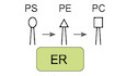
Spt5 is the only factor associated with RNA polymerase that is universally conserved in bacteria (where it is known as NusG, for N-utilisation substance G), archaea and eukaryotes (Werner & Grohmann, 2011). The NGN domain of Spt5/NusG (NGN = NusG N-terminal Domain) forms a stable lid over the active centre of RNA polymerase, thereby trapping nucleic acids into the cleft on the polymerase surface and ensuring processive elongation (Klein et al, 2011; Martinez-Rucobo et al, 2011; Bernecky et al, 2016, 2017; Liu & Steitz, 2017). However, eukaryotic Spt5 helps RNA polymerase progress through nucleosomes (Crickard et al, 2017; Ehara et al, 2019; Farnung et al, 2021) and is unique in having an acidic and largely unstructured tail at the amino terminus of the protein (Spt5N), immediately before the NGN domain (Fig 1A). Recent cryoEM structures of the transcribing yeast Pol II on a nucleosome substrate indicated that Spt5N, together with the NGN domain of Spt5, is sandwiched between the downstream nucleosome and the upstream DNA that emerges from Pol II (Ehara et al, 2019; Farnung et al, 2021). Therefore, Spt5N is ideally positioned to contribute to the capture and local retention of nucleosomal histones that are released from DNA during transcription. Such a role would be consistent with previous data showing that Spt5 is important to repress cryptic transcription within gene bodies across the yeast genome (Cheung et al, 2008). Here, we show that Spt5N contains a conserved histone binding motif, which is essential for cell viability and preserves the integrity of nucleosomes during transcription by Pol II.

Figure 1. Eukaryotic Spt5N binds histones H3–H4 via an evolutionarily conserved motif
Domain structure of Spt5/NusG proteins. The enlarged inset shows a conserved region of Spt5N from Saccharomyces cerevisiae (S.c.), Schizosaccharomyces pombe (S.p.), Aspergillus nidulans (A.n.), Homo sapiens (H.s.), Xenopus laevis (X.l.) and Arabidopsis thaliana (A.t.). Spt5N contains three invariant residues that were mutated in Spt5-3A (indicated by red asterisks), whilst residues with conservative substitutions are denoted by blue asterisks. GST, GST-MCM2N (human) and GST-Spt5N (yeast) were mixed as indicated with yeast histones H3–H4, in the presence of glutathione agarose beads (see STAR method for further details). The input and pull-down samples were analysed by SDS–PAGE before staining with colloidal Coomassie blue. The asterisks denote truncations of GST-Spt5N. Similar experiment to (B) but with histones H2A–H2B. Similar experiment to (B) to compare histone binding by GST-MCM2N and GST-MCM2N-2A (with Y81A Y90A mutations of conserved residues). Analogous experiment to (D), comparing histone binding by GST-Spt5 and GST-Spt5N-3A (with F180A E184A V187A mutations of conserved residues). Budding yeast Spt5N and Spt5N-3A were expressed as fusions to Protein A in budding yeast cells. Cell extracts were treated with DNase as indicated, to release nucleosomal histone complexes from chromatin, before immunoprecipitation (IP) of Spt5N/Spt5N-3A on magnetic beads coated with IgG. The bound proteins were eluted and then analysed by mass spectrometry (the panel indicates the detected spectral counts). Samples from an analogous experiment to that shown in (F) were analysed by immunoblotting of the indicated factors. The asterisk indicates a non-specific band in the immunoblot for histone H2B. Budding yeast cells expressing fusions of Spt5N and Spt5N-3A to Protein A were grown in parallel with cells expressing the equivalent wild type and mutant versions of fission yeast Spt5N (Sp Spt5N and Sp Spt5N-3A – the latter contained the F146A, E150A and V153A mutations). See also Fig EV1. Results and Discussion Eukaryotic Spt5N contains a conserved motif that binds to histones H3–H4To assay directly for histone binding by Spt5N via in vitro pull-down experiments, we purified yeast histones and a GST-tagged version of budding yeast Spt5N from Escherichia coli cell extracts, together with GST-tagged human MCM2N as a positive control (Fig 1B and C, input). Consistent with previous reports (Huang et al, 2015; Richet et al, 2015), MCM2N bound preferentially to histones H3–H4 (Fig 1B and C, GST pulldowns), dependent upon two conserved hydrophobic residues in the histone binding motif (Fig 1D) that are essential for histone binding (Foltman et al, 2013; Huang et al, 2015). Strikingly, Spt5N also bound preferentially to histones H3–H4 (Fig 1B and C, GST pulldowns) in similar experiments. Moreover, histone binding was impaired by mutation of conserved residues in Spt5N (Fig 1E, Spt5N-3A). To address further the specificity of these observations, we expressed a tagged version of Spt5N in budding yeast cells, incubated cell extracts in the presence or absence of nuclease to release nucleosomal histones from DNA and then purified tagged Spt5N before analysing the associated proteins by mass spectrometry. When purified from yeast cell extracts that had not been treated with nuclease, no partners of Spt5N were detected (Fig 1F and G, Spt5N −DNase). However, upon release of nucleosomal histones into the extract, Spt5N formed a specific complex with histones and FACT in vitro, which was largely abrogated upon mutation of the conserved residues in Spt5N that we showed above are important for histone binding (Fig 1F and G, Spt5N/Spt5N-3A +DNase; Fig EV1A shows that histone binding was retained by a fragment containing amino acids 112–233). Taken together, these findings define a histone binding motif in Spt5N that shows preference for H3–H4. Moreover, Spt5N and FACT can bind simultaneously to histone complexes released from nucleosomes, analogous to the Mcm2N-histone-FACT complexes that were reported previously (Foltman et al, 2013).
Click here to expand this figure.
Figure EV1. Truncation and mutation analysis of the histone binding activity of Spt5N from budding and fission yeasts
Each of the indicated versions of Spt5N was expressed in budding yeast cells as a fusion to Protein A and then analysed as in Fig 1H. The association of each variant of Spt5 with nucleosome-derived histones is summarised as shown (graded high to low as +++, + and −). In a similar experiment, the ability of the indicated variants of fission yeast Spt5N to bind nucleosome-derived histones, together with FACT, was analysed as in Fig 1H.Finally, we investigated whether the histone binding motif of budding yeast Spt5N has been functionally conserved during eukaryotic evolution. A tagged version of fission yeast Spt5N was expressed in budding yeast cells and then purified from extracts that had been treated with DNase to release nucleosomal histones from chromatin. In this way, fission yeast Spt5N was seen to form ternary complexes with histones and FACT in vitro, dependent upon the equivalent conserved residues to those described above for budding yeast Spt5 (Figs 1H and EV1B). These findings indicated that histone binding activity is a conserved feature of eukaryotic Spt5N.
The histone binding activity of Spt5N is essential for yeast cell viabilityAs a first step towards exploring the functional significance of histone binding by Spt5, we tried to introduce the spt5-3A mutations into one of the two copies of SPT5 in a diploid yeast cell, with the aim of subsequently generating haploid spt5-3A cells via sporulation. Intriguingly, this proved to be impossible despite repeated attempts, suggesting that mutation of the Spt5N histone-binding motif might cause dominant lethality, when co-expressed in cells together with wild type Spt5. We explored this possibility further in two ways. Firstly, we generated integrative vectors that targeted the yeast ura3 locus, expressing either wild type SPT5 or mutant spt5-3A from the endogenous SPT5 promoter, together with an empty vector control. These vectors were then transformed into cells expressing a tagged version of Spt5 from the endogenous SPT5 locus, so that expression of untagged Spt5 from the integrated plasmids could be distinguished by immunoblotting. Transformed cells expressing a single copy of wild type Spt5 from the ura3 locus were readily obtained (Fig 2A). In contrast, attempted integration of the plasmid containing spt5-3A yielded very few colonies and subsequent analysis showed that these all lacked expression of Spt5-3A protein (either due to a failure to express the plasmid encoded Spt5, or via a gene conversion that led to expression from the integrated plasmid of untagged wild type Spt5). Secondly, we generated haploid yeast strains expressing variants of Spt5 from the inducible GAL1,10 promoter. Whereas expression of wild type Spt5 did not affect cell growth, induction of Spt5-3A was lethal in wild type cells, as was expression of a truncated Spt5 protein that lacked Spt5N (Fig 2B). All three versions of Spt5 associated equivalently with Pol II (Fig 2C), whereas the isolated Spt5N domain did not bind Pol II (Fig 2C) and expression of Spt5N-3A did not induce lethality (Fig 2B, Spt5 1–285 3A). Taken together, these findings indicated that the association of Spt5-3A with Pol II produces a dominant lethal phenotype in wild type yeast cells, even when the wild type and mutant proteins are expressed at similar levels.

Figure 2. Mutation of the histone-binding motif of Spt5N is lethal, even in the presence of wild type Spt5
Linearised versions of the indicated plasmids were transformed into SPT5-9MYC budding yeast cells and integrated at the ura3 locus. For each transformation, four colonies were analysed by immunoblotting and DNA sequencing as shown. Colonies transformed with plasmid expressing Spt5-3A either lost the plasmid (1), contained the SPT5-3A DNA but failed to express Spt5-3A protein (2 + 4) or experienced a gene conversion and thus expressed wild type Spt5 from the integrated plasmid DNA (3). Serial dilutions of yeast cells expressing the indicated fragments of wild type Spt5 or Spt5-3A from the GAL1,10 promoter were grown on the indicated medium, together with a control of wild type cells. Yeast cells were generated that expressed Protein A-tagged versions of wild type Spt5 (full length or the indicated truncations), or full length Spt5-3A, under the control of the GAL1,10 promoter. Cells were grown in medium containing galactose and the ProteinA-tagged Spt5 proteins were isolated from cell extracts on magnetic beads coated with IgG. Association of the tagged Spt5 proteins with the elongating form of Pol II was monitored by immunoblotting, using antibodies specific to the indicated phosphorylation sites in the C-Terminal Domain repeats of the Rpb1 subunit of Pol II. Control and spt5-AID strains were grown at 30°C in rich medium, before addition of auxin (0.5 mM 3-indoleacetic acid or IAA) for 1 h. Spt5 protein was monitored in cell extracts by immunoblotting. Serial dilutions of control cells or spt5-AID cells, expressing the indicated versions of Spt5 from the GAL1,10 promoter, were grown on the indicated medium.To examine the phenotype of SPT5-3A in the absence of wild type Spt5, we generated yeast cells in which endogenous Spt5 was tagged with the auxin-inducible degron (Nishimura et al, 2009). Spt5-AID protein was degraded in the presence of auxin and the ubiquitin ligase OsTIR1 (Fig 2D). Correspondingly, spt5-aid ADH-OsTIR1 cells grew similarly to wild type cells under permissive conditions but were inviable on medium containing auxin (Fig 2E). Importantly, the lethality of spt5-AID in the presence of auxin was rescued by expression of wild type Spt5 protein, but not by expression of Spt5-3A (Fig 2E; isolated Spt5N and Spt5 lacking Spt5N also failed to rescue spt5-AID). These data indicate that the histone binding activity of Spt5N is essential for cell viability, though it is dispensable for the binding of Spt5 to Pol II.
Mutation of the Spt5N histone-binding motif does not block progression of the Pol II elongation complexSince Spt5 is an essential processivity factor for Pol II during transcription, we used ChIP-Seq to monitor the progression of the Pol II elongation complex across the budding yeast genome, in cells that either lacked Spt5, expressed wild type Spt5, or else expressed the Spt5-3A mutant. Cells corresponding to spt5-AID, spt5-AID GAL-SPT5 and spt5-AID GAL-SPT5-3A were synchronised in G1-phase to exclude changes in gene expression during the experiment that might otherwise reflect different stages of the cell cycle. Subsequently, cells were transferred to medium containing galactose to express wild type Spt5 or Spt5-3A, before addition of auxin for 60 min to degrade Spt5-AID (Fig 3A and B). In contrast to recent reports (Aoi et al, 2021; Hu et al, 2021), rapid depletion of Spt5-AID did not destabilise Rpb1 (Fig EV2A). Moreover, transient depletion of Spt5-AID was reversible and did not lead to a permanent loss of cell viability (Fig EV2B, spt5-AID), though expression of GAL-SPT5-3A reduced viability (Fig EV2B, spt5-AID GAL-SPT5-3A), as predicted by the data described above.

Figure 3. The histone binding activity of Spt5N is dispensable for progression of the Pol II elongation complex through transcription units
Experimental procedure, based on the yeast strains YCE281, YCE350 and YCE356. Immunoblot analysis at the indicated times for the samples described in (A). Cells from the end of the experiment described in (A) were fixed and processed for ChIP-Seq, using antibodies specific for phosphorylated Serine 5 in the C-Terminal Domain of the Pol II catalytic subunit Rpb1 (CTD-Ser5P). For each of the three indicated strains, the histograms represent the normalised read density (DNA sequence read count per million reads, or RPM), across a sample gene body. Average of the normalised read density (RPM) for the CTD-Ser5P ChIP-Seq data from two independent experiments, for regions spanning 1kb upstream and downstream of the transcription start site (TSS) of actively transcribed genes across the yeast genome (the analysis focussed on 821 actively transcribed genes, as described in Materials and Methods). For each transcribed gene, an RNA Pol II travelling ratio was calculated as indicated. Log2 values of the calculated Pol II travelling ratios for each actively transcribed gene were arranged in order from low to high and then plotted against percentile. Principal component analysis of two independent experiments, for the Pol II CTD-Ser5P ChIP-Seq signal across the yeast genome in the three indicated strains. Click here to expand this figure.
Figure EV2. Protein levels and cell viability after transient inactivation of Spt5-AID and expression of GAL-SPT5 or GAL-SPT5-3A
Cells were grown as in Fig 3A and auxin (0.5 mM IAA) was then added to the cultures for the indicated times. The indicated proteins were monitored by immunoblotting of cell extracts. At the indicated timepoints in analogous experiments to that described in Fig 3A and B, cells were sonicated and plated on rich medium lacking auxin and with glucose as the carbon source, before growth at 30°C for 2 days. Colony formation was then counted and the data represent the means and standard deviations from three biological replicates.Phosphorylation of Serine 5 in the C-terminal domain (CTD) repeats of the Rpb1 RNA polymerase subunit was then monitored in ChIP-Seq experiments, as a proxy for the Pol II elongation complex. Consistent with previous studies (Buratowski, 2009), CTD-Ser5P was detected across transcription units in cells expressing wild type Spt5 (the analysis focused on 821 actively transcribed genes, as described in Materials and Methods), being particularly enriched in the first few hundred nucleotides at the 5′ end of genes (Figs 3C and D, and EV3A and D, spt5-AID GAL-SPT5). However, the CTD-Ser5P signal in cells lacking Spt5 was markedly decreased in the central portion of gene bodies and towards the 3′ end of genes (Figs 3C and D, and EV3A and D, spt5-AID), consistent with defective progression of the Pol II elongation complex. As an alternative way of representing these data, we compared globally the CTD-Ser5P signal in the first 500 bp and the last 500 bp of transcription units (Fig 3E), leading to the calculation of a “travelling ratio” for Pol II (Shetty et al, 2017). This confirmed the decrease of Pol II towards the 3′ ends of genes across the budding yeast genome in the absence of Spt5 (Fig 3F). These data reflect the essential role of budding yeast Spt5 as a processivity factor for the Pol II elongation complex. Furthermore, the data closely resemble the defect in Pol II elongation that was previously observed in fission yeast cells upon rapid depletion of Spt5 (Shetty et al, 2017).
Click here to expand this figure.
Figure EV3. Confidence intervals and correlation coefficients for ChIP-Seq and MNase-Seq experiments
Normalised read density (RPM) for the CTD-Ser5P ChIP-Seq data are shown for two biological replicates, for regions spanning 1kb upstream and downstream of the transcription start site (TSS) of actively transcribed genes across the yeast genome. The right panel shows the mean values that are depicted in Fig 3D, together with the associated confidence intervals (see Materials and Methods). Similar analysis for the MNase-Seq data from Fig 4B. Analogous analysis for the H3K4me3 ChIP-Seq data in Fig 4D. Pearson correlation coefficients (see Materials and Methods) of the two biological replicates for the experiments in Figs 3D and 4B and D.In contrast, progression of Pol II was detected across entire gene bodies in cells expressing Spt5-3A, resembling the situation in control cells that expressed wild type Spt5 (Figs 3C and D, and EV3A and D, spt5-AID GAL-SPT5). Correspondingly, the Pol II travelling ratio for actively transcribed genes was similar across the genome of cells expressing Spt5-3A or wild type Spt5 (Fig 3F). Furthermore, a principal component analysis of the CTD-Ser5P datasets, which are composed of two independent repeats for each strain, indicated that the distribution of CTD-Ser5P was similar in cells expressing wild type Spt5 or Spt5-3A, whereas the equivalent distribution in the absence of Spt5 was distinct (Fig 3G). Therefore, these data indicated that unlike Spt5 depletion, mutation of the essential Spt5N histone-binding motif does not block the processive transcription of Pol II at transcription units across the budding yeast genome.
Spt5N histone binding activity preserves transcribed chromatinFinally, we monitored the integrity of transcribed chromatin in analogous experiments to those described above, using two complementary approaches. In the first of these, we monitored nucleosome occupancy by digestion of chromatin with micrococcal nuclease (MNase) followed by high throughput DNA sequencing (MNase-Seq). This indicated a severe drop in nucleosome occupancy within transcription units in cells that did not express wild type Spt5 (Figs 4A and B and EV3B and D; Fig EV4A illustrates that the defect was greatest for highly expressed genes). Strikingly, the reduction in nucleosome occupancy was observed both in cells lacking all forms of Spt5 (Figs 4A and B, EV3B and D, and EV4A, spt5-AID) and also in cells expressing Spt5-3A (Figs 4A and B, EV3B and D, and EV4A, spt5-AID GAL-SPT5-3A). These findings indicated that the histone binding activity of Spt5N is important to preserve nucleosome density in transcribed regions. In the second approach, we focussed specifically on the inheritance of H3–H4 from transcribed nucleosomes, rather than studying bulk nucleosome occupancy that is also influenced by the deposition of newly synthesised histones. To do this, we used ChIP-Seq to monitor the occupancy of nucleosomes containing trimethylation of histone H3 lysine 4 (H3K4me3). Previous studies showed that H3K4me3 is a stable mark of transcribed chromatin that persists on chromatin for several generations in yeast cells (Ng et al, 2003; Shilatifard, 2008; Yu et al, 2018; Jeronimo et al, 2019). Interestingly, the maintenance of H3K4me3 in transcription units was profoundly defective, both in cells lacking Spt5 and also in cells expressing Spt5-3A (Figs 4C–E and EV3C and D). Moreover, a meta-analysis of the data for cells expressing Spt5-3A or wild type Spt5 indicated that the defect in the retention of H3K4me3 correlated with gene expression levels, with the greatest loss of the H3K4me3 signal in the presence of Spt5-3A corresponding to highly expressed genes (Fig 4F; examples are provided in Fig EV4B). Therefore, these findings indicate that the histone binding activity of Spt5N is essential in budding yeast cells for the preservation of chromatin integrity during Pol II transcription.

Figure 4. Spt5N histone binding activity preserves nucleosome integrity during transcription
Samples from the experiment described in Fig 3A were also processed for MNase-Seq. For each of the three indicated strains, the histograms represent the normalised DNA sequence read count per million reads (RPM), across a sample gene body. Average of the normalised read density (RPM) for the MNase-Seq data from two independent experiments, for regions spanning 1kb upstream and downstream of the transcription start site (TSS) of actively transcribed genes across the yeast genome (as above, the analysis focussed on 821 active genes, as described in Materials and Methods). Samples from the same experiment were further processed for ChIP-Seq using antibodies to histone H3K4me3. For each of the three indicated strains, the normalised read count per million reads is presented across a sample gene body. Average of the normalised read density (RPM) for the H3K4me3 ChIP-Seq datasets from two independent experiments, for regions spanning 1kb upstream and downstream of the transcription start site (TSS) of actively transcribed genes. Principal component analysis of two independent experiments, for the H3K4me3 ChIP-Seq signals across the yeast genome in the three indicated strains. Ratio of the H3K4me3 ChIP-Seq signal (TSS to +500 bp) in cells expressing Spt5-3A and cells expressing wild type Spt5. The boxes extend from the 25th to 75th percentiles whilst the “whiskers” illustrate the minimum and maximal values and the lines within each box correspond to the median values. The data are expressed as a function of the gene expression level, based on the read density of the ChIP-Seq data for CTD-Ser5P (from TSS to TTS) in Fig 3, ordered from lowest to highest values in four quartiles (colour coded as indicated). The correlation between H3K4me3 in cells expressing Spt5-3A (spt5-AID GAL-SPT5-3A) and control cells (spt5-AID GAL-SPT5) is progressively worse from Q1 to Q4, for genes expressed at progressively higher levels. Click here to expand this figure.
Figure EV4. Spt5N histone binding activity is most important at highly expressed genes
Further examples of MNase-Seq data for the experiment presented in Fig 4A and B. The data correspond to two highly expressed genes (COR1 and KAR2), two low expressed genes (PKC1 and VPS72) and two inactive genes (PHO89 and YOR389W), based on the ChIP-Seq data for Rpb1 CTD-Ser5P in Fig 3. Analogous data for the same six genes, from the H3K4me3 ChIP-Seq experiment in Fig 4C and D.Based on the data in this study, together with the recent observation that Spt5N and the Spt5 NGN domain are sandwiched between the downstream nucleosome and the upstream DNA during Pol II transcription (Farnung et al, 2021), we propose that Spt5 couples progression of the Pol II elongation complex to the local restoration of chromatin on upstream DNA, by combining two essential activities. Firstly, the NGN domain of Spt5 promotes the processivity of the Pol II elongation complex as shown previously (Bernecky et al, 2016, 2017; Farnung et al, 2021), reflecting the universal role of Spt5/NusG as a processivity factor for RNA polymerase in all living cells (Werner & Grohmann, 2011). Secondly, we propose that the newly defined histone-binding motif in Spt5N helps to capture histones from the downstream nucleosome, to facilitate subsequent histone transfer to the adjacent upstream DNA. We show that Spt5N binds in vitro to histones H3–H4 and can form histone-dependent complexes together with FACT. Such ternary complexes are likely to represent intermediates in histone transfer, which help to ensure that the H3–H4 tetramer from the downstream nucleosome is retained transiently by the Pol II elongation complex before local transfer to the upstream DNA. In future studies, it will be important to determine the structure of Spt5N in association with histones H3–H4, together with the structure of intermediates in the process of histone transfer during transcription by the Pol II elongation complex. In this context, it will be important to explore how the histone binding activities of Spt5 and FACT cooperate with Spt6 and other non-essential histone chaperones to ensure the efficient transfer of nucleosomal histones from the downstream nucleosome to the upstream DNA. The retention of the H3–H4 tetramer during transcription is particularly important, since the tails of H3 and H4 carry post-translational modifications that contribute to epigenetic gene regulation (Bannister & Kouzarides, 2011). However, it is possible that the H3–H4 tetramer is transferred in association with at least one H2A–H2B dimer, and we note that both FACT and Spt6 have been reported to bind all four histones under various experimental conditions (Bortvin & Winston, 1996; Belotserkovskaya et al, 2003; McCullough et al,
留言 (0)