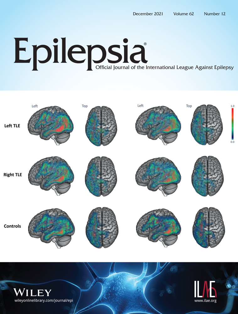
Abnormal functional connectivity profiles predict drug responsiveness in patients with temporal lobe epilepsy
Objective
This work was undertaken to study the functional connectivity differences between non-seizure-free and seizure-free patients with temporal lobe epilepsy (TLE) and to identify imaging predictors for drug responsiveness in TLE.
Methods
In this prospective study, 52 patients with TLE who presented undetermined antiseizure medication responsiveness and 55 demographically matched healthy controls were sequentially recruited from Xiangya Hospital. Functional magnetic resonance imaging data were acquired during a Chinese version of the verbal fluency task. The patients were followed up until the outcome could be classified. The subject groups were compared in terms of activation profile, task-residual functional connectivity (trFC), and generalized psychophysiological interaction (gPPI) analyses. Moreover, we extracted imaging characteristics for logistic regression and receiver operating characteristic evaluation.
Results
With a mean follow-up of 1.1 years, we identified 27 non-seizure-free patients and 19 seizure-free patients in the final analyses. The Chinese character verbal fluency task successfully activated the language network and cognitive control network (CCN) and deactivated the default mode network (DMN). In the non-seizure-freedom group, the trFC between the hippocampus and bilateral brain networks was attenuated (p < .05, familywise error corrected). For the gPPI analysis, group differences were mainly located in the precuneus, middle frontal gyrus, and inferior parietal lobule (p < .001, uncorrected; k ≥ 10). The regression model presented high accuracy when predicting non-seizure-free patients (area under the curve = .879, 95% confidence interval = .761–.998).
Significance
In patients with TLE who would not achieve seizure freedom with current antiseizure medications, the functional connectivity between the hippocampus and central nodes of the DMN, CCN, and language network was disrupted, leading to language decline. Independent of hippocampal sclerosis, abnormalities, especially the effective connectivity from the hippocampus to the DMN, were predictive biomarkers of drug responsiveness in patients with TLE.


留言 (0)